
System and method for quantifying pigmented lesion using oct

a technology of optical coherence tomography and pigmented lesion, which is applied in the field of system and method for quantifying pigmented lesion using optical coherence tomography, can solve the problems of difficult to remove perfectly pigmented lesion, severe thermal damage to the skin, and difficult to achieve perfect removal of pigmented lesion, etc., and achieve the effect of increasing the measurement rang

Active Publication Date: 2016-08-18
KOREA PHOTONICS TECH INST
View PDF4 Cites 0 Cited by
  • Summary
  • Abstract
  • Description
  • Claims
  • Application Information

AI Technical Summary

Benefits of technology

The patent is about a system and method for using OCT to calculate the size of a pigmented lesion. The invention increases the measurement range in the axial direction to which rays are irradiated. This leads to more accurate and reliable results when measuring the size of the pigmented lesion.

Problems solved by technology

A nanosecond pulsed laser, in the existing treatment process of pigmented lesions, may cause severe thermal damage to the skin due to its deep heat penetration.
This may cause thermal damage and complications, making it difficult to remove perfectly a pigmented lesion.
However, there is no existing diagnostic system that can be coordinated with the laser treatment technology, making it impossible to monitor a change of the lesion under treatment in real time.

Method used


Image

  • System and method for quantifying pigmented lesion using oct
  • System and method for quantifying pigmented lesion using oct
  • System and method for quantifying pigmented lesion using oct

Examples


Embodiment Construction

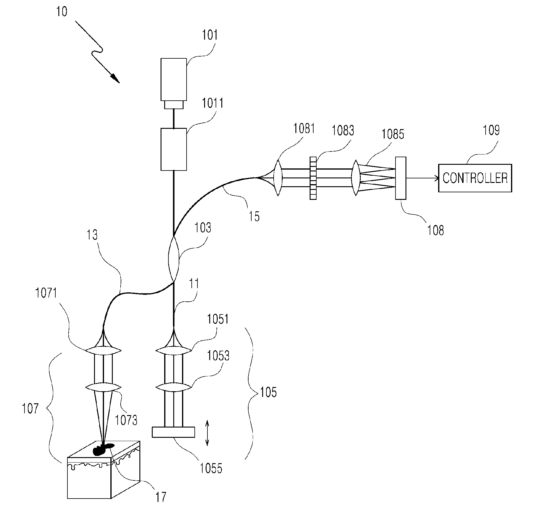
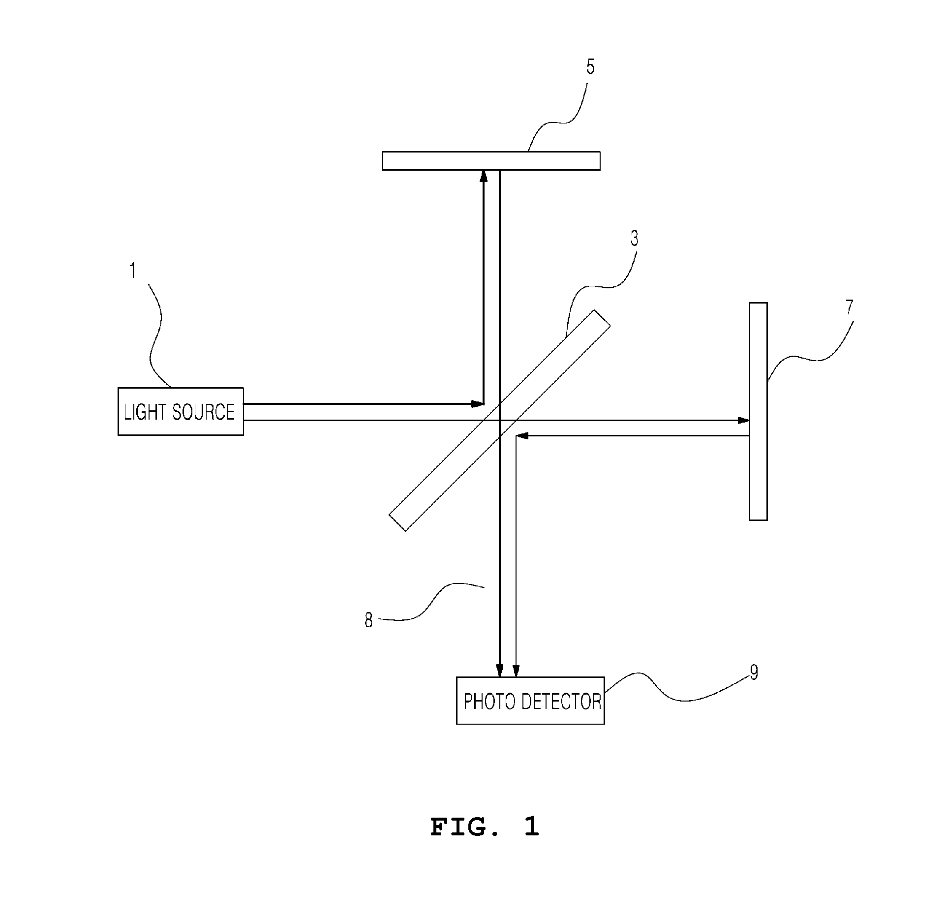
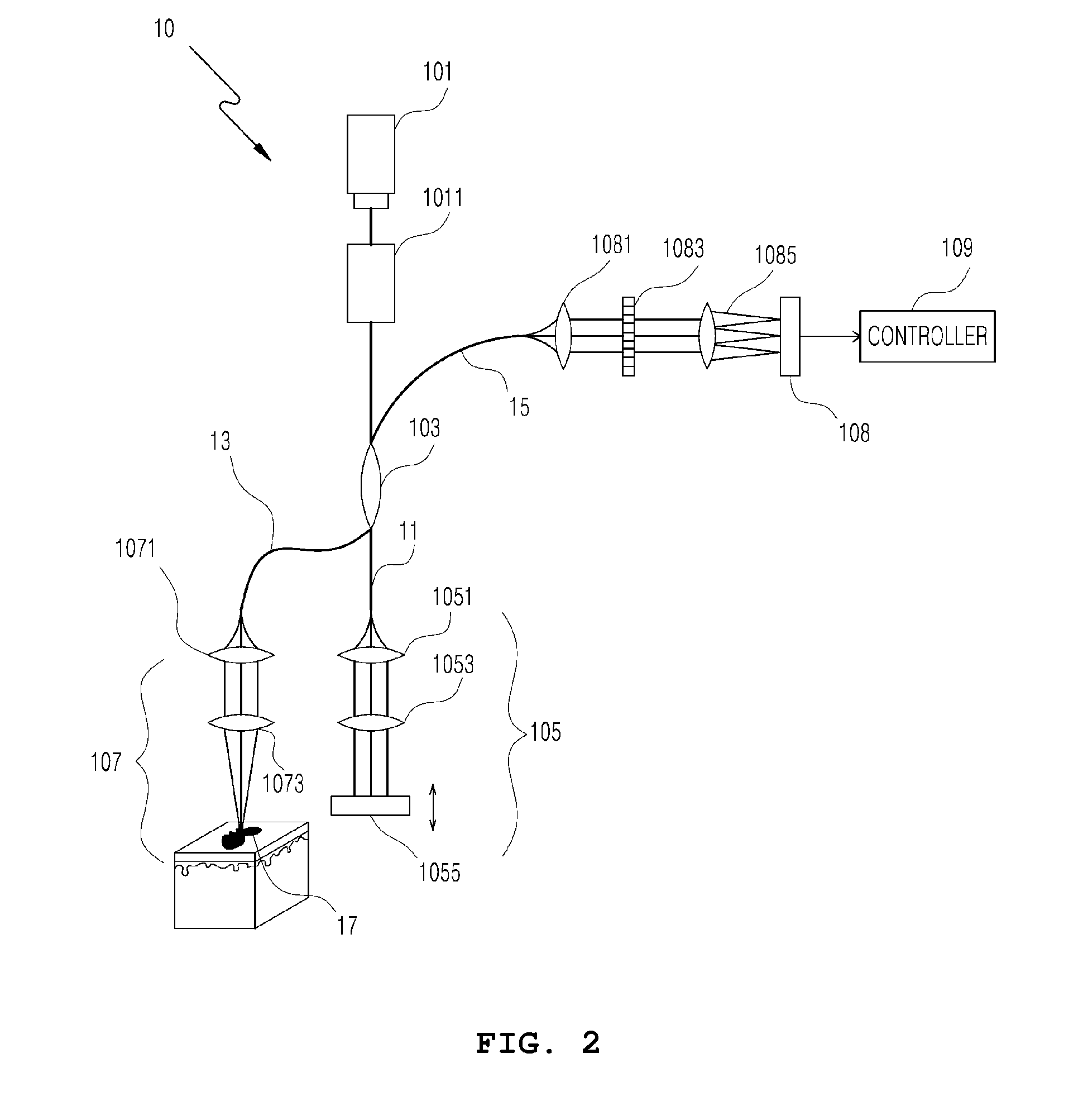
[0032]The present invention will be described in detail below with reference to the accompanying drawings. Repeated descriptions and descriptions of known functions and configurations that have been deemed to make the gist of the present invention unnecessarily obscure will be omitted below. The embodiments of the present invention are intended to fully describe the present invention to a person having ordinary knowledge in the art to which the present invention pertains. Accordingly, the shapes, sizes, etc. of components in the drawings may be exaggerated to make the description clearer.

[0033]FIG. 1 shows a Michelson interferometer for explaining a concept of an interference signal. Referring to FIG. 1, a Michelson interferometer may include a light source 1, a beam splitter 3, a dynamic mirror 5, a fixed mirror 7, and a photo detector 9. Light emitted from the light source 1 is vertically split by the beam splitter 3. The split beams are incident on the dynamic mirror 5 and fixed ...


PUM


Abstract

Disclosed herein is a method for quantifying a pigmented lesion using Optical Coherence Tomography (OCT). The method includes (a) irradiating light and receiving an interference signal produced by reflection of the light from first and second boundary layers of a pigmented lesion; and (b) calculating size information of the pigmented lesion using phase information of the interference signal. According to embodiments of the present invention, there is an advantage of allowing calculation of size information of a pigmented lesion using OCT, by increasing a measurement range in the axial direction to which beams are irradiated.

Description

BACKGROUND OF THE INVENTION[0001]1. Field of the Invention[0002]The present invention relates generally to a system and method for quantifying a pigmented lesion using optical coherence tomography (OCT) and, more particularly, to a system and method for quantifying the extent of decomposition of a pigmented lesion, which is difficult to observe with the naked eye, to allow monitoring of skin treatments.[0003]2. Description of the Related Art[0004]As well known to those skilled in the art, there are various forms of pigmented lesions appearing on the skin, such as moles, freckles, blemishes, warts, blotches, OTA nevus, tattoos, etc. Pigmented legions generally develop when prolonged exposure to ultraviolet radiation causes the skin to produce excessive pigment melanin or aggregate nevus cells that produce the pigment melanin. Tattooing is the practice of creating wounds in the skin and intentionally inserting color pigments into the wounds.[0005]In a medical context, tattoos may be d...

Claims


Application Information

Patent Timeline
IPC IPC(8): A61B5/00
CPCA61B5/4848A61B5/445A61B5/7257A61B5/0066A61B5/7278A61B5/444A61B5/441
Inventor LEE, BYEONG-ILSHIN, IN HEEEOM, JOO BEOMSONG, WOOSUB
Owner KOREA PHOTONICS TECH INST
Learn More