Devices and methods for delivering an endocardial device

a technology of endocardial devices and endocardial valves, which is applied in the field of medical/surgical devices and methods, can solve the problems of reducing the effectiveness of the heart, back-up of pressure in the vascular system behind the ventricle, and millions of hospital visits internationally, so as to reduce the total volume of the heart chamber, improve the blood ejection fraction thereof, and reduce the stress applied to the heart

Active Publication Date: 2016-10-20
EDWARDS LIFESCIENCES CORP
View PDF3 Cites 13 Cited by
  • Summary
  • Abstract
  • Description
  • Claims
  • Application Information

AI Technical Summary

Benefits of technology

[0016]The present invention relates to a ventricular partitioning device and a method of employing the device in the treatment of a patient with heart failure (HF). Embodiments of the device are adapted to span a chamber of the heart, typically the left ventricle, and partition the chamber into a main productive portion and a secondary non-productive portion. This partitioning reduces the total volume of the heart chamber, reduces the stress applied to the heart and, as a result, improves the blood ejection fraction thereof.
[0018]The ribs of the expandable frame have distal ends secured to the central hub and free proximal ends. The distal ends are preferably secured to the central hub to facilitate radial self expansion of the free proximal ends of the ribs away from a centerline axis. The distal ends of the ribs may be pivotally mounted to the hub and biased outwardly or fixed to the hub. The ribs may be formed of material such as superelastic NiTi alloy that permits compression of the free proximal ends of the ribs toward a centerline axis into a contracted configuration, and when released, allows for their self expansion to an expanded configuration.
[0019]The free proximal ends of the ribs are configured to engage and preferably penetrate the tissue lining a heart chamber, typically the left ventricle, to be partitioned so as to secure the peripheral edge of the partitioning component to the heart wall and to fix the partitioning component within the chamber so as to partition the chamber in a desired manner. The tissue-penetrating proximal tips are configured to penetrate the tissue lining at an angle approximately perpendicular to a centerline axis of the partitioning device. The tissue penetrating proximal tips of the ribs may be provided with attachments such as barbs or hooks that prevent withdrawal of the tips from the heart wall.
[0041]Described herein are systems, devices, and methods for partitioning a heart. The systems may include a partitioning device or implant, applicators for inserting, repositioning and / or removing them, and methods of positioning, deploying and removing them. The implants described herein are cardiac implants that may be inserted into a chamber of a patient's heart, particularly the left ventricle. The implant may support the heart wall, or in some variations the implant is a ventricular partitioning device for partitioning the ventricle into productive and non-productive regions, and / or for reducing the volume of the ventricle.
[0052]The systems described herein may also include a steering mechanism that bends the distal end region of the delivery catheter. The steering mechanism may include tendons or pull wires that pull one or more sides of the catheter to bend the catheter for steering. In some variations, described in greater detail below, the catheter is adapted to be steered by bending selectively in one or more directions. In some variations, the catheter includes hinge-points or cut-out regions that allow for column strength (allowing pushing / pulling of the catheter axially), while making the catheter flexible in one or more directions. The catheter may also be formed of multiple layers; for example, a guide catheter may include an outer catheter formed of a metal or other appropriate material providing column strength and having a lumen in which an inner catheter resides. The inner catheter may also include one or more lumen (e.g., an inflation lumen, a perfusion lumen, etc.). The catheter may also include a pullwire and / or a torque wire.

Problems solved by technology

Heart failure annually leads to millions of hospital visits internationally.
In certain pathological conditions, the ventricles of the heart become ineffective in pumping the blood, causing a back-up of pressure in the vascular system behind the ventricle.
The reduced effectiveness of the heart is usually due to an enlargement of the heart.
A myocardial ischemia may, for example, cause a portion of a myocardium of the heart to lose its ability to contract.
Once this tissue dies, it no longer functions as a muscle and cannot contribute to the pumping action of the heart.
As this situation worsens, the local area of compromised myocardium may in fact bulge out as the heart contracts, further decreasing the heart's ability to move blood forward.
As the heart begins to fail, diastolic pressures increase, which stretches the ventricular chamber prior to contraction and greatly increases the pressure in the heart.
CHF is a major cause of death and disability in the United States and elsewhere.
As a patient's heart enlarges, it pumps less efficiently and, in time, the heart becomes so enlarged that it cannot adequately supply blood to the body.
A congestive heart failure patient typically has an ejection fraction of 40% or less, and as a consequence, is chronically fatigued, physically disabled, and burdened with pain and discomfort.
Further, as the heart enlarges, heart valves lose the ability to close adequately.
An incompetent mitral valve allows regurgitation of blood from the left ventricle back into the left atrium, further reducing the heart's ability to pump blood.
Prolonged ischemia and occlusion of coronary arteries can result in myocardial tissue in the ventricular wall dying and becoming scar tissue.
As the disease progresses, a local area of compromised myocardium can bulge during the heart contractions, further decreasing the heart's ability to pump blood, and further reducing the ejection fraction.
Drug therapy typically treats the symptoms of the disease and may slow the progression of the disease, but it does not cure the disease.
Presently, the only treatment considered curative for heart failure is heart transplantation, but these procedures are high risk, invasive, and costly.
Further, there is a shortage of hearts available for transplant, many patients fail to meet transplant-recipient qualifying criteria.
As is the case with heart transplant, these procedures are invasive, risky, and costly, and many patients do not qualify medically for the procedure.
While these various procedures and devices have been found to be successful in providing some relief from heart failure symptoms and in slowing disease progression, none has been able to stop the course of the disease.
Delivery of a partitioning device may be made complicated by the anatomy of a patient and by aspects or characteristics of the delivery device or partitioning device itself.

Method used


Image

  • Devices and methods for delivering an endocardial device
  • Devices and methods for delivering an endocardial device
  • Devices and methods for delivering an endocardial device

Examples


first embodiment

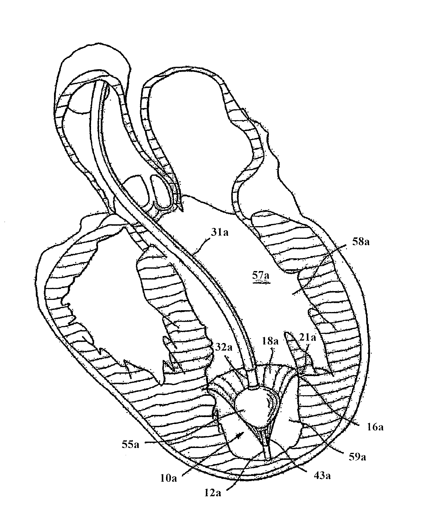
[0160]In some embodiments, as the guide catheter 31a, 31b is withdrawn, it begins to bend as it is withdrawn through the vascular anatomy of the patient, through the aortic arch, for example. In some instances, this bend may drive the distal tip of the delivery catheter, and therefore the partitioning device, out of position. For example, the guide catheter may drive the device towards the center of the heart, i.e. towards the ventricular septum. In some instances, it may be preferred that the delivery catheter and / or partitioning device are not moved or repositioned by the guide catheter as it is withdrawn. This may be accomplished in one of several embodiments. In a first embodiment, as shown in FIG. 10, a ring 60b is added to the distal end of the delivery catheter 32b. A wire 62b may be coupled to the ring 60b. The wire may be disposed along the length of the delivery and / or guide catheter, and may be configured to maintain the position of the distal end of the delivery catheter...

third embodiment

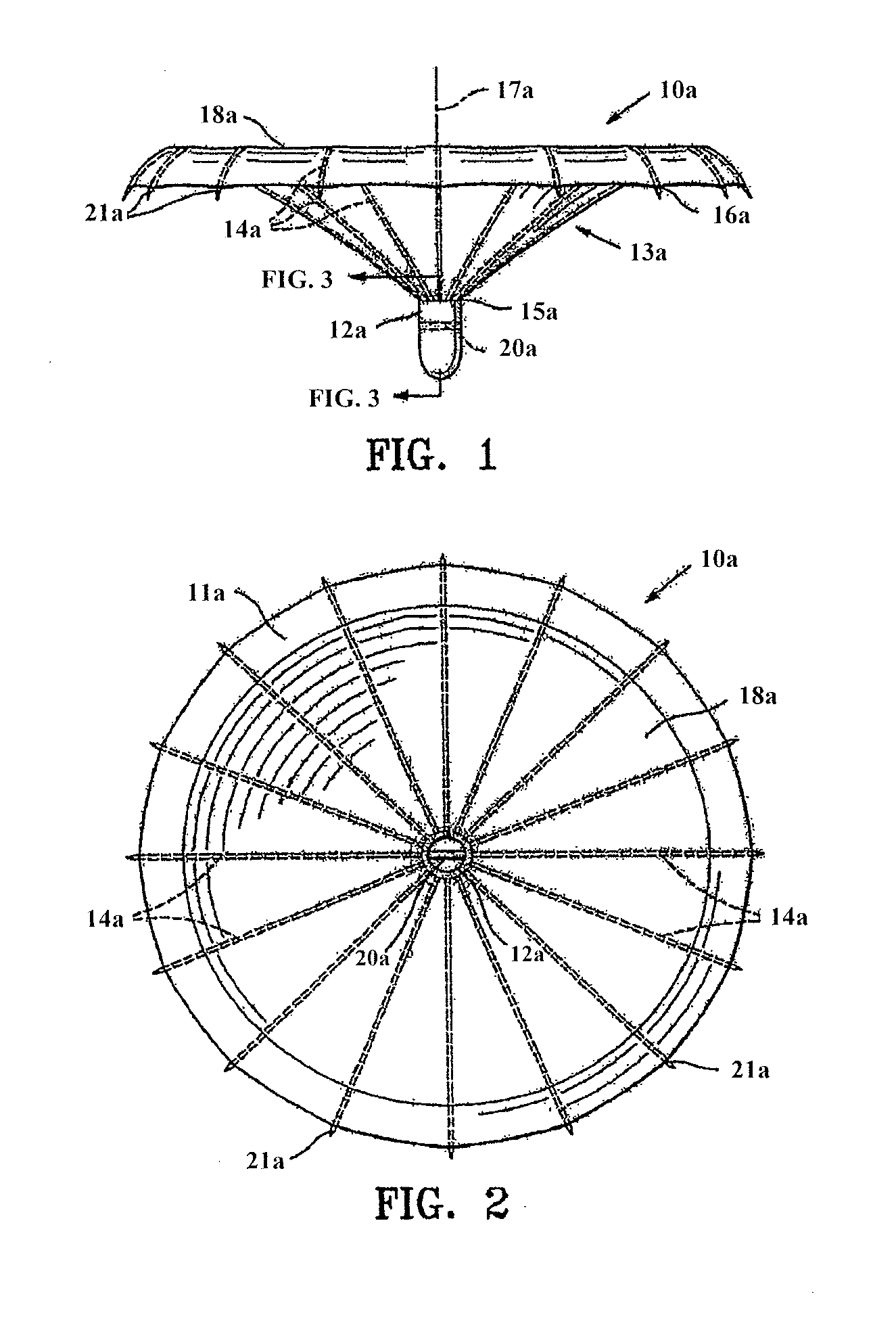
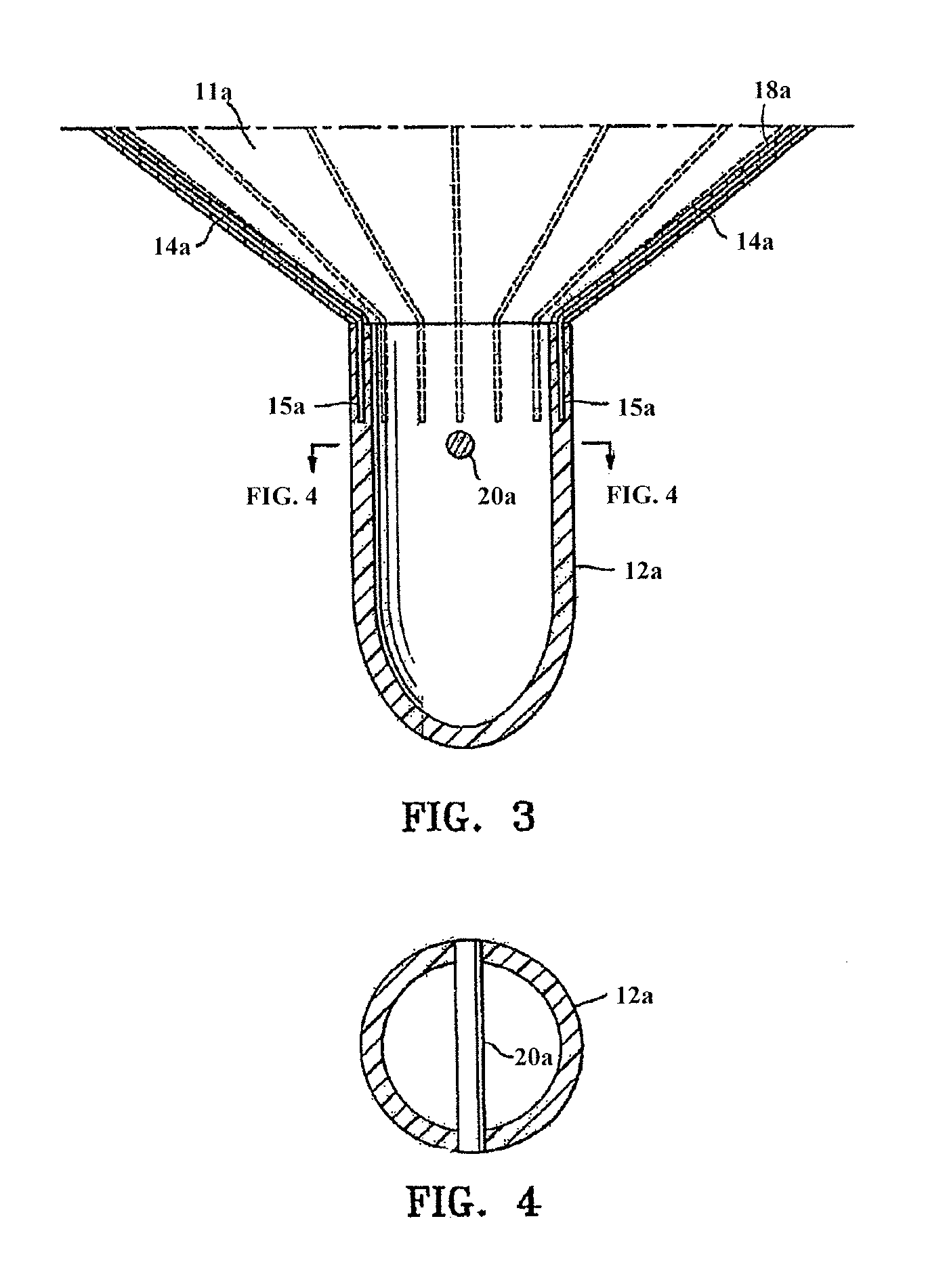
[0221]As described above, embodiments of the partitioning device 10a, both unilaminar and bilaminar embodiments, are conveniently formed by placing a thermoplastic tube 60, e.g. polyethylene or high density polyethylene (HDPE), over the ribs 14a of the frame 13a as shown in FIG. 62A until the proximal ends 16a of the ribs 14a extend out the ends of the thermoplastic tubes to form thermoplastic-encased ribs. FIGS. 62A-62C illustrate a first, second, and third embodiment showing the frame of the device described herein having sleeves, or more specifically thermoplastic tubes 60. As shown, the device may include full sleeves 60 disposed along the full length of the struts (FIG. 62A), partial sleeves 60′ staggered along the length of the struts (FIG. 62B), or shortened sleeves 60″ (FIG. 62C). As shown in FIG. 62B, by reducing the amount of tubing used, and by staggering the positioning of the tubing along the length of the struts 14a, the implants collapsed profile may be reduced. As sh...


PUM

PropertyMeasurementUnit
pressureaaaaaaaaaa
pressureaaaaaaaaaa
pressureaaaaaaaaaa

Abstract

Systems for partitioning a ventricle of a heart include a partitioning device or implant, and an applicator for inserting, repositioning and / or removing the partitioning device. The implant may support the ventricle wall and may reduce the volume of the ventricle. The delivery system for delivering and deploying a partitioning device into a ventricle may include a catheter having a distal coupling element for coupling to a partitioning device in a collapsed configuration; the catheter may also have an expansion member for applying force to the partitioning device to fully expand it into a deployed configuration and to secure or seal it against the ventricle wall.

Description

CROSS-REFERENCE TO RELATED APPLICATIONS[0001]This patent application claims priority as a continuation-in-part of U.S. patent application Ser. No. 13 / 827,927, filed on Mar. 14, 2013, now U.S. Patent Application Publication No. 2013-0270735, and as a continuation-in-part of U.S. patent application Ser. No. 13 / 828,184, filed Mar. 14, 2013, now U.S. Patent Application Publication No. 2013-0274595, both of which are continuation-in-parts of U.S. patent application Ser. No. 12 / 893,832, filed on Sep. 29, 2010, now U.S. Pat. No. 9,078,660, which is a continuation-in-part of U.S. patent application Ser. No. 11 / 860,438, filed on Sep. 24, 2007, now U.S. Pat. No. 7,897,086, which is a continuation-in-part of U.S. patent application Ser. No. 10 / 913,608, filed on Aug. 5, 2004, now abandoned. Each of these patent applications is herein incorporated by reference in their entirety.[0002]U.S. patent application Ser. No. 12 / 893,832 also claims priority as a continuation-in-part of U.S. patent applica...

Claims


Application Information

Patent Timeline
Patent Type & Authority Applications(United States)
IPC IPC(8): A61F2/24A61B90/00A61M25/09
CPCA61F2/2487A61B2090/3966A61B90/39A61M25/09A61B17/12022A61B17/12122A61B17/12172B29C43/18A61B2017/00243A61B2017/00867A61B2017/12054A61B2017/12095A61B2017/00526B29L2031/753B29C65/18B29C70/74B29C66/1122B29C66/532B29C66/53261B29C66/545B29C66/636B29C66/71B29C66/712B29C66/727B29C66/729B29C66/81429B29C66/81431B29C66/8322B29K2027/18B29K2023/06B29K2023/065
Inventor BOUTILLETTE, MICHAEL P.KERMODE, JAMES R.ALEXANDER, MILES D.KHAIRKHAHAN, ALEXANDERNIKOLIC, SERJAN D.
Owner EDWARDS LIFESCIENCES CORP
Why Eureka
Try Eureka