Systems and methods for in-operating-theatre imaging of fresh tissue resected during surgery for pathology assessment

a fresh tissue and pathology technology, applied in the field of systems and methods, can solve the problems of no tumor cells left behind, no life-saving treatment opportunities, and both patient death rate and overall treatment cost can dramatically increase, so as to facilitate in-operating-theater analysis of tissue samples, the time necessary for preparing and analyzing samples is reduced.

Active Publication Date: 2016-10-20
SAMANTREE MEDICAL SA
View PDF0 Cites 11 Cited by
  • Summary
  • Abstract
  • Description
  • Claims
  • Application Information

AI Technical Summary

Benefits of technology

[0010]The disclosed technology brings histopathology into the operating theatre, to enable real-time intra-operative digital pathology. The disclosed technology utilizes a confocal imaging device that can analyze, in the operating theatre, “optical slices” of fresh ti

Problems solved by technology

Current surgical technology cannot precisely define the margins of a tumor, resulting in missed opportunities for life-saving treatment.
Tumor residues may remain undetected and untreated until they grow to advanced stages, at which point both patient death rate and overall treatment costs can dramatically increase.
However, a complete resection requires “tumor-free margins” of the resected tissue, meaning that no tumor cells are left behind in the patient.
Currently, there is no reliable means available to guide the resection of solid tumors.
For certain indications, this results in the need to re-operate on many patients days after the initial operation when an analysis is obtained from the histo-pathology laboratory.
Such follow-up surgical procedures usually lead to less favorable outcomes for the patient, psychological stress, and can roughly double treatment and hospitalization costs.

Method used


Image

  • Systems and methods for in-operating-theatre imaging of fresh tissue resected during surgery for pathology assessment
  • Systems and methods for in-operating-theatre imaging of fresh tissue resected during surgery for pathology assessment
  • Systems and methods for in-operating-theatre imaging of fresh tissue resected during surgery for pathology assessment

Examples


Embodiment Construction

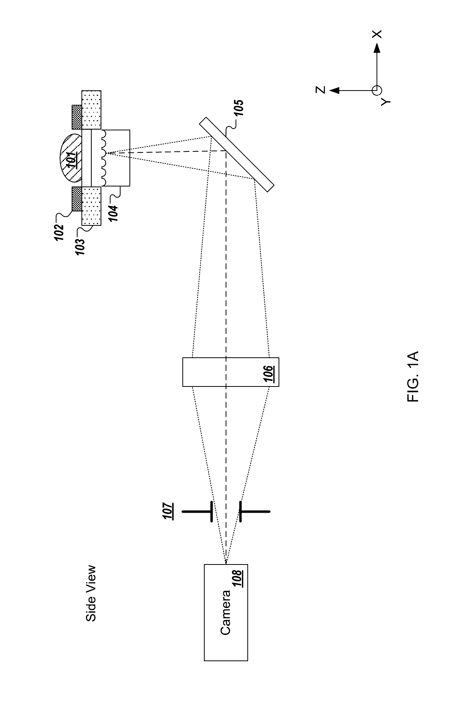
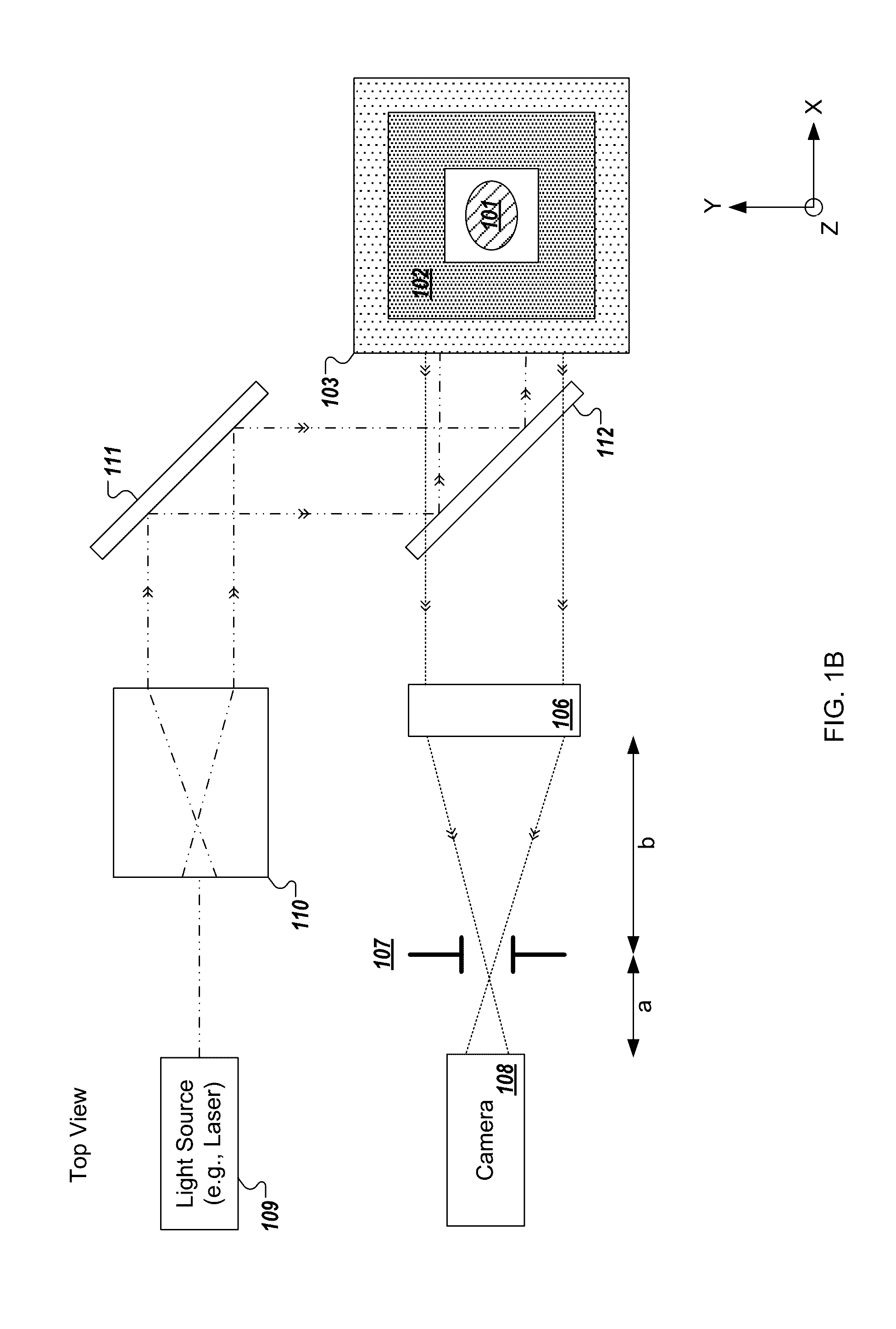
[0240]In the present text the expression “micro optical element” is used to describe a miniaturized focusing element with a cross sectional diameter of less than 1 mm (e.g., between 10 micrometers and 500 micrometers) that focuses light. In some implementations, the micro optical element is a micro lens having a paraxial radius of curvature that is in the order of magnitude of its diameter. In some implementations, the micro optical element is a refractive lens, Fresnel zone plate, GRIN lens, or micro reflective objective. The term “micro optical element array” is used to describe a structure composed of a plurality of micro optical elements positioned in a grid which may be, but is not necessarily, periodic. While the description may describe embodiments of the disclosed technology implemented with a micro lens array, similar embodiments may be implemented with micro optical elements.

[0241]The expression “fresh tissue” is generally used herein to describe tissue resected or otherwi...


PUM


Abstract

The disclosed technology brings histopathology into the operating theatre, to enable real-time intra-operative digital pathology. The disclosed technology utilizes confocal imaging devices image, in the operating theatre, “optical slices” of fresh tissue—without the need to physically slice and otherwise process the resected tissue as required by frozen section analysis (FSA). The disclosed technology, in certain embodiments, includes a simple, operating-table-side digital histology scanner, with the capability of rapidly scanning all outer margins of a tissue sample (e.g., resection lump, removed tissue mass). Using point-scanning microscopy technology, the disclosed technology, in certain embodiments, precisely scans a thin “optical section” of the resected tissue, and sends the digital image to a pathologist rather than the real tissue, thereby providing the pathologist with the opportunity to analyze the tissue intra-operatively. Thus, the disclosed technology provides digital images with similar information content as FSA, but faster and without destroying the tissue sample itself.

Description

RELATED APPLICATIONS[0001]This application claims priority to U.S. Provisional Patent Application Ser. No. 62 / 141,223, entitled Systems and Methods for In-Operating-Theatre Imaging of Fresh Tissue Resected During Surgery for Pathology Assessment by Rachet et al., the content of which is incorporated herein by reference in its entirety.BACKGROUND OF THE INVENTION[0002]Solid epithelial cancers account for over 10% of all deaths each year world-wide. This figure is expected to increase. Early-stage cancer diagnosis and subsequent complete surgical removal of the tumor offers the best chance for complete cancer cure. With early-stage tumor screening and diagnostics becoming more efficient, the bottleneck lies in efficient surgical management. Current surgical technology cannot precisely define the margins of a tumor, resulting in missed opportunities for life-saving treatment. Tumor residues may remain undetected and untreated until they grow to advanced stages, at which point both pati...

Claims


Application Information

Patent Timeline
IPC IPC(8): G01N33/483G01N21/64G02B21/16
CPCG01N33/4833G02B21/16G01N21/6458G01N2201/068G01N2021/6463G01N2021/6439G01N2201/0633G01N21/6428G02B21/0012G02B21/0032G02B21/004G02B21/0076G02B21/0088G02B21/26G02B21/34G02B21/361G02B21/0044G02B21/24G01N21/77G01N2021/7786
Inventor RACHET, BASTIENKOSANIC, DAVORSHAFFER, ETIENNE
Owner SAMANTREE MEDICAL SA
Why Eureka
Try Eureka