
Biomarker assay using microparticle aggregation

Inactive Publication Date: 2016-11-17
THE UNIVERSITY OF AKRON
View PDF3 Cites 2 Cited by
  • Summary
  • Abstract
  • Description
  • Claims
  • Application Information

AI Technical Summary

Benefits of technology

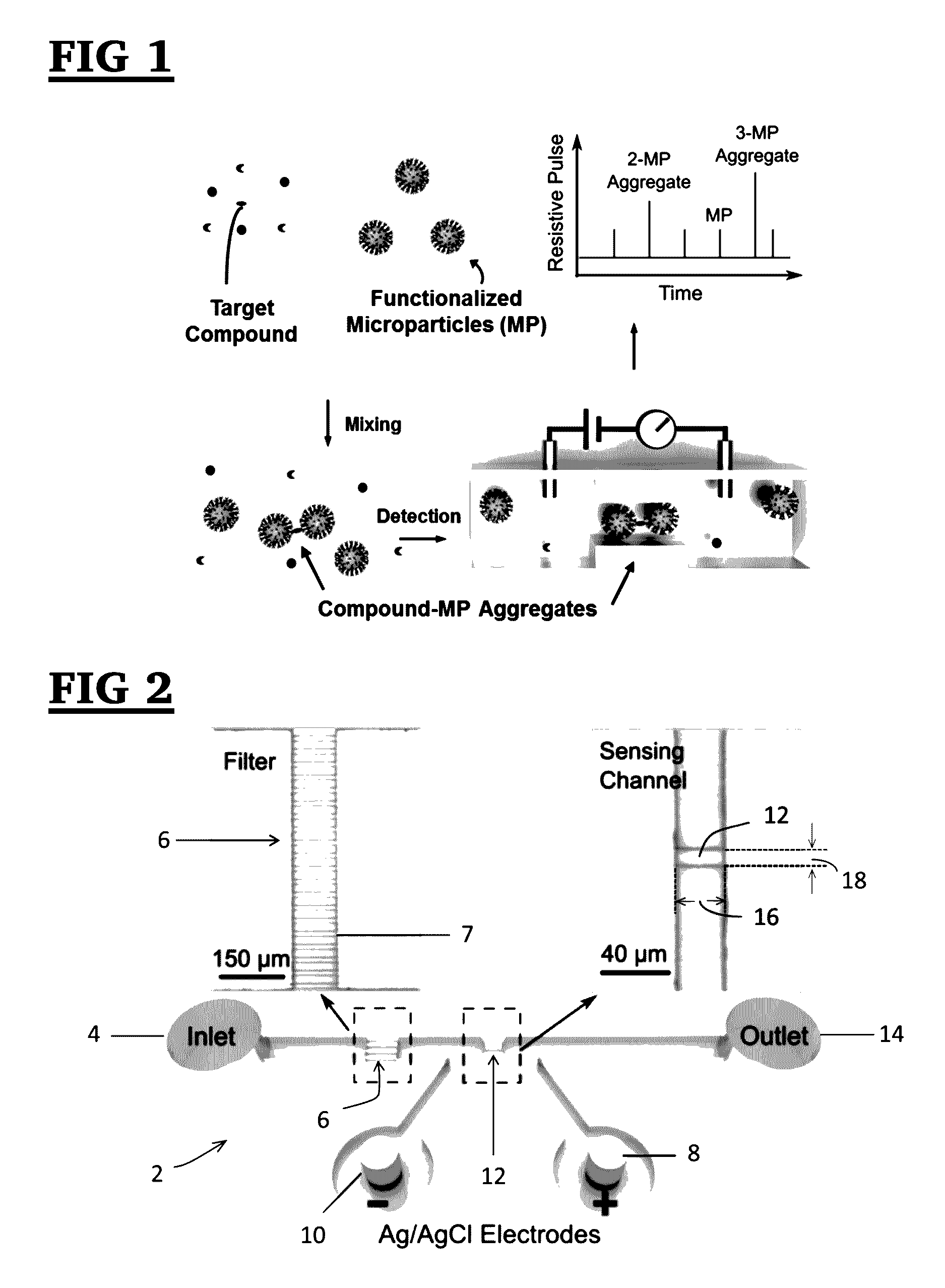
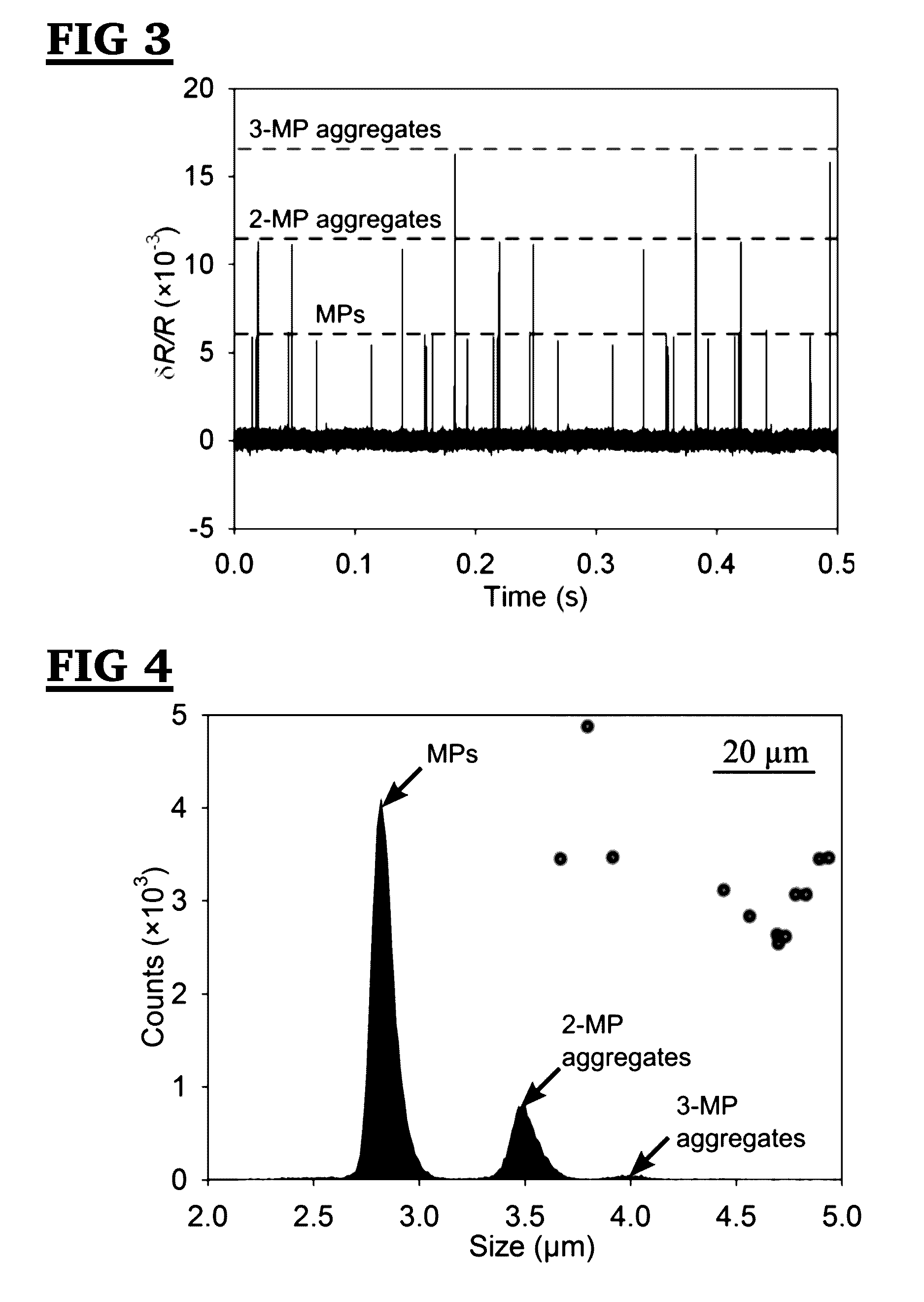
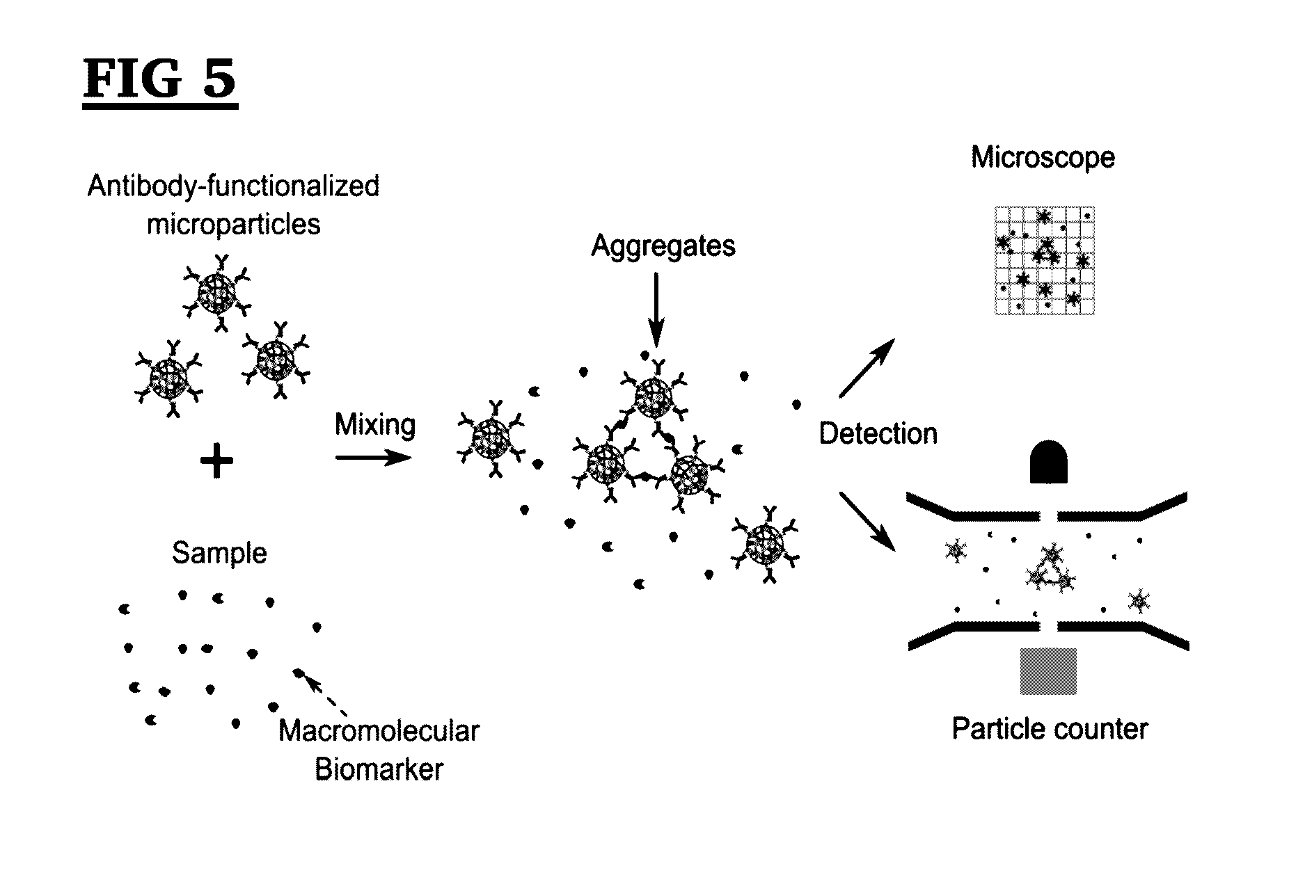
The present invention is a method for detecting biomarkers and other compounds in a fluid sample using functionalized microparticle aggregates. The method is sensitive, cost-effective, versatile, and label-free. It can be adapted for detecting any biomacromolecules or other compounds as long as there are high affinity capture probes available. The detection range of biomarkers can be accurately tuned by changing the concentration of functionalized microparticles. Compared to conventional methods, the present invention is faster, more sensitive, and requires no complex measurement setup and sample preparations.

Problems solved by technology

However, conventional immunoassays such as enzyme-linked immunosorbent assay (ELISA), require labelling antibodies, long assay time, and bulky, complicated detection instruments.
Most of these methods require labelling of a detection probe or optical detection or the modifications of sensing surfaces, and typically employ bulky, expensive and complicated detection instruments.
However, these methods are usually preformed in nanoscale sensing channels, which is problematic because: (i) fabrication of nanoscale sensing channels has been found to be very difficult, requiring an expensive and complex nanofabrication facility; and (ii) nanoscale devices have a very low throughput, i.e. each nanoscale channel can only handle a very small amount of sample at a time.
None of these methods are of much use for laboratories and clinics lacking immediate access of analytical instruments.

Method used


Image

  • Biomarker assay using microparticle aggregation
  • Biomarker assay using microparticle aggregation
  • Biomarker assay using microparticle aggregation

Examples


example 1

Resistive Pulse Sensor Fabrication and Testing Procedure

[0118]The resistive pulse sensor was fabricated using the standard soft lithography method. It consists of 1) a sensing channel with a width of 10 μm and a length of 30 μm to detect aggregates; 2) an on-chip filter with a pore width of 10 μm; and 3) a pair of Ag / AgCl electrodes to measure the resistive pulses. A two-layer SU8 mold, consisting of patterns for the sensing channel and the filter (with a thickness of 10 μm) and patterns for reservoirs (with a thickness of 40 μm), was created by a two-step photolithography process (See, FIG. 20). Microchannel, filters and reservoirs were formed by pouring polydimethylsiloxane (PDMS) onto the two-layer SU8 mold followed by degassing and curing. The filter blocks background particles larger than the sensing channel to ensure a continuous detection. This two-layer structure offers a higher sensitivity of the sensing channel without increasing the flow resistance of reservoirs.

[0119]Nex...

example 2

Sample Preparation

[0121]To prepare antibody-functionalized MPs, firstly, streptavidin functionalized magnetic MPs with an average diameter of 2.80 μm (dynabeads M280, Life Technologies, USA) were diluted 1 / 65 in phosphate buffer saline (PBS, pH 7.4, Sigma-Aldrich, USA) containing 0.1% bovine serum albumin (BSA, Sigma-Aldrich, USA). The biotinylated goat anti-human ferritin antibody (anti-ferritin Ab, 6.5 mg / ml, US Biological, USA) was diluted 1 / 780 in PBS with 0.1% BSA. Next, 166.7 μl of diluted M280 MP solution was mixed with 166.7 μl diluted anti-ferritin Ab solution for 30 min in a thermal mixer at a speed of 650 rpm at room temperature. M280 MPs conjugated with biotinylated anti-ferritin Ab through the streptavidin-biotin binding. It has been found that further increasing the incubation time has little effect on the volume fraction of aggregates. The MP solution was then placed on a magnet to separate MPs from the solution; next the supernatant containing unconjugated anti-ferri...

example 3

Materials and Methods for Analysis of Sample

[0123]Streptavidin-functionalized Microparticle (MP) (Dynabeads M-280 with a diameter of 2.8 μm), biotinylated polyclonal rabbit anti-goat IgG (rAb) antibodies and goat anti-rabbit IgG (goat IgG) antibodies (labeled with Alexa Fluor 488) were bought from Life Technologies (Carlsbad, Calif., USA). Goat anti-human ferritin polyclonal antibody (gAb) and human ferritin were purchased from United States Biological (Salem, Mass., USA). NHS-Fluorescein, NHS-PEG4-Biotinyltion and Zeba spin desalting column were purchased from Thermo Scientific (Waltham, Mass., USA). Dimethyl sulfoxide (HPLC grade) was bought from Alfa Aesar (USA). Phosphate buffer saline (PBS, pH 7.4), and bovine serum albumin (BSA) were obtained from Sigma-Aldrich (St Louis, Mo., USA).

[0124]To prepare the immunoaggregation sample, MP and biotinylated rAb were diluted to 0.16 mg / mL and 6.4 ng / mL separately in PBS containing 0.1% BSA. Equal volumes of 166.7 μL of diluted MP solutio...


PUM


Abstract

In various aspects and embodiments, the present invention is directed to versatile, label-free method for the quantitative and qualitative detection of biomarkers and / or other compounds in a fluid sample using functionalized microparticle aggregates. In these methods, micron-scale particles are functionalized to specifically interact with the biomarker being measured and added to the sample to form aggregates, the size and number of which are counted to find a volume fraction and / or number fraction of aggregates in the sample. There is a direct correlation between the volume fraction and number fraction of these aggregates and the concentration of the corresponding biomarker. By comparing the measured volume fraction and / or number fraction of aggregates in the sample to a calibration curve, the concentration of that biomarker may be determined even for biomarkers or other target compounds in samples at very low concentrations, without the need for fluorescence and enzyme labelling of antibodies.

Description

CROSS REFERENCE TO RELATED APPLICATIONS[0001]This application claims the benefit of U.S. provisional patent application Ser. No. 62 / 160,014 entitled “Label-free Biomarker Assay in a Micro Resistive Pulse Sensor via Immunoaggregation,” filed May 12, 2015, and incorporated herein by reference in its entirety.REFERENCE TO GOVERNMENT SUPPORT[0002]The invention was developed at least in part with the support of National Science Foundation grant number CMMI-1129727. The government may have certain rights in the invention.FIELD OF THE INVENTION[0003]One or more embodiments of the present invention relates to a method for detecting and / or quantifying biomarkers, other compounds or microoganisms in a fluid. In certain embodiments, the present invention relates to methods for measuring the concentration of one or more biomarkers and / or other target molecule using aggregation of functionalized microparticles.BACKGROUND OF THE INVENTION[0004]Biomarker detection represents an important task for ...

Claims


Application Information

Patent Timeline
IPC IPC(8): G01N33/543G01N15/06G01N15/02
CPCG01N33/54326G01N15/0227G01N2015/0065G01N2015/0092G01N15/0656G01N15/0266G01N2015/0681G01N2015/1006G01N2015/1486G01N2015/1493G01N15/1459G01N15/1031G01N15/1484G01N33/543G01N33/54313G01N15/1023G01N15/01G01N15/075G01N15/1433
Inventor CHENG, GANGZHE, JIANGHAN, YUWU, HAIYAN
Owner THE UNIVERSITY OF AKRON
Learn More