Eye disease treatment agent, screening method therefor, and method for predicting rejection response associated with retinal pigment epithelial cell transplant

Active Publication Date: 2017-01-12
HEALIOS KK
View PDF1 Cites 1 Cited by
  • Summary
  • Abstract
  • Description
  • Claims
  • Application Information

AI Technical Summary

Benefits of technology

The present invention allows for the use of RPE cells that have a specific genotype for patients with ophthalmic diseases. By matching only one of the three gene loci of the RPE cells with those of the patient, rejection responses can be reduced, and in compatible cells can be distinguished and selected. This method also helps in determining whether immunosuppressants are necessary for treatment. Overall, this invention improves treatment efficiency and reduces preparation costs.

Problems solved by technology

However, an allogeneic transplantation strategy in consideration of HLA compatibility has not existed to date in the transplantation to retina disease patients by using a retinal pigment epithelial cell (hereinafter to be indicated as “RPE cell”).

Method used


Image

  • Eye disease treatment agent, screening method therefor, and method for predicting rejection response associated with retinal pigment epithelial cell transplant
  • Eye disease treatment agent, screening method therefor, and method for predicting rejection response associated with retinal pigment epithelial cell transplant
  • Eye disease treatment agent, screening method therefor, and method for predicting rejection response associated with retinal pigment epithelial cell transplant

Examples


preparation example 1

Preparation of Human iPS Cell-Derived RPE Cells

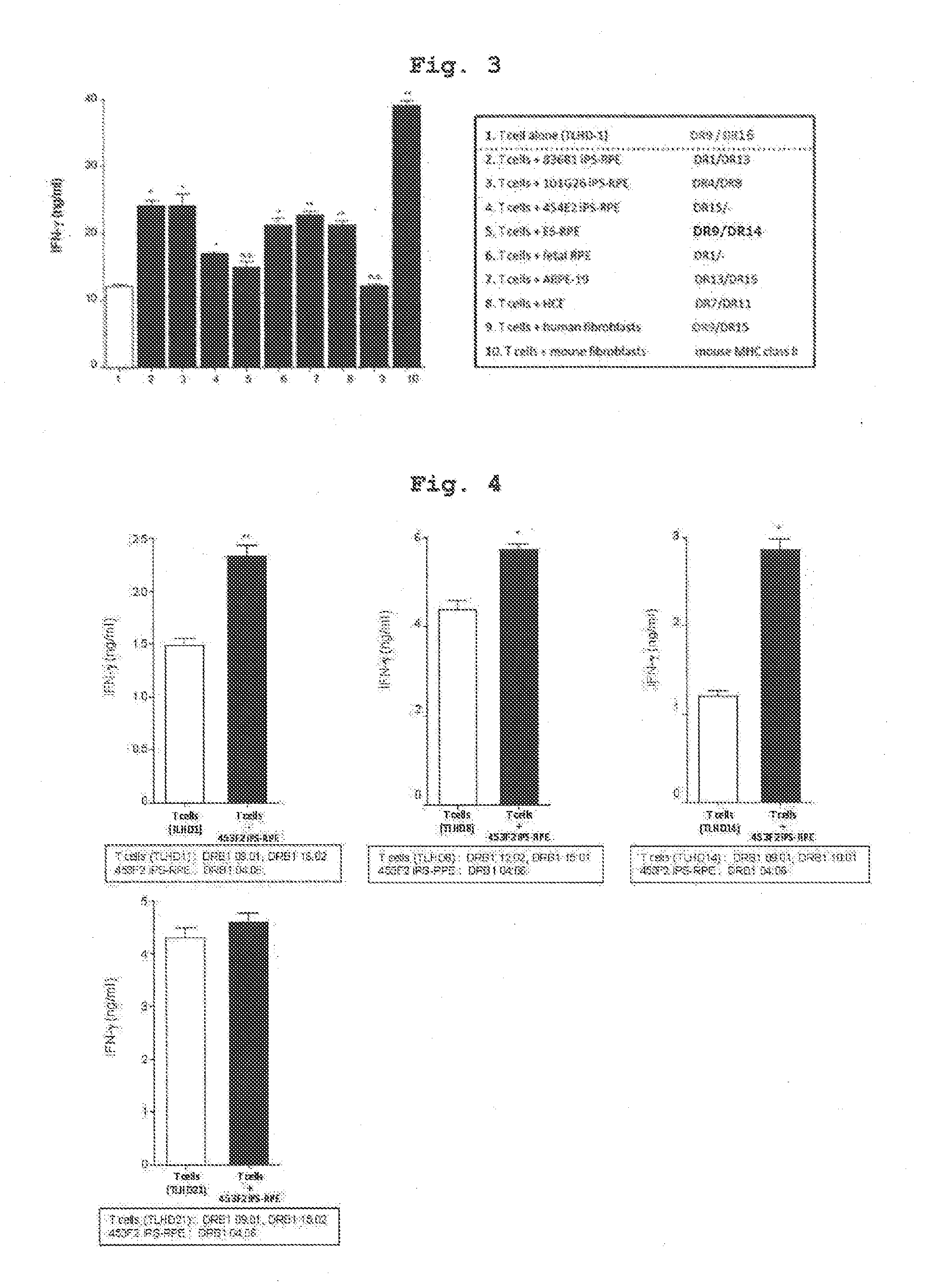
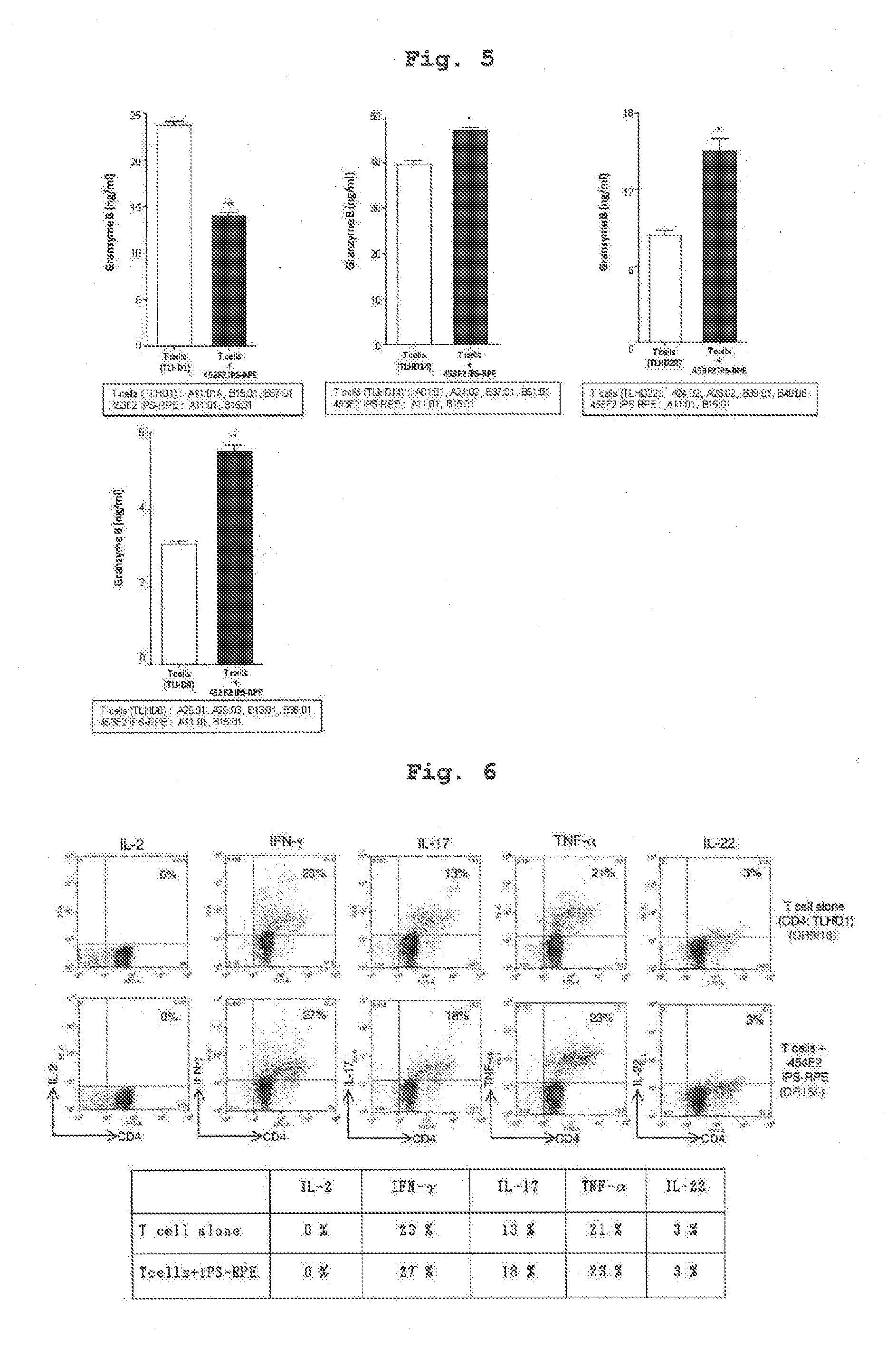
[0104]Using IPS cell line 454E2 or 453F2 (provided by Kyoto University) disclosed in Nature Methods 8, 409-412 2011 as iPS cells, and according to of the differentiation induction method described in J Cell Sci (2009)122: 3169-3179, 454E2-iPS cell-derived RPE cells and 453F2-iPS cell-derived RPE cells (both cells have homozygous genotype at HLA-A, -B, -DR3 gene loci) were prepared.

[0105]In addition, using, as iPS cell line, iPS cells established from human skin-derived fibroblast of a test subject name TLHD-1 according to the method described in Nature (2011) 474: 225-229 instead of 454E2 or 453F2, TLHD-1-iPS cell-derived RPE cells were prepared by a similar method.

preparation example 2

Preparation of Human T Cell Suspension

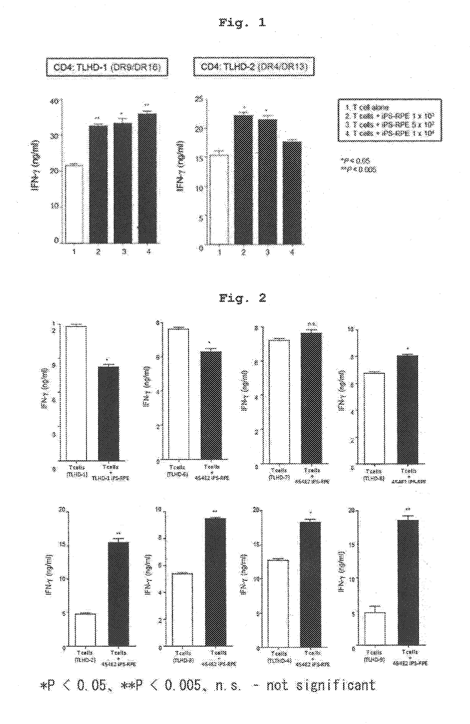
[0106]CD4 positive T cells and CD8 positive T cells were separated from human peripheral blood (test subject names; TLHD-1-TLHD-22) by using a MACS magnetic cell sorter, suspended in 100U / ml rhIL-2-containing RPMI culture medium (RPMI 1640 445 ml, FBS 50 ml, penicillin-streptomycin 5 ml) to prepare each cell suspension.

(Reference 1) HLA Typing

[0107]The results of HLA typing of the cells used in Examples 1-8 by a conventional method are shown in the following Table. The results of HLA typing of respective T cells of test subjects obtained in Preparation Example 2 are shown in Tables 1-1, 1-2, and the results of HLA typing of the human iPS-derived RPE cells of Preparation Example 1 and those induced from other iPS cells are shown in Table 2.

[0108]Relative to 454E2-iPS cell-derived RPE cells (homozygous at 3 gene loci of HLA-A, HLA-B, HLA-DR), the test subject names TLHD-10 and TLHD-21 had the same alleles in the genotype of 3 gene loci of HLA-A, H...

preparation example 3

Preparation of Monkey iPS Cell-Derived RPE Cells

[0110]iPS cells were established from the fibroblast derived from the skin of Macaca fascicularis according to the method described in Nature (2011) 474: 225-229 (GLISluse), and monkey iPS cell-derived RPE cells were prepared according to the differentiation induction method described in J Cell Sci (2009)122: 3169-3179 from each of normal monkeys (A, B) and a disease model monkey (C) prepared by forming a retinal photocoagulation spot in one eye by retinal photocoagulation procedure.


PUM


Abstract

The present invention provides an agent for treating ophthalmic diseases and a screening method for an agent for treating ophthalmic diseases and the like. The present invention also provides a method for predicting rejection associated with transplantation of retinal pigment epithelial cell to patients with ophthalmic diseases.

Description

TECHNICAL FIELD[0001]The present invention relates to an agent for treating ophthalmic diseases, a screening method therefor and a method for predicting a rejection response associated with retinal pigment epithelial cell transplantation.BACKGROUND ART[0002]Major histocompatibility complex (MHC) is known as HLA (human leukocyte antigen) in human and is expressed in almost all cells and tissues. HLA mainly contains 6 gene loci antigens of A, B, C, DR, DQ and DP, each of which is constituted of complicated combinations of several dozen kinds of different types (allele) in tens of thousands of combinations. HLA is responsible for an important immune mechanism in the human body and the main role thereof is presentation of antigen to recognize self or non-self. When a person receives transplantation of a cell or tissue derived from other person (allogeneic transplantation), HLA is recognized as the most important antigen (foreign substance) by immunocompetent cells such as T cells and th...

Claims


Application Information

Patent Timeline
IPC IPC(8): C12Q1/68G01N33/68A61L27/36
CPCC12Q1/6883A61L27/3666A61L27/3604G01N33/6866G01N33/6863G01N2500/10C12Q2600/136C12Q2600/156G01N2333/57G01N2333/96433G01N2800/245A61L2430/16A61K35/30G01N33/6893A61P27/02A61P37/06A61P43/00
Inventor TAKAHASHI, MASAYOSUGITA, SUNAO
Owner HEALIOS KK
Why Eureka
Try Eureka