Automated segmentation of organ chambers using deep learning methods from medical imaging

a technology of deep learning and organ chambers, applied in image analysis, image enhancement, instruments, etc., can solve the problems of inhomogeneity of intensity, dynamic motion, time-consuming and laborious manual segmentation, etc., and achieve the effect of reducing misalignmen

Active Publication Date: 2017-04-20
RGT UNIV OF CALIFORNIA
View PDF4 Cites 99 Cited by
  • Summary
  • Abstract
  • Description
  • Claims
  • Application Information

AI Technical Summary

Benefits of technology

[0057]align stack images of the body chamber by performing conto...

Problems solved by technology

However, manual segmentation is tedious, time consuming and prone to intra- and inter-observer variability (Frangi, A. F. et al.
To summarize, there are several challenges in the automated LV segmentation from cardiac MRI datasets: heterogeneities in the brightness of LV cavity due to blood flow; presence of papillary muscles with signal intensities similar to that of the myocardium; complexity in segmenting the apical and basal slice images; partial volume effects in apical slices due to the limited resolution of cardiac MRI; inherent noise associated with cine cardiac MRI; dynamic motion of the heart and inhomogeneity of intensity; considerable variability in shape and intensity of the heart chambers across patients, notably in pathological cases, etc.
Due to these technical barriers, the task of automatic segmentation of the heart chambers from cardiac MRI is still a challenging problem (Petitjean, C. and Dacher, J.-N.
Pixel classification, image-based and deformable methods suffer from a low robustness and accuracy and require extensive user interaction (Petitjean, C. and Dacher, J.-N.
However, it is very difficult to build a model that is general enough to cover all possible shapes and dynamics of the heart chambers (Petitjean, C. and Dacher, J.-N.
Small datasets lead to a large bias in the segmentation, which makes these methods inefficient when the heart shape is outside the learning set (e.g., congenital heart defects, post-surgical remodeling, etc.).
Furthermore, current learning-based approaches for LV segmentation have certain limitations.
These methods employ multiple stages of intensity standardization, estimation and normalization, which are computationally expensive and affect the success of further steps.
As such, their performance is rather medio...

Method used


Image

  • Automated segmentation of organ chambers using deep learning methods from medical imaging
  • Automated segmentation of organ chambers using deep learning methods from medical imaging
  • Automated segmentation of organ chambers using deep learning methods from medical imaging

Examples


example 1

A Combined Deep-Learning and Deformable-Model Approach to Fully Automatic Segmentation of the Left Ventricle in Cardiac MRI

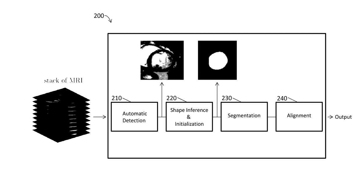
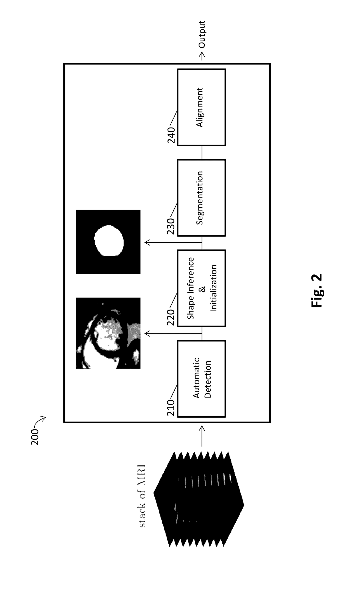
[0112]As reported in Avendi et al. 2016 Medical Image Analysis 30: 108-119, segmentation of the left ventricle (LV) from cardiac magnetic resonance imaging (MRI) datasets is an essential step for calculation of clinical indices such as ventricular volume and ejection fraction. In this work, we employ deep learning algorithms combined with deformable models to develop and evaluate a fully automatic LV segmentation tool from short-axis cardiac MRI datasets. The method employs deep learning algorithms to learn the segmentation task from the ground true data. Convolutional networks are employed to automatically detect the LV chamber in MRI dataset. Stacked autoencoders are used to infer the LV shape. The inferred shape is incorporated into deformable models to improve the accuracy and robustness of the segmentation. We validated our method using 45 cardiac MR datase...

example 2

Automatic Segmentation of the Right Ventricle from Cardiac MRI Using a Learning-Based Approach

[0158]This study accurately segments the right ventricle (RV) from cardiac MRI using a fully automatic learning-based method. The method utilizes deep learning algorithms, i.e., convolutional neural networks and stacked autoencoders, for automatic detection and initial segmentation of the RV chamber. The initial segmentation is then combined with the deformable models to improve the accuracy and robustness of the process. We trained our algorithm using 16 cardiac MRI datasets of the MICCAI 2012 R V Segmentation Challenge database and validated our technique using the rest of the dataset (32 subjects).

[0159]Results: An average Dice metric of 82.5% along with an average Hausdorff distance of 7.85 mm were achieved for all the studied subjects. Furthermore, a high correlation and level of agreement with the ground truth contours for end-diastolic volume (0.98), endsystolic volume (0.99) and eje...


PUM


Abstract

Systems and methods are disclosed for automatically segmenting a heart chamber from medical images of a patient. The system may include one or more hardware processors configured to: obtain image data including at least a representation of the patient's heart; obtain a region of interest from the image data; organize the region of interest into an input vector; apply the input vector through a trained graph; obtain an output vector representing a refined region of interest corresponding to the heart based on the application of the input vector through the trained graph; apply a deformable model on the obtained output vector representing the refined region of interest; and identify a segment of a heart chamber from the application of the deformable model on the obtained output vector.

Description

BACKGROUND[0001]Field[0002]The invention relates to image segmentation devices and related methods of automatically segmenting organ chambers using deep learning methods to compute clinical indices from imaging data.[0003]Description of the Related Art[0004]Cardiac magnetic resonance imaging (MRI) is now routinely being used for the evaluation of the function and structure of the cardiovascular system (Yuan, C. et al. 2002 J Magn Reson Imaging 15(1): 62-67; Lima, J. C. and Desai, M. Y. 2004. J Am Coll Cardiol 44(1): 1164-1171; Frangi, A. F. et al. 2001 IEEE Trans Med Imag 20(1): 2-5; Petitjean, C. and Dacher, J.-N. 2011 Med Image Anal 15(2): 169-184; Tavakoli, V. and Amini, A. A. 2013 Comput Vis Image Underst 117(9); 966-989; Heimann, T. and Meinzer, H.-P. 2009 Med Image Anal 13(4): 543-563; Suinesiaputra, A. et al. 2014 Med Image Anal 18(1): 50-62). Segmentation of the left ventricle (LV) from cardiac MRI datasets is an essential step for calculation of clinical indices such as ven...

Claims


Application Information

Patent Timeline
IPC IPC(8): G06T7/00G06T3/00G06V10/764
CPCG06T7/0012G06T7/0081G06T7/0089G06T7/0038G06T2207/20124G06T2207/20081G06T2207/10088G06T2207/10081G06T2207/30048G06T3/0006G06T7/11G06T7/149G06T7/162G06V20/64G06V10/454G06V2201/031G06V10/82G06V10/764G06T7/38
Inventor AVENDI, MICHAEL RASHIDIJAFARKHANI, HAMIDKHERADVAR, ARASH
Owner RGT UNIV OF CALIFORNIA
Why Eureka
Try Eureka