Method for acquiring retina structure from optical coherence tomographic image and system thereof

a tomographic image and optical coherence technology, applied in the field of optical imaging, can solve the problems of inability to meet the doctors' real-time capability and accuracy requirements, and the existing segmentation method is usually slow, so as to simplify the active contour model, reduce the complexity of calculation, and simplify the calculation process

Active Publication Date: 2017-05-11
SHENZHEN INST OF ADVANCED TECH
View PDF1 Cites 9 Cited by
  • Summary
  • Abstract
  • Description
  • Claims
  • Application Information

AI Technical Summary

Benefits of technology

[0008]The present disclosure further provides a method for acquiring a retina structure from an optical coherence tomographic image, comprising: calculating a Region of Interest (ROI) image of a retina optical coherence tomographic source image of a first frame of three-dimensional image volume data; performing a Gaussian filtering on the ROI image of the first frame; acquiring a segmentation position of a first layer boundary and a segmentation position of a second layer boundary in the ROI image of the first frame, according to a multi-resolution method and a simplified active contour model; acquiring segmentation positions of rest layer boundaries in the ROI image of the first frame according to the segmented position of the first layer boundary and the segmented position of the second layer boundary, to obtain segmentation positions of all the layer boundaries in the ROI image of the first frame; determining segmentation positions of layer boundaries in a second frame image according to the segmented positions of all the layer boundaries in the ROI image of the first frame, determining segmentation positions of layer boundaries in a third frame image according to the segmented positions of the layer boundaries in the second frame image, and orderly making iterations to obtain segmentation positions of layer boundaries in all the rest frame images, wherein the first, second and third frames are orderly adjacent frames.
[0009]The present disclosure utilizes the layered structure characteristics of the retina image and converts the two-dimensional image segmentation into the one-dimensional positioning, thereby simplifying the calculation process. The present disclosure proposes a customized active contour model for an initial positioning to reduce the calculation complexity, and in conjunction with the subsequent edge curve smoothing based on the Savitzky-Golay algorithm, it is beneficial to obtain a smooth boundary. The present disclosure models adjacent frames using the correlation between adjacent OCT images through the Kalman filtering, which avoids the repetitive initialization beginning from the second frame. At the same time, the filtering algorithm can improve robustness to the image distortion caused by the occurrence of blood vessel shade in the image, the motion, or the non-uniform illumination. The method in the embodiment of the present disclosure can ensure that each layer boundary of the retina image is quickly obtained while ensuring the segmentation accuracy.

Problems solved by technology

But the existing segmentation methods are usually slow, and cannot meet the doctors' requirements of real-time capability and accuracy at the same time.

Method used


Image

  • Method for acquiring retina structure from optical coherence tomographic image and system thereof
  • Method for acquiring retina structure from optical coherence tomographic image and system thereof
  • Method for acquiring retina structure from optical coherence tomographic image and system thereof

Examples


Embodiment Construction

[0034]In order that the objectives, technical solutions and advantages of the embodiments of the present disclosure are clearer, the embodiments of the present disclosure will be further described in details as follows with reference to the drawings. Here the exemplary embodiments of the present disclosure and descriptions thereof are just used to explain, rather than limiting, the present disclosure.

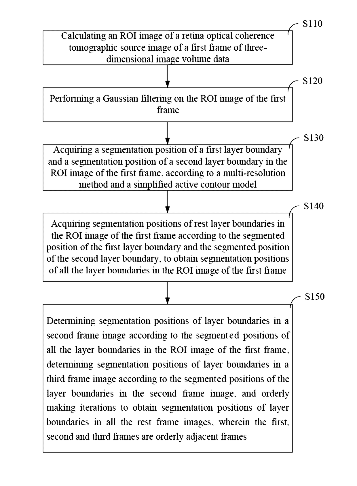
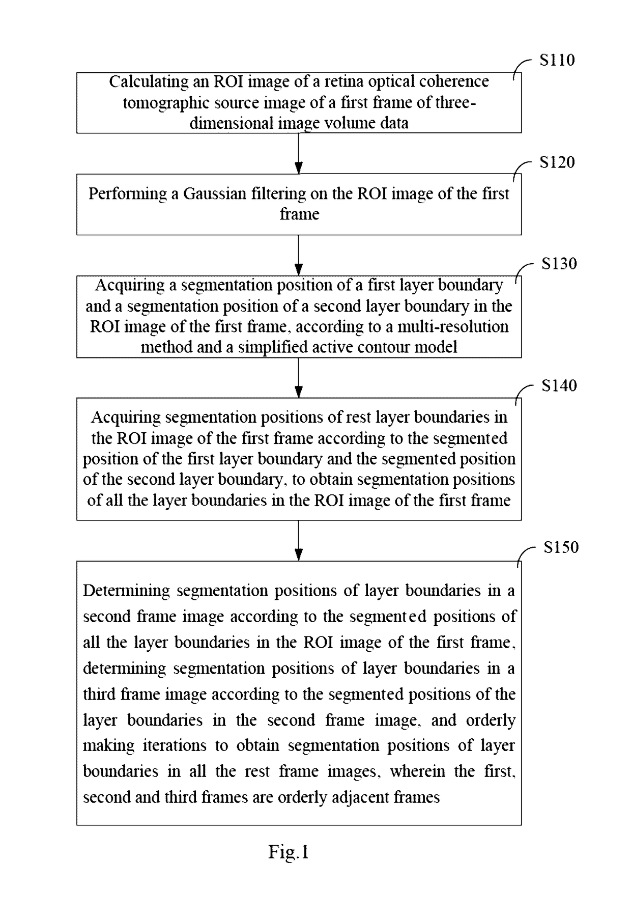
[0035]The present disclosure provides a method for acquiring a retina structure from an optical coherence tomographic image. The method utilizes correlation between adjacent OCT images, models adjacent frames through Kalman filtering, orderly calculates the position of the retina layer boundary in each frame, and finally obtains the retina structure. In which, the retina layer boundary includes Inner Segment-Outer Segment (IS-OS), Vitreous Body-Retinal Nerve Fiber Layer (Vitreous-NFL), Outer Segment-Retinal Pigment Epithelium (OS-RPE), Retinal Pigment Epithelium-Choroid (RPE-Choroid), O...


PUM


Abstract

The present disclosure provides a method for acquiring a retina structure from an optical coherence tomographic image and a system thereof. The method comprises: calculating a region of interest (ROI) image of a source image; performing a Gaussian filtering of the ROI image; calculating a first estimated boundary position of a first layer boundary and a second estimated boundary position of a second layer boundary using a multi-resolution method; refining the first layer boundary and the second layer boundary respectively using a simplified active contour model according to the first estimated boundary position and the second estimated boundary position, to obtain a first initial boundary position of the first layer boundary and a second initial boundary position of the second layer boundary; smoothing the first initial boundary position and the second initial boundary position using a filtering method; acquiring segmentation positions of rest layer boundaries in the ROI image according to a segmented position of the first layer boundary and a segmented position of the second layer boundary. The present disclosure can improve the calculation speed and reduce the calculation complexity while maintaining high location accuracy.

Description

TECHNICAL FIELD[0001]The present disclosure relates to the technical field of optical imaging, and particularly, to a method for acquiring a retina structure from an optical coherence tomographic image and a system thereof.BACKGROUND[0002]Optical Coherence Tomography (OCT) is an optical signal acquiring and processing method without invasion to the organization structure of an imaging surface. In recent years, OCT has become a main retina disease diagnosis technique.[0003]Regarding the retina disease diagnosis, doctors need to partition, recognize and diagnose the images of normal and diseased regions of the retina in real time. OCT can achieve a higher spatial resolution (several microns) than other imaging means such as ultrasonic imaging, X-ray imaging and magnetic resonance imaging, thus attracts extensive attentions recently.[0004]The quantitative analyses of different layers of the retina can promote the diagnosis of the visual function change and help doctors to diagnose glau...

Claims


Application Information

Patent Timeline
Patent Type & Authority Applications(United States)
IPC IPC(8): G06T7/12G06T7/00G06T7/11G06T5/20
CPCG06T7/12G06T5/20G06T7/0012G06T7/11G06T2207/20024G06T2207/30041G06T2207/20104G06T2207/20016G06T2207/20116G06T2207/10101G06T7/149
Inventor HU, QINGMAOZHANG, TIANQIAO
Owner SHENZHEN INST OF ADVANCED TECH
Why Eureka
Try Eureka