Measurement of body fluid volumes

a body fluid and volume measurement technology, applied in the field of body fluid volume measurement, can solve the problems of albumin leakage and distribution to the interstitial fluid, limited use of icg method, laborious method,

Inactive Publication Date: 2017-06-22
PHARMACOPHOTONICS INC
View PDF2 Cites 1 Cited by
  • Summary
  • Abstract
  • Description
  • Claims
  • Application Information

AI Technical Summary

Problems solved by technology

However, utility of the ICG method has been limited by many of the same factors as the iodine-based testing.
Therefore, it is a very labor intensive method.
Another drawback in the use of labeled albumin, in the dilution technique to measure plasma volume, is that albumin also “leaks” and distributes to the interstitial fluid.
However, this technique is too difficult and impractical to perform.

Method used


Image

  • Measurement of body fluid volumes

Examples


example 1

Measurement of TVPV and ECFV in Bilaterally Anephric Rats

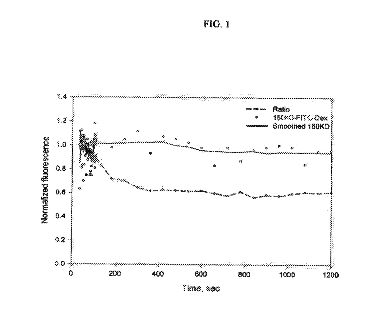
[0042]The example shown here was a test conducted on a bilaterally anephric rat, which was infused with a mixture of 3 kDa TEXAS RED®-dextran and 150 kDa FITC-dextran. The dynamic plasma fluorescence intensity was obtained by in vivo two-photon liver imaging of vascular plasma. Only the vascular plasma containing regions in each image were included for calculation. The decay curve of the fluorescence intensity of the 150-kDa FITC-dextran as well as the decay curve of the ratio of the fluorescence intensity of the TEXAS RED®-dextran to that of the FITC-dextran after the infusion is shown in FIG. 1. Using the ratio rather than the 3 kDa TEXAS RED®-dextran or the 150 kDa FITC-dextran signal directly helped reduce the signal fluctuation caused by focus movement during imaging since the same fluctuation showed up in both channels.

[0043]To test if the volumes determined by this method agree with expected values we injected a mixture...

example 2

Anticipated Minimally Invasive Method for Measuring Fluid Volumes in a Patient with Renal Failure

[0046]A minimally invasive method for measuring TVPV, ECFV and TV in a patient with renal failure uses a small dextran (molecule size of about 1 kDa to about 20 kDa) labeled with a first fluorescent dye to distribute to the vascular and interstitial spaces and a large dextran (molecule size of about 70 kDa to about 500 kDa) labeled with a second fluorescent dye to distribute only to the vascular space of the animal The molecules can be simultaneously detected in vivo using a dual channel fluorescence detection device and a proprietary fiber optic catheter. The fluorescence device and the fiber optic catheter have both been disclosed in a pending U.S. patent application Ser. No. 12 / 425,827, the disclosure of which is hereby incorporated by reference as if fully set forth herein and, more specifically, for this specific subject matter disclosed at Paragraphs [0077] to [0093], and FIGS. 1 a...

example 3

Measurement of TVPV and ECFV in Bilaterally Anephric Rats

[0049]Determination of plasma volume is accomplished using a 150 kDa dextran conjugated to a 2-SulfhydroRhodamine (2SHR) fluorescent dye. A bolus injection or alternatively a rapid infusion of the molecule is given to the subject. A blood sample is taken approximately 10 to 15 minutes after the molecule enters the subjects blood stream. The sample is analyzed to determine the concentration of the molecule in the blood plasma. This analysis can be advantageously accomplished using an ELISA colorimetric immunoassay containing monoclonal antibodies directed against 2SHR. Calculation of the plasma volume (PV) is determined as follows:

PV=Dose / PC,

where the Dose is the concentration per m in the dose solution, and PC is the concentration of the fluorescent molecule contained in the plasma per ml.

[0050]The ELISA colorimetric assay is incorporated into a module that allows determination of the plasma volume at the patient's bedside. A ...


PUM

PropertyMeasurementUnit
extracellular fluid volumeaaaaaaaaaa
concentrationaaaaaaaaaa
molecular sizeaaaaaaaaaa

Abstract

The present invention is related generally to measurement of body fluid volumes in an animal subject. The body fluid volumes of interest include extracellular fluid volume (ECFV), total vascular plasma volume (TVPV) and interstitial fluid volume (IFV). The methods are especially beneficial for subjects suffering from renal failure and particularly those undergoing renal dialysis. ECFV can be measured by administering a first molecule which is non-metabolized and permeable to vessel walls of the vascular system wherein the first molecule is distributed within the total vascular space as well as the interstitial space. TVPV can be measured by administering a second molecule which is non-metabolized and impermeable to vessel walls of the vascular system wherein the second molecule is distributed within only the vascular space. IFV can then be calculated using the equation IFV=ECFV−TVPV.

Description

CROSS-REFERENCE TO RELATED APPLICATIONS[0001]The present invention is a continuation-in-part of U.S. patent application Ser. No. 13 / 318,097, filed Apr. 18, 2012, which is a 371 filing claiming priority to PCT / US2010 / 032997, filed Apr. 29, 2010, and claims the benefit of U.S. Provisional Patent Application No. 61 / 174,100 filed Apr. 30, 2009, and the contents of which are incorporated herein by reference in their entirety.BACKGROUND OF THE INVENTION[0002]The present invention is related generally to measurement of body fluid volumes in an animal subject. The body fluid volumes of interest include extracellular fluid volume (ECFV), total vascular plasma volume (TVPV) and interstitial fluid volume (WV), with TVPV being the preferred indicator. The methods are especially beneficial for subjects suffering from renal failure, and particularly those undergoing renal dialysis.[0003]Body fluid volume status is a critical metric in the management of many chronic and acute medical conditions. V...

Claims


Application Information

Patent Timeline
Patent Type & Authority Applications(United States)
IPC IPC(8): A61K49/00A61B5/145G01N33/58A61B5/1455A61K9/00G01N33/68
CPCA61K49/0054A61K9/007A61K49/0043A61K49/0041A61B5/1451G01N33/6893G01N33/582A61B5/1455A61K9/0019A61K49/0052G01N2800/347A61K49/0002
Inventor MOLITORIS, BRUCE A.WANG, EXINGSANDOVAL, JR., RUBEN M.
Owner PHARMACOPHOTONICS INC
Why Eureka
Try Eureka