Method of Detecting an Endotoxin Using Limulus Amebocyte Lysate Substantially Free of Coagulogen
a technology of amebocyte lysate and endotoxin, which is applied in the field of detecting endotoxins, can solve the problems of multiple organ failure, death, and great danger for the infected organism
- Summary
- Abstract
- Description
- Claims
- Application Information
AI Technical Summary
Benefits of technology
Problems solved by technology
Method used
Image
Examples
example 1
Purification of LAL Using Tangential Flow Filtration
[0072]Limulus amebocyte lysate (LAL) extracted from blood cells (amoebocytes) of horseshoe crab was obtained from Lonza (Basel, Switzerland). The LAL was subjected to hollow fiber modified polyethersulfone (mPES) membrane filters (SpectrumLabs) for tangential flow filtration (TFF) to remove the coagulogen. The TFF system was set up using a 30 kDa MWCO mPES filter, Cole Parmer Masterflex Pump Tubing and the appropriate pump and backpressure valve. The system was flushed with LAL reagent water (LRW), then depyrogenated using IN NaOH for 1 hr at room temperature. LRW was used to rinse the system after depyrogenation then the system was equilibrated using TFF buffer.
[0073]The LAL was diluted at a ratio of 1:10 to 1:8 with TFF buffer (50 mM Tris with 77 mM NaCl at pH 7.4-7.5 at room temperature) before being fed into the filter at a flow rate of 430 ml / min while maintaining a transmembrane pressure (TMP) of 5-8 psi until 90% of the tota...
example 2
Quantification of Coagulogen
[0076]Semi-quantitative evaluation of the remaining coagulogen in the LAL was conducted by performing SDS-PAGE / protein gel stain on the LAL retentate and comparing the band density of the 20 kD coagulogen band in the original LAL to the coagulogen band, if any, in the TFF retentate. See, e.g., FIG. 7A.
[0077]Additionally, an α-coagulogen antibody was used to perform a Western blot then compare the band density of the 20 kD coagulogen bands. See, e.g., FIG. 7B.
[0078]The identity of coagulogen was confirmed by running the LAL permeate (containing the coagulogen) under reducing conditions on an SDS-PAGE gel, and then sequencing the three bands. See FIG. 9. Each of the three bands was confirmed as a coagulogen fragment by protein sequencing.
[0079]Both quantitative methods confirm the present invention removed a substantial percentage of coagulogen from the LAL.
[0080]The amount of coagulogen in LAL was determined using Western blot analysis. FIG. 8A. The result...
example 3
Stability of LAL Substantially Free of Coagulogen
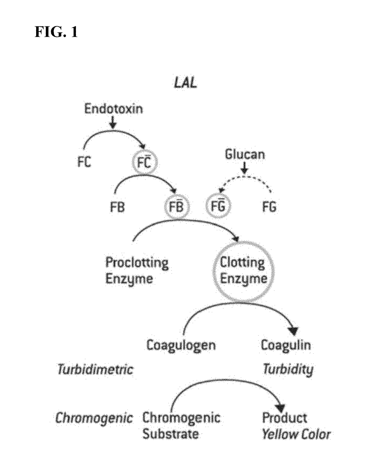
[0081]The stability of the LAL substantially free of coagulogen was investigated. LAL substantially free of coagulogen samples were stored for 1, 2, 3, 4, 5, 6, 7, 8, 9 and 10 weeks. At the end of the indicated time period, the LAL substantially free of coagulogen sample is mixed with the LAL chromogenic reagent in a 96-well plate and placed in an incubating plate reader that measures absorbance at 405 nm. The reaction was automatically monitored over time the appearance of a yellow color.
[0082]In the presence of endotoxin, the lysate will cleave the chromogenic substrate, causing the solution to become yellow. The time required for the change is inversely proportional to the amount of endotoxin present. Time required to reach 50 mOD for each of the samples is found in FIG. 10. The data suggests LAL substantially free of coagulogen is stable for at least 10 weeks.
PUM
| Property | Measurement | Unit |
|---|---|---|
| flow rate | aaaaa | aaaaa |
| flow rate | aaaaa | aaaaa |
| flow rate | aaaaa | aaaaa |
Abstract
Description
Claims
Application Information
 - R&D
- Intellectual Property
- Life Sciences
- Materials
- Tech Scout
- Unparalleled Data Quality
- Higher Quality Content
- 60% Fewer Hallucinations
Browse by: Latest US Patents, China's latest patents, Technical Efficacy Thesaurus, Application Domain, Technology Topic, Popular Technical Reports.
© 2025 PatSnap. All rights reserved.Legal|Privacy policy|Modern Slavery Act Transparency Statement|Sitemap|About US| Contact US: help@patsnap.com


