Photoacoustic and ultrasonic endoscopy system including a coaxially configured optical and electromagnetic rotary waveguide assembly and implementation method thereof

a photoacoustic and ultrasonic technology, applied in the field of medical tomographic endoscopy apparatus, can solve the problems of inability to meet both technical requirements, inability to achieve commercially successful or clinically applicable pae systems, and inability to meet the requirements of both technical requirements, so as to achieve the effect of decreasing the quality of the imag

Active Publication Date: 2018-03-01
UNIST ULSAN NAT INST OF SCI & TECH
View PDF7 Cites 13 Cited by
  • Summary
  • Abstract
  • Description
  • Claims
  • Application Information

AI Technical Summary

Benefits of technology

[0035]Existing PAE systems (e.g., in Prior Documents 4, 7, and 8) using a torque coil-based proximal actuation mechanism have problems in that, since the optical fiber and the signal wire are simply arranged in a parallel structure inside a torque coil, mechanical torque cannot be uniformly transmitted from the proximal part to the scanning tip of the probe. The problem becomes even worse when a rotational scanning is performed in conditions u

Problems solved by technology

After the first conceptual suggestion of PAE by Oraevsky et al. in 1997 as described in Prior Document 5, in which the imaging probe was referred to as optoacoustic endoscope (OAE), a number of PAE probes were developed to address such technical requirements as “probe miniaturization” and “specifying a device configuration or operation principle for endoscopy.” However, no commercially successful or clinically applicable PAE system that satisfies both of the technical requirements has been developed yet due to many underlying technical challenges.
The most well-known and toughest challenge is that to successfully create a working PAE probe, all the optical and acoustic elements should be effectively integrated and arranged in a small and restricted space; an adequate scanning mechanism, through which a tomographic image can be produced, should also be developed and integrated into the device.
However, the main drawback of the electronic scanning mechanism is that, since it is relatively more difficult to reduce the size of the related endoscopic probe than that of the mechanical scanning mechanism, such problems as crosstalk or signal interference between channels may occur; the costs of implementing the system may also be high.
First, its major drawback is that, since a single-element ultrasonic transducer that can receive the signals bounced back only from the aiming direction of the transducer surface is mounted on the scanning tip of an endoscopic probe, in order to obtain a 2D or 3D image, a series of processes that emit a laser pulse and then detect the generated photoacoustic waves should be repeatedly performed by changing the phys

Method used


Image

  • Photoacoustic and ultrasonic endoscopy system including a coaxially configured optical and electromagnetic rotary waveguide assembly and implementation method thereof
  • Photoacoustic and ultrasonic endoscopy system including a coaxially configured optical and electromagnetic rotary waveguide assembly and implementation method thereof
  • Photoacoustic and ultrasonic endoscopy system including a coaxially configured optical and electromagnetic rotary waveguide assembly and implementation method thereof

Examples


Embodiment Construction

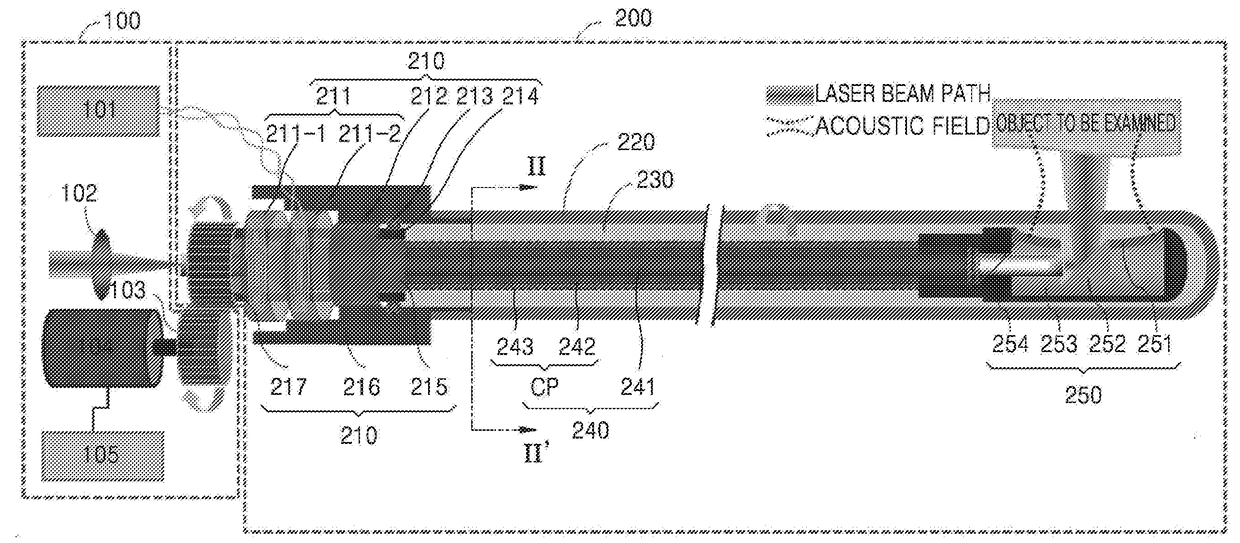
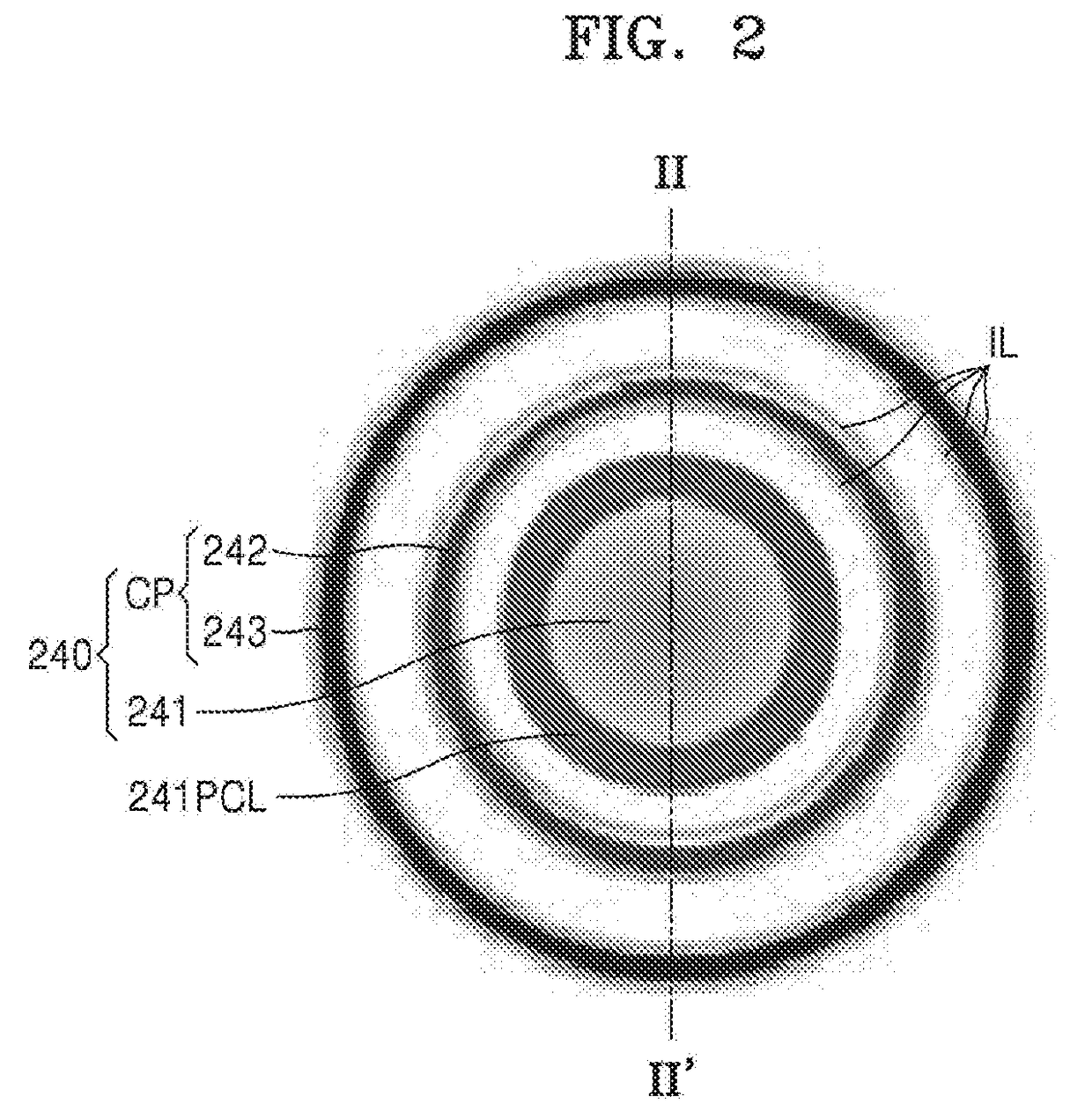
[0068]The present disclosure may include various embodiments and modifications, and embodiments thereof will be illustrated in the drawings and will be described herein in detail. The advantages and features of the present disclosure and methods of achieving the advantages and features will be described more fully with reference to the accompanying drawings, in which embodiments are shown. The present disclosure may, however, be embodied in many different forms and should not be construed as being limited to the embodiments set forth herein.

[0069]Reference will now be made in detail to embodiments, examples of which are illustrated in the accompanying drawings. In the drawings, the same elements are denoted by the same reference numerals, and a repeated explanation thereof will not be given.

[0070]It will be understood that although the terms “first”, “second”, etc. may be used herein to describe various elements, these elements should not be limited by these terms. These elements ar...


PUM


Abstract

A photoacoustic-ultrasonic dual-mode endoscope includes: a probe and a probe driving unit, wherein the probe includes: a coaxially configured optical and electromagnetic rotary waveguide assembly including an optical fiber, the optical fiber including a core and a cladding, and a conductive path coaxially arranged with the optical fiber; a scanning tip located at an end of the coaxially configured optical and electromagnetic rotary waveguide assembly and configured to deliver a laser beam to an object to be examined and detect a photoacoustic signal and an ultrasonic signal generated from the object to be examined; and a plastic catheter surrounding outer surfaces of the coaxially configured optical and electromagnetic rotary waveguide assembly and the scanning tip, wherein the conductive path includes: a first conductive path including a portion coaxially arranged with the optical fiber; and a second conductive path including a portion coaxially arranged with the optical fiber and insulated from the first conductive path.

Description

CROSS-REFERENCE TO RELATED APPLICATION[0001]This application claims the benefit of Korean Patent Application No. 10-2016-0107773, filed on Aug. 24, 2016, in the Korean Intellectual Property Office, the disclosure of which is incorporated herein in its entirety by reference.BACKGROUND1. Field[0002]One or more of the embodiments described in the present disclosure relate to a medical tomographic endoscopic apparatus that has a long and slender probe shape, like the current endoscopic ultrasound (EUS) probes utilized in clinics, wherein the endoscopic apparatus is inserted into the object to be examined and provides a tomographic image of the interior thereof. The key feature of the proposed endoscopic system is the ability to provide both a high-quality photoacoustic image and a typical ultrasonic endoscopic image based on the improved probe flexibility and rotational uniformity compared to existing photoacoustic endoscopic systems. The inventive concept of the present disclosure may ...

Claims


Application Information

Patent Timeline
IPC IPC(8): A61B1/00A61B8/12A61B5/00A61B1/04
CPCA61B1/00117A61B8/12A61B5/6852A61B5/0066A61B1/04A61B5/0095A61B1/00165A61B8/4416A61B8/445A61B8/4461A61B2562/247A61B5/0035A61B8/4444
Inventor YANG, JOON-MOKIM, CHAE UN
Owner UNIST ULSAN NAT INST OF SCI & TECH
Why Eureka
Try Eureka