
Ray-tracing Methods for Realistic Interactive Ultrasound Simulation

a simulation and interactive technology, applied in the field of virtual reality ultrasound image modeling, can solve the problems of unsolved theoretical problems, requiring a long training period for ultrasound specialists, and learning the proper execution

Active Publication Date: 2018-03-08
VIRTAMED
View PDF8 Cites 13 Cited by
  • Summary
  • Abstract
  • Description
  • Claims
  • Application Information

AI Technical Summary

Benefits of technology

This patent describes a method for simulating ultrasound images using a computer-implemented method. It involves identifying specific properties of the anatomical surface and calculating multiple paths for light to reach the surface. These paths are then combined to create the final image. This method can be used for medical training purposes and allows for more realistic and accurate imaging.

Problems solved by technology

Learning the proper execution of this modality thus requires a lengthy training period for ultrasound specialists.
However, simulating all possible ultrasound-tissue interaction phenomena is still an unsolved theoretical problem.
For instance, the ultrasound texture (speckles) is a result of constructive and destructive interference of ultrasonic waves mainly scattered by sub-wavelength particles, such as cell nuclei, other organelles, etc.
However, no known method can observe a sufficiently large tissue region (40-150 mm for OB / GYN ultrasound examination) in such fine detail with cellular structures.
243-250, but this wave simulations for ultrasound are still not fast enough to handle the complexity of clinical ultrasound in real-time.
The computational complexity of such wave simulations restricts their use to offline simulation, as used in specific applications e.g., for transducer design and image processing algorithm development and validation.
In the context of training simulators however these approaches are not applicable, as the generation of US images at interactive rates is essential and hence of high interest.
However, scatterer-based methods do not inherently take into account reflection and distortion of the ultrasound beam at the interfaces between large-scale structures, the source of artifacts such as acoustic shadowing, ghosting, and multiple reflections.
This may be true for numerical phantoms and artificial tissue-mimicking material; however, in actual anatomy perfect reflections and refractions are never the case, hence these methods often produce artificial, toy-like images not comparable to actual patient ultrasound.
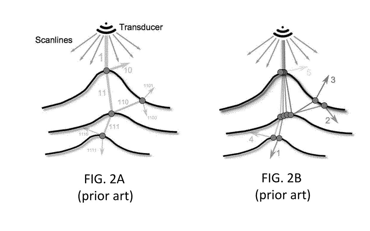
For instance, the state-of-the-art methods [BBRH13,SAP*15] evaluate a diffuse reflection term only locally (similar to specular reflections in computer graphics) but do not take non-specular indirect contributions into account, hence suffering from “hard” shadows, sharp reflections, and too crisp structure borders—atypical in real US imaging.
[SAP*15] further attempts to address these issues with a post-processing step to make images visually closer to expected; this, nevertheless, does not deal with the main problem of the wrong initial assumptions and can only partially improve the realism, while requiring additional post-processing at the expense of the overall computation efficiency and complexity.
One major limitation of these deterministic ray-tracing approaches is that deeper recursion levels require summing over an exponentially-growing number of rays, although these contribute less and less to the final image.
Therefore, these algorithms quickly become computationally inefficient.
Furthermore, such method exhibits poor parallelism, since multiple rays on subsequent levels are dependent on the single parent rays.
Introduced in the late seventies, this deterministic method has long been shown to be inferior to stochastic techniques for natural scenes, such as Monte-Carlo path tracing methods.
Surprisingly, however, stochastic techniques have not been investigated in the literature for the purpose of ultrasound image simulation.
However, the purpose of the latter application is not to emulate the image formation process of ultrasound machines, and since therapy ultrasound frequencies cannot be used for image generation, these methods cannot directly be used to simulate radio-frequency or B-mode images.
In particular the specific issues of generating realistic ultrasound images with the proper reflection and refraction for different anatomy component properties has not been investigated in the state of the art works for ultrasound simulation.

Method used


Image

  • Ray-tracing Methods for Realistic Interactive Ultrasound Simulation
  • Ray-tracing Methods for Realistic Interactive Ultrasound Simulation
  • Ray-tracing Methods for Realistic Interactive Ultrasound Simulation

Examples


Embodiment Construction

Ultrasound Imaging Simulation System

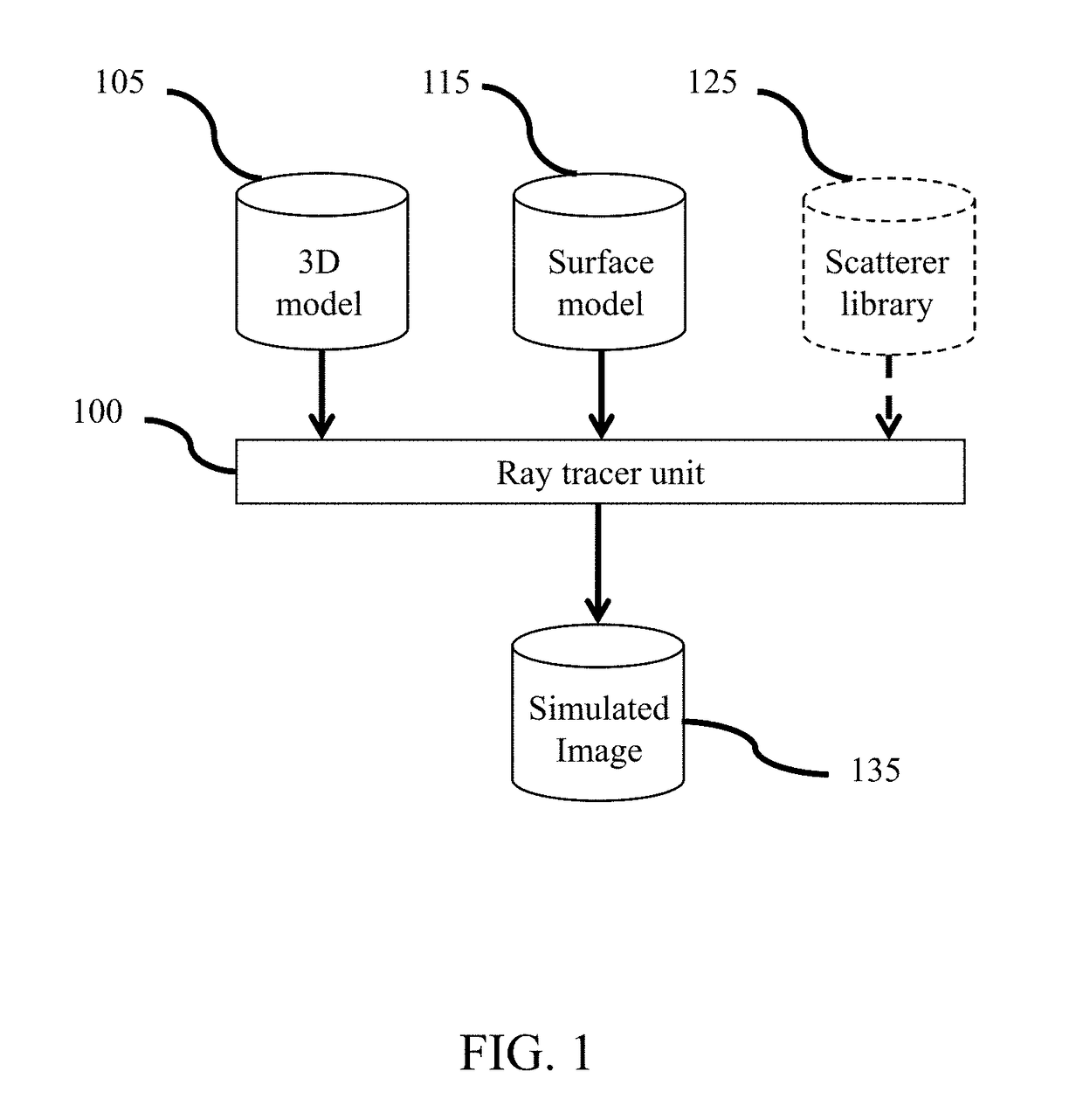
[0030]FIG. 1 represents an ultrasound imaging simulation system comprising a ray tracer unit 100 in connection with a 3D model 105 comprising a diversity of anatomy objects and the surface model parameters 115 characterizing different ultrasound tissue properties for the anatomy objects in the ultrasound volume model 105. The ray tracer unit 100 may comprise at least one central processing unit (“CPU”) circuit, at least one memory, controlling modules, and communication modules to reconstruct simulated ultrasound images 135 corresponding to different views of the ultrasound volume model. In a possible embodiment, the ray tracer unit may be part of the rendering unit in an ultrasound simulation system (not represented) and the simulated ultrasound images may be displayed in real time on a screen of the ultrasound simulation system (not represented). Such an exemplary ultrasound system is for instance described in patent application WO 2017 / 064249, ...


PUM


Abstract

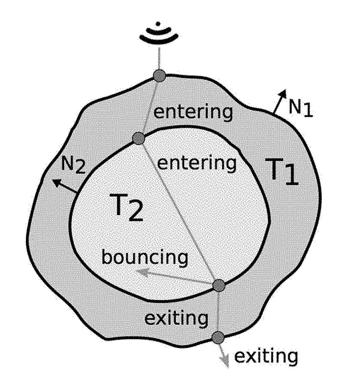
Stochastic ray-tracing methods such as Monte-Carlo ray-tracing may be adapted to provide more efficient and more realistic ultrasound imaging systems and methods. Many random ray paths that are perturbed according to a probability distribution may be generated until they converge to the correct solution. New surface thickness and roughness models enable to reproduce complex ultrasound interactions such as multiple reflections and refractions. The contribution of each ray path may be further weighted to better simulate a beamformed ultrasound signal. Tracing many individual rays per transducer element is easily parallelizable on modern GPU computing architectures.

Description

CROSS REFERENCE TO RELATED APPLICATIONS[0001]This application claims the benefit of U.S. Provisional Application No. 62 / 383,829, filed Sep. 6, 2016, and U.S. Provisional Application No. 62 / 448,661, filed Jan. 20, 2017, which is incorporated by reference in their entireties.FIELD OF THE INVENTION[0002]Methods described herein relate to ultrasound image simulation in general, and more specifically to virtual reality ultrasound image modeling for ultrasound practice training simulation purposes, for example in the medical field.BACKGROUND OF THE INVENTIONMedical Imaging Simulators[0003]Application of ultrasound requires a high level of expertise both in manipulating imaging devices and also in analyzing and interpreting resulting images, for instance in the medical field for accurate diagnosis and intervention guidance. Learning the proper execution of this modality thus requires a lengthy training period for ultrasound specialists.[0004]To facilitate the training of medical students a...

Claims


Application Information

Patent Timeline
IPC IPC(8): G09B23/28G09B9/00G06F17/10G06T15/08G06T15/06G06T11/00
CPCG09B23/286G09B9/00G06F17/10G06T15/08G06T15/06G06T11/005G06T2210/41G06T2215/16
Inventor MATTAUSCH, OLIVERGOKSEL, ORCUNMAKHINYA, MAXIM
Owner VIRTAMED
Learn More