
Tissue stain and use thereof

Active Publication Date: 2018-08-16
GI SUPPLY
View PDF3 Cites 0 Cited by
  • Summary
  • Abstract
  • Description
  • Claims
  • Application Information

AI Technical Summary

Benefits of technology

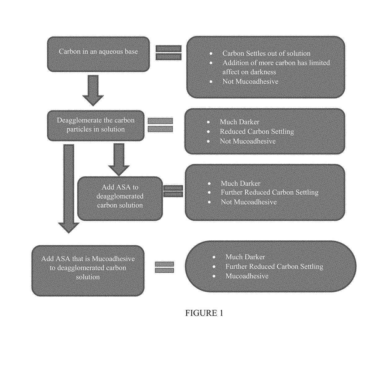
The patent text describes a new tissue staining composition that is easy to use, can be observed easily during injection into tissue, and provides a durable, reliable mark that can be visualized over time. The composition uses carbon particles with a small particle size, which enhances the darkness of the staining agent. The carbon particles can remain in suspension for extended periods without sedimentation, and the composition includes an anti-settling agent and / or a mucoadhesive agent. The composition is also heat stable and can be thermally sterilized. Overall, the tissue staining composition described in this patent is more effective and reliable than other commercially available options.

Problems solved by technology

However, existing stains (e.g., methylene blue, indigo carmine, indocyanine green-based stains) may be difficult to visualize endoscopically even at the time of injection into tissue of interest because the stain may leak or diffuse out of the tissue at the point of entry and thus visually obscure the area being marked.
Furthermore, even after injection, it may become difficult to identify the marked regions months to years later because the marks may fade or even disappear over time.
In other words, such stains may not facilitate permanent marking of tissue to accommodate accurate, long term tracking of tissue of interest.
As a result, it can be difficult or even impossible for a physician or other medical practitioner to monitor with confidence abnormal tissue, pre-cancerous or cancerous lesions over time.
Furthermore, it has been observed that with certain tissue stains, the staining agents, if particulate, may settle out of solution over time.
However, this step may not be performed adequately, if at all, prior to injection, and as a result differing amounts of staining agent may be delivered during different marking procedures or when different areas are marked during the same procedure, resulting in inconsistent marks or marks where insufficient staining agent has been introduced to create a durable mark.
These impurities can be associated with complications such as inflammatory reactions or may even be carcinogens.

Method used


Image

  • Tissue stain and use thereof
  • Tissue stain and use thereof
  • Tissue stain and use thereof

Examples


example 1

Carbon Particle Size on Darkness of Tissue Stain

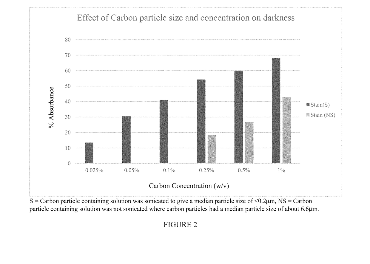
[0127]This Example shows that carbon particle size has a profound impact on the darkness of a tissue staining composition.

[0128]Various test solutions were prepared containing different concentrations of carbon particles ranging from 0.025%-1.0% (w / v) of carbon particles (Monarch 4750 from Cabot Corp., Billerica Mass.) that were either not deagglomerated and thus contained carbon particles having a mean particle diameter of about 6.6 μm or deagglomerated by sonication to produce carbon particles having a mean diameter less than 0.2 μm. The balance of each test solution included 15% (w / v) glycerol, 1% (w / v) Tween® 80, 1% (w / v) benzyl alcohol, 0.01% (w / v) simethicone, and sterile water for injection (SWFI).

[0129]The test solutions were prepared as follows.

[0130]A first (2×) pre-mix was prepared by adding the following ingredients to SWFI at 80-100° C., one at a time, and mixing for 15 minutes before adding the next ingredient, to give th...

example 2

f Carbon Particle Size on Particle Sedimentation (Settling)

[0138]This example demonstrates that the size of the carbon particles can have a profound effect on the settling of the carbon particles.

[0139]In this example, two tissue stains were prepared that were the same (containing 1% (w / v) Tween® 80, 15% (w / v) glycerol, 1% (w / v) benzyl alcohol, and 0.01% (w / v) simethicone), and containing 0.27% (w / v) carbon particles (Monarch 4750 from Cabot Corp., Billerica Mass.) that were either not deagglomerated and thus contained carbon particles having a mean particle diameter of about 6.6 μm or deagglomerated by homogenization to produce carbon particles having a mean diameter less than 0.2 μm. The resulting particles were analyzed for particle size and stability.

[0140]The tissue stain containing the deagglomerated carbon particles were prepared from a carbon particle pre-mix containing the ingredients set forth in TABLE 2A, and a Tween® 80 premix set forth in TABLE 2B.

TABLE 2ACarbon Particl...

example 3

rticle-Based Tissue Stains with Desirable Anti-Settling and Mucoadhesive Properties

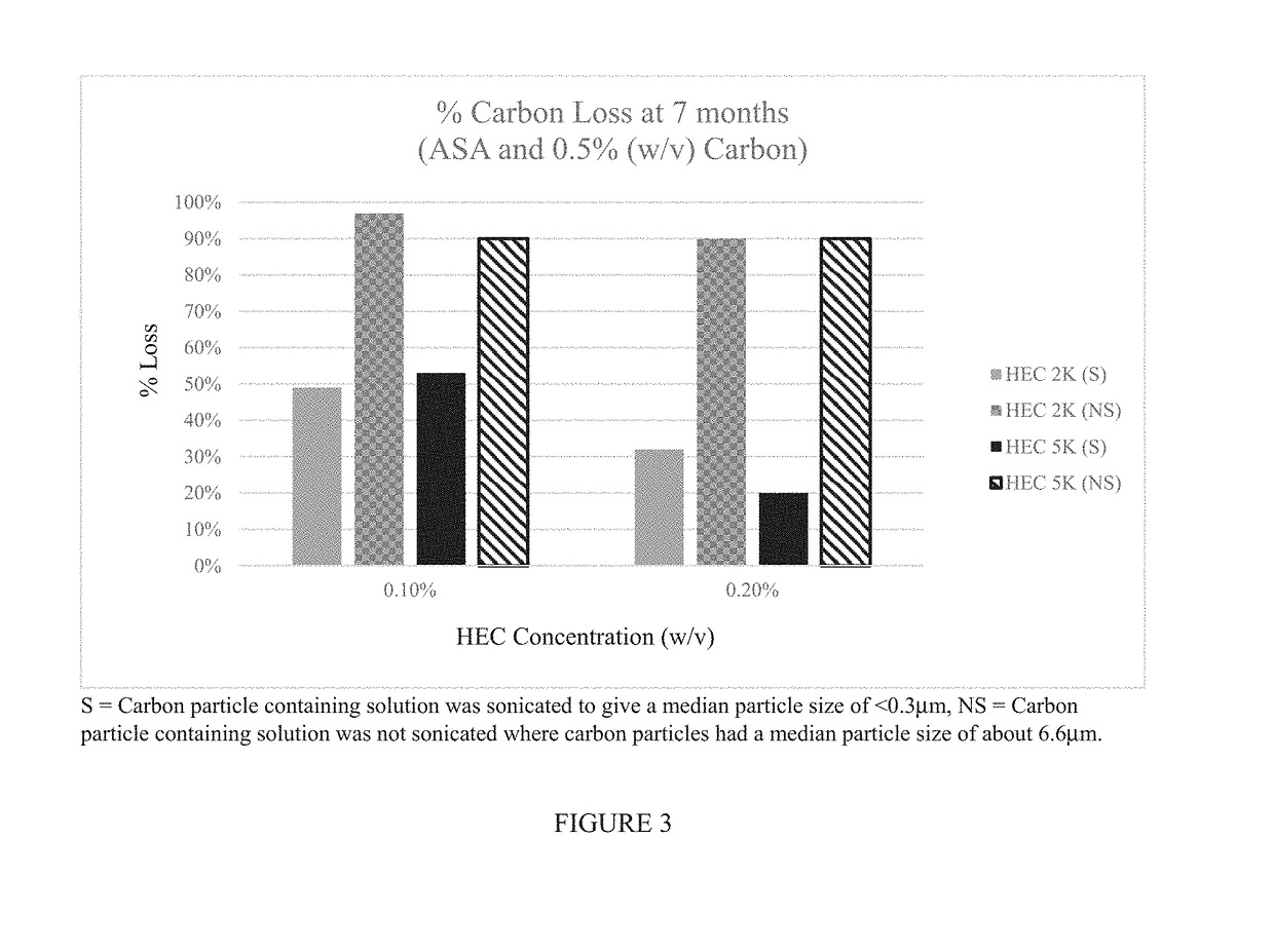
[0152]This example demonstrates that it is possible to create a carbon particle-based tissue staining composition that is more resistant to settling than commercially available tissue staining solutions. The size of the carbon particles has a profound effect on the settling of the particles. For example, carbon particles having a mean particle diameter less than 0.3 μm (for example, less than 0.2 μm) exhibit much lower sedimentation over time especially in the presence of an agent, for example, a surfactant, such as a non-ionic surfactant, that prevents reagglomeration of the carbon particles.

[0153]However, under certain circumstances, it may be desirable to combine the carbon particles (e.g., in the presence of a surfactant) with an anti-settling agent. This example also demonstrates that the particle size and / or the settling agent can be used to create a tissue staining composition that has the appr...


PUM


Abstract

Provided is a tissue staining composition containing carbon particles having a mean particle diameter less than 0.3 μm in diameter (optionally less than 0.2 μm in diameter) together with one or more agents that maintain the carbon particles in suspension (for example, an anti-settling agent and / or surfactant). In certain circumstances, the anti-settling agent may also have mucoadhesive properties. The tissue staining composition is visually dark and does not disperse rapidly when introduced into regions of tissue of interest making it ideal for marking the regions that can be visualized clearly and over prolonged periods of time via, for example, direct visualization, endoscopic or laparoscopic inspection. The invention also provides methods of making and using the tissue staining composition for marking regions of tissue of interest, for example, gastrointestinal tissue, as well as other tissues.

Description

CROSS REFERENCE TO RELATED APPLICATIONS[0001]This application claims the benefit of, and priority to, U.S. Provisional Patent Application Ser. No. 62 / 458,805 filed Feb. 14, 2017, which is hereby incorporated by reference herein in its entirety.FIELD OF THE INVENTION[0002]The invention relates generally to a tissue staining composition for marking regions of living tissue, for example, regions of the gastrointestinal tract, in order to identify and visualize the regions at a later point in time.BACKGROUND[0003]In certain medical procedures there is a need to mark regions of tissue (for example, cancerous or pre-cancerous lesions) so that they can then be later identified, for example, via endoscopic, laparoscopic, or open surgical procedures, weeks, months or years later. Such markings can identify tissue to be surgically excised via a subsequent procedure or permit a physician or other medical provider to assess whether cytological or morphological changes have occurred over time th...

Claims


Application Information

Patent Timeline
IPC IPC(8): A61K49/00
CPCA61K49/006A61B90/39A61K49/0093A61B2090/395G01N1/30G01N2001/302
Inventor TIMM, MARY JOWHALEN, ROBERT G.CAMERON, AMY A.LEE, PATRICK
Owner GI SUPPLY
Learn More