In general, methods sections of research reports provide sufficient detail to replicate a specific oculomotor paradigm, although the concept of
standardization in oculomotor research and testing has not been as widely adopted as in other clinical visual measurements (e.g., visual perimetry,
visual acuity, contrast sensitivity) that have achieved widespread use and implementation in commercial products.
Despite an extensive catalogue of oculomotor signs of injury and
disease known in the oculomotor literature (Leigh & Zee, 2006), the neurological
community lacks standardized clinically-relevant tools to assess various aspects of
neural processing (Pelak & Hoyt, 2005) especially deployable eye-tracking solutions that can be used in operational, field settings (Liston, Simpson, Wong, Rich, & Stone, 2016; Port, Madsen, Means, T., & Wicks, 2015) such as forward operating hospitals, clinics, and
athletic training rooms.
Limitations of the general-purpose approach include: setup time, complexity, expense, and use of hardware and
software with unnecessary
degrees of freedom.
Translating the results of clinical oculomotor research into a clinically-relevant oculometric
neurological examination has been limited by the availability of trained personnel, resources, and proper
eye movement equipment.
Furthermore,
gaze computations based upon 3d measurements (Hennessey & Lawrence, 2013) necessary for head-free tracking yield noisier eye-position measurements than 2d approaches, due to the larger number of measurements of eye features (e.g., multiple corneal reflections, ellipsoidal fit to
pupil outline), all subject to independent sources of
noise.
Thus, head-free trackers that include one or more cameras embedded in a housing detachable from the visual display (e.g., Tobii Pro X2-30, LC Technologies The EyeFollower®) reduce
degrees of freedom relative to trackers with multiple independent cameras (e.g., Smart Eye), and decrease difficulty of mechanical setup somewhat, but retain the disadvantages of head-free tracking (e.g., a larger number of tracked features, computational burden, eye-position
noise) for use in collecting high-precision eye-position measurements from clinical populations.
Moreover, the complexity of head-free tracking is unnecessary for a clinical device which assumes a known
head position and introduces
noise needlessly into eye-position traces.
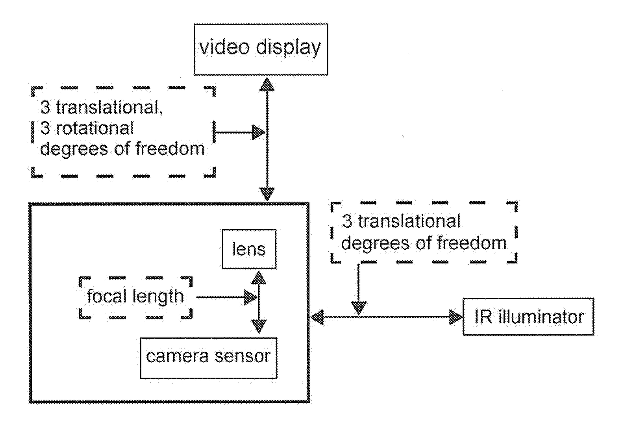
For head-free tracking, the fields of use of general-purpose eye-trackers have been too broad to allow for purpose-built trackers, except in very specialized cases.
Prior art in commercial head-fixed eye-tracking solutions (e.g., Arrington, ISCAN, SMI) are designed for general-purpose tracking (e.g., no predefined camera position, no specified
display size, no predefined illuminator position, no fixed distance between the subject and the camera), and thus introduce unnecessary complexity for constrained head-fixed tracking (e.g., specified
display size, known camera position, known viewing distance).
In these systems, cameras and illuminators are all positioned independently from the visual display (for example, see U.S. Pat. No. 8,226,574 FIG. 1), all subject to independent sources of vibration and
mechanical noise, and which can all be inadvertently misaligned, degrading the precision of the
tracking system and possibly invalidating and / or disturbing the calibration, a drawback also present in eye-tracking systems built for research purposes (Liston, Simpson, et al., 2016).
These large number of mechanical
degrees of freedom increase the complexity of setting up the tracker, make it less robust and less portable, and require data-collection by an operator with some training in eye-tracking.
In many cases, setup and optimization of a new standalone eye tracker can take a day or more, making them unworkable in field settings.
Thus, the day or more it may require to set up the eyetracker, the complexity associated many mechanical degrees of freedom, and expense associated with general-purpose hardware (e.g., positioners, mounts,
zoom lenses,
optics hardware) become insignificant compared to the years of use that the hardware may provide in a fixed laboratory setting.
Although purpose-built neurological devices that measure a specific oculomotor sign (e.g., pupillometers may measure
pupil diameter and dynamics) or a small set of signs (e.g.,
pupil diameter and saccadic
peak velocity), no single purpose-built device may
record the entire set of oculometric signs that may occur in a given neurological condition.
Thus, a large number of such purpose-built devices may be required to measure the entire set of oculometric signs that may occur in a given neurological condition.
Furthermore, prior art in collection of oculometric data requires extensive expertise for setup,
programming, and interpretation of
data records, all of which may depend on the particular type of equipment being used.
These issues have prevented
standardization of oculomotor technology and wide application in medical domains by non-experts.

