Minimally invasive tissue harvesting device

a tissue harvesting device and minimally invasive technology, applied in the field of tissue transplantation, can solve the problems of small areas of sheet grafts lost after placement, skin tissue may be subject to many forms of damage, and expensive and complex cultivation procedures, so as to reduce the time taken and discomfort, less invasive

Active Publication Date: 2018-11-15
SKOG MARTEN +7
View PDF3 Cites 25 Cited by
  • Summary
  • Abstract
  • Description
  • Claims
  • Application Information

AI Technical Summary

Benefits of technology

[0017]By the rotary motion of the cutting tool, a plurality of soft tissue micro grafts may be harvested and drawn into the collection container. The harvested soft tissue micro grafts collected in the collection container will be in a size and shape ready to be used in subsequent tissue grafting procedures. At the same time, the tissue harvesting device of the present invention minimises the damage caused to the donor site, which reduces the healing time and minimises the creation of new wounds at the donor sites whilst reducing the time needed for the procedure as well as increasing the amount of harvested soft tissue micro grafts compared to known tissue grafting devices. The disclosed device is simple and inexpensive when compared to existing tissue grafting systems and it can be used in a clinical setting not requiring cell cultures, enzymes, or specialised laboratory facilities.
[0031]An object of the present invention is to provide new method for less invasive soft tissue grafting procedures which enable the operator to collect autologous soft tissue micro grafts ready for direct transplantation without further processing in particular but not limited to, autologous skin grafting procedures such as when treating severe wounds.

Problems solved by technology

Skin tissue may be subject to many forms of damage, including burns, trauma, depigmentation and the like.
However, grafting techniques generally involve a large amount of tissue being removed from a donor site, preferably from the patient, and / or expensive and complex cultivation procedures using laboratory facilities to form larger portions of graft tissue from smaller donor samples.
Small areas of a sheet graft can be lost after placement because a build-up of fluid (e.g., a hematoma) can occur under the sheet graft following placement of the sheet graft.
Split-thickness grafts may be preferable to full-thickness grafts because removing large amounts of full-thickness skin tissue from the donor site can lead to scarring and extensive healing times at the donor site, as well as an increased risk of infection.
However, skin tissue removed from the donor site for a split-thickness skin auto graft may include both the epidermis and a portion of the dermis, which can lead to some scarring and / or depigmentation (e.g., hyper-or hypo-pigmentation) at the donor site.
Large areas of skin may be damaged by burns, which generally require removal of larger areas of tissue from donor sites to repair them.
A commonly encountered problem in larger wounds (e.g., burns etc.) is lack of healthy skin surfaces, e.g., skin from another part of the patient that is not damaged and can be donated to the wound area.
The holes that are formed in the mesh are usually healing with the formation of scar tissue which causes a “fish scale” pattern on the patient which is a big part of the problem of aesthetic outcome associated with skin grafting.
Frequently the skin grafts fail and have to be removed, and new skin grafts need to be harvested further worsening the extent and aesthetical appearance of both the donor sites and engraftment sites.
Cultured cells are expensive and come with risks and are not always working in a satisfying way.
Feeder layer cells are often of murine origin and compose an inherent risk in the form of transfer of pathogens.
Furthermore, culturing and expansion of cells is believed to enrich for rapid dividing cells that might have an increased risk of becoming cancerous cells.
However, this device creates considerable sized wounds at the donor site and also limits the depth at which tissue can be harvested.
Another problem during skin grafting procedures is the creation of new wounds on the donor sites.
A study in Linköping, Sweden showed that 40% of the donor sites got problems during the healing.
The main drawbacks with this method is the amount of time required and the size and site limitations of the donor tissue.

Method used


Image

  • Minimally invasive tissue harvesting device
  • Minimally invasive tissue harvesting device
  • Minimally invasive tissue harvesting device

Examples


Embodiment Construction

[0040]In the following, a detailed description of a tissue harvesting device according to the present invention is presented. In the drawing figures, like reference numerals designate identical or corresponding elements throughout the several figures. It will be appreciated that these figures are for illustration only and are not in any way restricting the scope of the invention.

[0041]In the context of the present invention, it is understood that the terms “distal” and “distally” refer to a position or direction (furthest) away from the operator when using the tissue harvesting device according to the present invention. Correspondingly, the terms “proximal” and “proximally” refer to a position or direction closest to or towards the operator when using the tissue harvesting device according to the present invention.

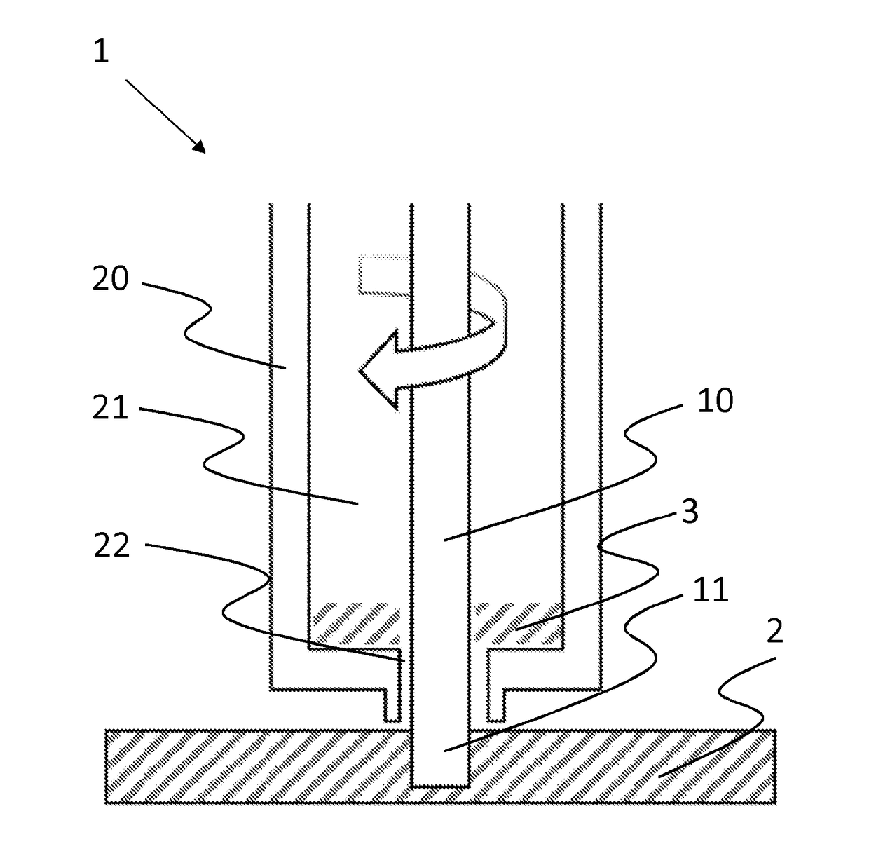
[0042]In FIG. 1, a schematic cross-sectional view of a tissue harvesting device 1 is shown. Here, only the distal portion intended to make contact with soft tissue is show...


PUM


Abstract

A tissue harvesting device (1), comprising a rotary cutting tool (10) adapted to cut soft tissue and a collection container (20) including a central bore (21) extending in a longitudinal direction and with a distal opening (22), wherein the cutting tool is arranged in the central bore of the collection container and adapted to pass through the distal opening and draw harvested soft tissue fragments into the collection container, and a housing (30) accommodating the cutting tool and the collection container, wherein the collection container is axially movable with respect to the housing.

Description

TECHNICAL FIELD[0001]The present invention relates generally to the field of tissue transplantation, and more specifically, to devices and methods for replacing or regenerating soft tissue by autotransplantation (autograft), allotransplantation (allograft) or xenotransplantation (xenograft). An autograft is a tissue transplant donated from the same individual, usually from another area of the body to heal injured tissue. An allograft is a tissue transplant from a genetically non-identical donor of the same species, whereas a xenograft is a tissue transplant from a different species. The apparatus of the present invention facilitates the collection of soft tissue fragments from one or more donor sites, e.g. for use in transplantation of micro grafts for wound treatment, such as deeper burns, pressure ulcers etc.[0002]The invention is not limited to use only in wound care and its related uses, but may be used to collect soft tissue fragments from any other site in a human or animal bo...

Claims


Application Information

Patent Timeline
Patent Type & Authority Applications(United States)
IPC IPC(8): A61B17/322A61B17/32
CPCA61B17/322A61B17/32002A61B2017/00969A61B2017/320064A61B17/3494A61B2017/0023A61B2017/00734A61B2017/00761A61B10/0233A61B10/0283A61B2017/3225A61B2010/0225
Inventor SKOG, MARTENSIVLER, PETTERRAVICHANDRAN, RANJITHKUMARJUNKER, JOHANAILI, DANIELTHORFINN, JOHANELMASRY, MOUSTAFA MOHAMED HUSSEINHARRIS, JAMES
Owner SKOG MARTEN
Why Eureka
Try Eureka