Monoclonal antibody specific to pcv2 and method for diagnosing pmws using same

a technology of pcv2 and monoclonal antibody, which is applied in the field of monoclonal antibody specific to pcv2 and the method of diagnosing pmws using same, can solve the problem of difficult to accurately diagnose pcv2 simply

Inactive Publication Date: 2018-12-20
BIOPOA +1
View PDF0 Cites 2 Cited by
  • Summary
  • Abstract
  • Description
  • Claims
  • Application Information

AI Technical Summary

Benefits of technology

[0025]Accordingly, the monoclonal antibody according to the present invention can make it possible to determine whether the antibody with respect to PCV2 is a neutralizing antibody by a vaccine antigen or an antibody induced by immune decoy.

Problems solved by technology

However, it is difficult to accurately diagnose PCV2 simply based on the antibody titer because the replication of PCV2 is inhibited and a higher level of the neutralizing antibody is exhibited in pigs vaccine with a PCV2 VLP vaccine, whereas the titer of the antibody, which cannot neutralized PCV2, especially the antibody at the C-terminus of CP, is elevated (Trible B R et al., Vaccine 2012(30) 4079-85, Antibody responses following vaccination versus infection in a porcine circovirus-type 2 (PCV2) disease model show distinct differences in virus neutralization and epitope recognition).

Method used


Image

  • Monoclonal antibody specific to pcv2 and method for diagnosing pmws using same
  • Monoclonal antibody specific to pcv2 and method for diagnosing pmws using same
  • Monoclonal antibody specific to pcv2 and method for diagnosing pmws using same

Examples


example 1

on of PCV2 Antigen

[0078]1-1. Synthesis of Peptides

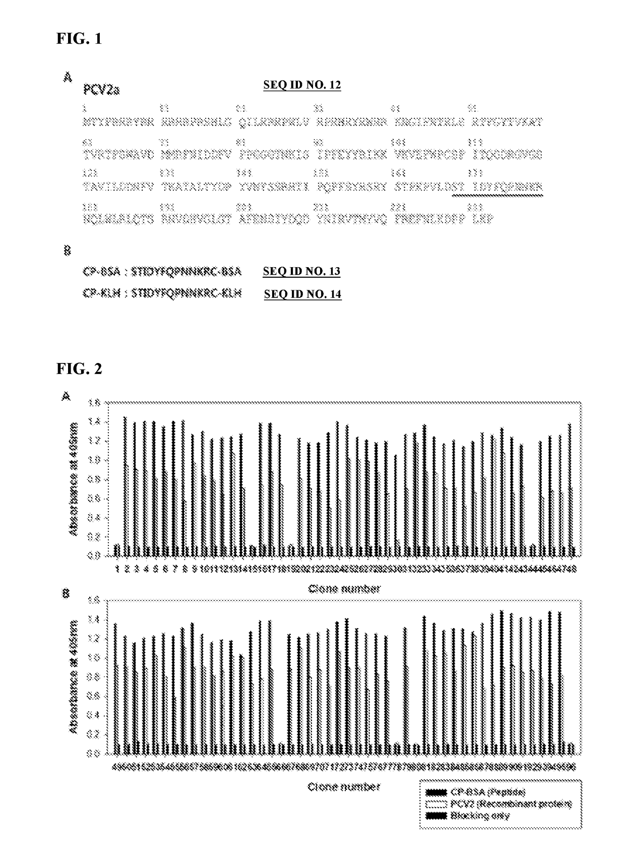
[0079]Cysteine was added to the end of the 12 amino acids positioned from the 169th to the 180th, which was known as a decoy epitope of porcine circovirus 2 (PCV2), to synthesize a peptide (Peptron, Korea). BSA and KLH were conjugated to the cysteine of the C-terminus and used it as an antigen for screening antibody and the immunity of chickens. The STIDYFQPMMKR peptide was named as CP-BSA or CP-KLH, short for the circovirus peptide.

[0080]1-2. PCV2 Recombinant Protein (Monomer)

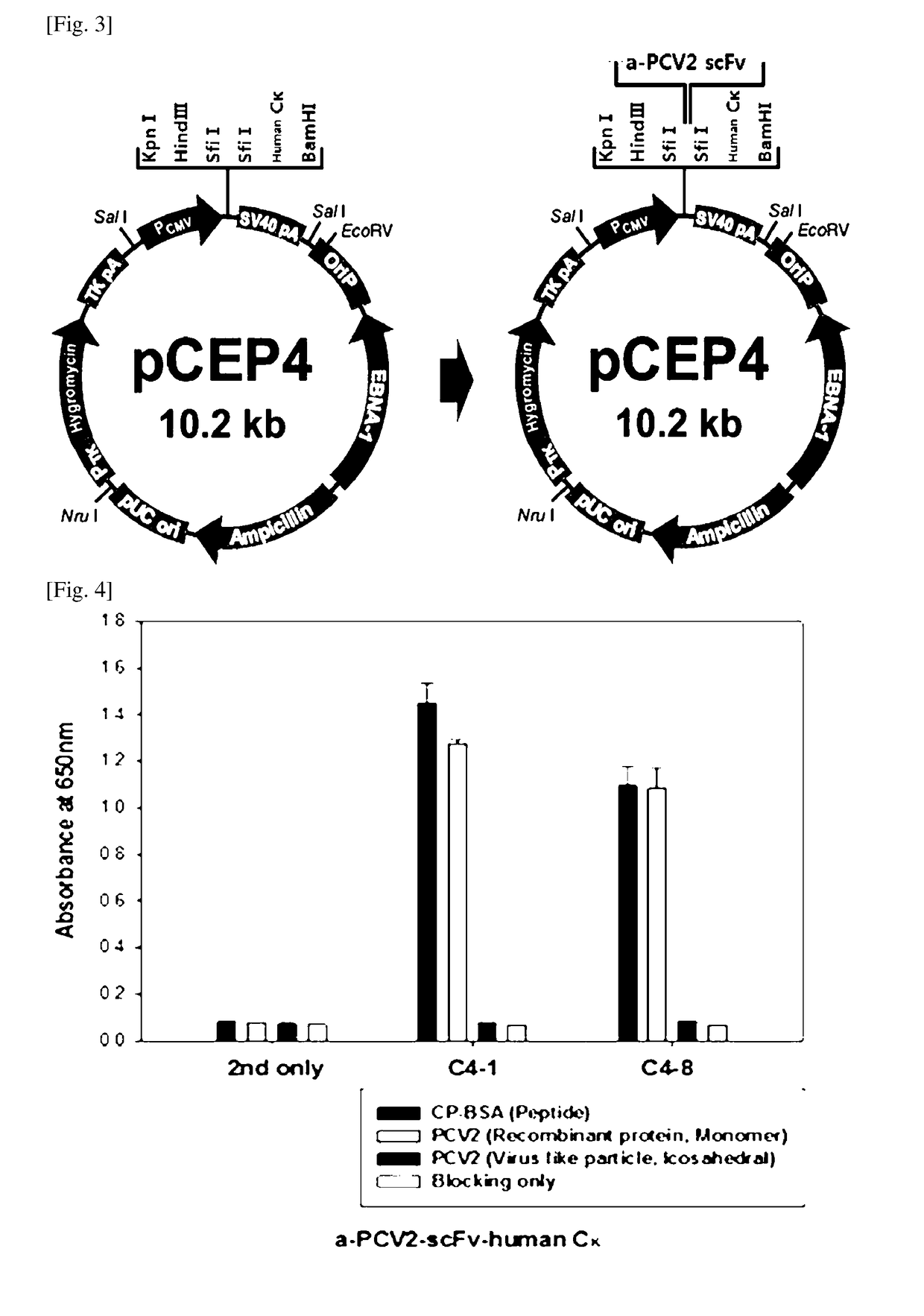
[0081]The amino acids at the N-terminus of PCV2 were deleted not to generate an icosahedral structure but to be present in a monomeric form to expose the CP (169 to 180) decoy epitope, thereby enabling a recombinant protein expressed in E. coli to include the amino acid residues of the CP of PCV2 from the 43rd to the 233rd (FIG. 1). The recombinant protein was expressed in E. coli using the method known in the art [see (Pileri E et al., Vet J. 2014, 201(3), 429...

example 2

on of Library of Immune Antibody Against PCV2

[0085]2-1. Immunization

[0086]The peptide KLH conjugate (CP-KLH; 50 μg) synthesized in Example 1, as an antigen, was mixed with phosphate buffered saline (PBS; 750 μL) and cultured at 37° C. for 30 minutes. Then, the resultant was emulsified in an adjuvant of water-in-oil emulsion (RIBI+MPL+TDM+CWS adjuvant, Sigma, St. Louis, Mo., USA) containing detoxified endotoxin (monophosphorylate lipid A species; MPL) and mycobacterial cell wall components (TDW, CWS) in 2% squalene, and then subcutaneously injected to 3 chickens. In the same manner, the chickens were further inoculated 3 weeks thereafter, and subsequently 2 weeks thereafter thereby performing a total of 3 immunizations. The antibody titer of the immunized chickens was determined by an enzyme-linked immunosorbent assay (ELISA) using the horseradish peroxidase (HRP) conjugated anti-chicken IgG (Y) polyclonal antibody (rabbit anti-chicken IgG(Y)-HRP, Millipore corporation, Billeria, Mas...

example 3

anning on Fixed Antigen (Bio-Panning)

[0096]Bio-panning was performed using magnetic beads (Dynabeads M-270 Epoxy, Invitrogen). An antigen was coated while rotating / stirring 3 μg of CP-BSA (peptide) and PCV2 recombinant protein (monomer) to the 1×10⁷ beads at room temperature for 20 hours. The coated beads were washed 4 times with PBS, blocked with PBS containing 3% BSA at room temperature for 1 hour, and cultured with the phage-displayed scFv obtained from Example 2-3 at room temperature for 2 hours. To remove the phage, which is not bound to the antigen coated to the beads, the beads were washed with 0.05% Tween 20 / PBS, and the bound phage was eluted using 50 μL of 0.1 M glycine-HCl (pH 2.2) and neutralized with 2 M Tris-HCl (pH 9.1). E. coli ER2738 was infected using the phage-containing supernatant and, for the overnight amplification of phage, was rescued using VCSM13 helper phage. Additionally, the culture broth infected with the phage was plated on an LB agar plate containing ...


PUM

PropertyMeasurementUnit
volumeaaaaaaaaaa
volumeaaaaaaaaaa
volumeaaaaaaaaaa

Abstract

The present invention relates to a monoclonal antibody specific to porcine circovirus 2 (PCV2) and a method for diagnosing post-weaning multi-systemic wasting syndrome (PMWS) using the same. More specifically, the present invention relates to monoclonal antibodies C4-1 and C4-8 of scFV-human Cκ fusion recombinant protein, which specifically binds to a decoy epitope of porcine circovirus 2, and to a method for diagnosing post-weaning multi-systemic wasting syndrome using the same. The monoclonal antibody of the present invention makes it possible to determine whether an antibody against PCV2 is a neutralizing antibody by a vaccine antigen or an antibody induced by immune decoy.

Description

BACKGROUND OF THE INVENTIONField of the Invention[0001]The present invention relates to a monoclonal antibody specific to porcine circovirus 2 (PCV2) and a method for diagnosing post-weaning multi-systemic wasting syndrome (PMWS) using the same. More specifically, the present invention relates to monoclonal antibodies C4-1 and C4-8 of an scFV-human Cκ fusion recombinant protein, which specifically binds to a decoy epitope of porcine circovirus 2, and a method for diagnosing post-weaning multi-systemic wasting syndrome (PMWS) using the same.Related Art[0002]Porcine circovirus 2 (PCV2) is a small non-enveloped icosahedral virus classified into the family of Circoviridae, which contains single-stranded circular DNG genome of about 1.76 kb and has two major open reading frames (ORFs). ORF1 produces viral replication protein (Rep) and ORF2 produces capsid protein (CP) (J. Gillespie et al. J Vet Intern Med, 2009, 23, 1151-6, Porcine circovirus type 2 and porcine circovirus-associated dise...

Claims


Application Information

Patent Timeline
Patent Type & Authority Applications(United States)
IPC IPC(8): C07K16/46C07K16/08G01N33/532
CPCC07K16/46C07K16/081G01N33/532C07K2317/622C07K2317/24G01N2800/26C07K2317/34C07K2317/52G01N33/56983G01N2333/01G01N2800/38C07K16/08G01N33/53
Inventor CHO, SUN HEEKIM, TAE EUNCHUNG, JUN HOKIM, HYO RIJIN, JUN YEONG
Owner BIOPOA
Why Eureka
Try Eureka