Preimplantation screening

a technology of preimplantation and screening, applied in the field of preimplantation screening, can solve the problems of limited success rate of vitro fertilization (ivf), low success rate, and difficulty in selecting good quality embryos for uterine implantation, and achieve the effect of assessing embryo viability

Inactive Publication Date: 2019-01-31
UNIVERSITY OF WARWICK
View PDF0 Cites 0 Cited by
  • Summary
  • Abstract
  • Description
  • Claims
  • Application Information

AI Technical Summary

Benefits of technology

[0013]The present inventors have identified two proteins, TMPRSS2 and PRSS8, in the culture media of IVF embryos, the levels of which can be used to predict embryo viability. The expression of both TMPRSS2 and PRSS8 is upregulated as the embryo progresses to the blastocyst stage. Importantly, these markers have been found to correlate with embryo viability as assessed by successful embry

Problems solved by technology

In vitro fertilization (IVF) has a limited success rate with less than 40% of IVF treatments resulting in live births (www.nhs.uk/conditions/IVF).
A major contributing factor to the low success rate is the difficulty in selecting good quality embryos for uterine implantation, particularly in cases of ‘single embryo transfer’ where only one embryo is transferred to a mother to avoid problems with multiple births.
Defective embryos of this nature are either unsuitable for implantation altogether, or in some cases will result in implantation followed by miscarriage of the unviable or “developmentally-impaired” embryo.
The development of a reliable method for assessing human embryo viability remains a significant challenge for improving pregnancy rates in IVF clinics.
However, there are disadvantages associated with these existing techniques (see review by Bolton et al.
Moreover, morphological examination is not always a reliable means by which to detect those chromosomal abnormalities that occur in unviable embryos.
The embryo is mechanically removed from its outer shell so that cells can be aspirated, which may result in physical damage to the embryo at the time of biopsy.
PGS is further complicated by a phenomenon known as mosaicism, where

Method used


Image

  • Preimplantation screening
  • Preimplantation screening
  • Preimplantation screening

Examples


example 1

Analysis of Human and Murine Embryonic Transcriptome for Serine Protease Genes Expressed Throughout Pre-Implantation Development

[0083]In silico analysis was performed on publicly available datasets from the Gene Expression Omnibus project, the GEO project, (Edgar et al. 2002 Nucleic Acids Research, 30:1, 207-210), accession number GSD3959 (homo sapiens) and GDS813 (mus musculus). Statistical analysis of the datasets obtained from the GEO project was performed using SPSS Software, IBM.

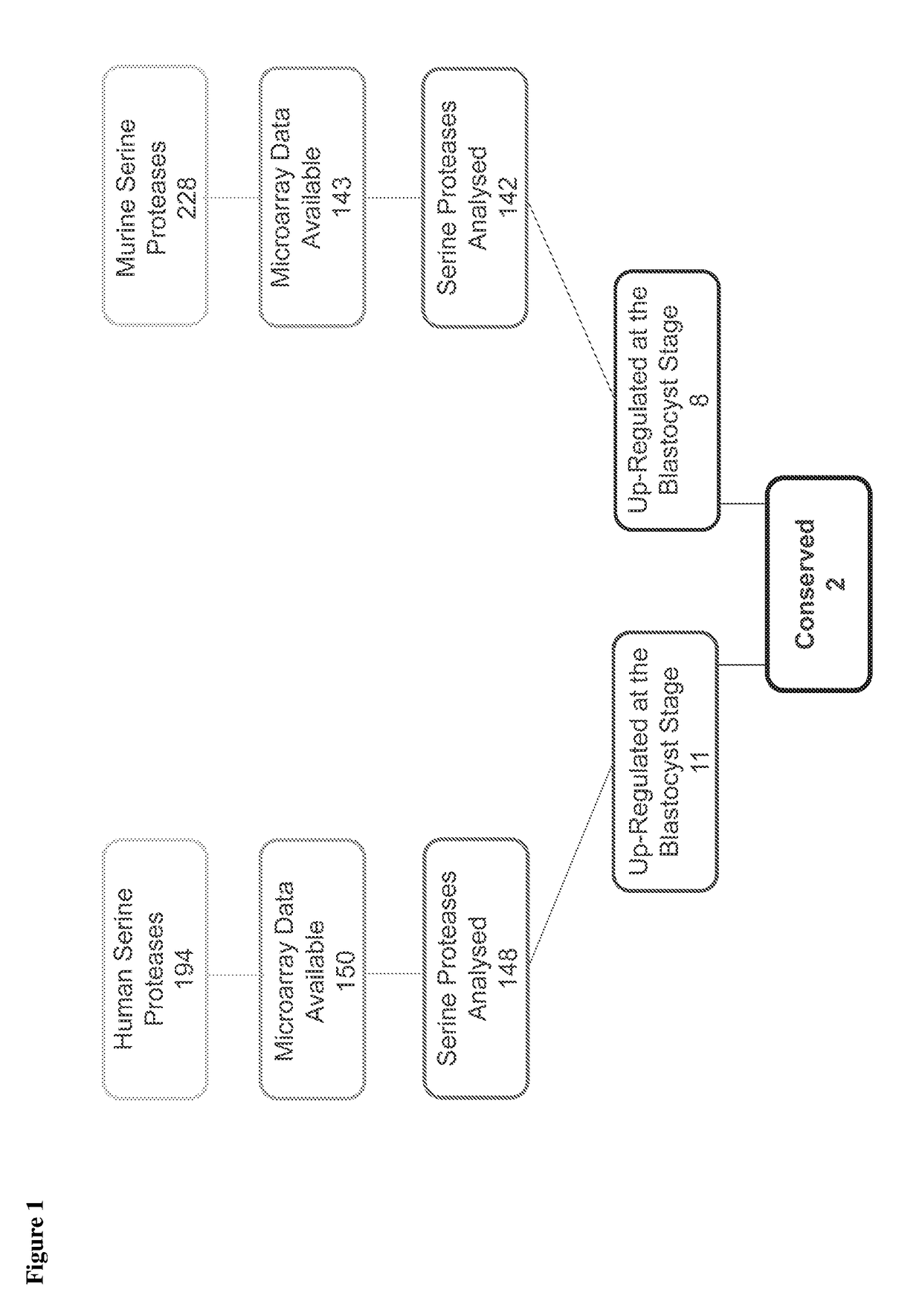
[0084]As shown in FIG. 1, a total of 194 human and 228 murine serine protease genes were identified in the available datasets, and microarray data on expression during pre-implantation embryo development was available for 150 and 143 of these genes, respectively. 11 human serine proteases and 8 mouse serine proteases were identified as being selectively up-regulated at the blastocyst stage and 2 of these proteases were found to be conserved between species. These two serine proteases are TMPRSS2 and PRS...

example 2

Measurement of Protease and Trypsin Activity in Embryo-Conditioned Medium (ECM)

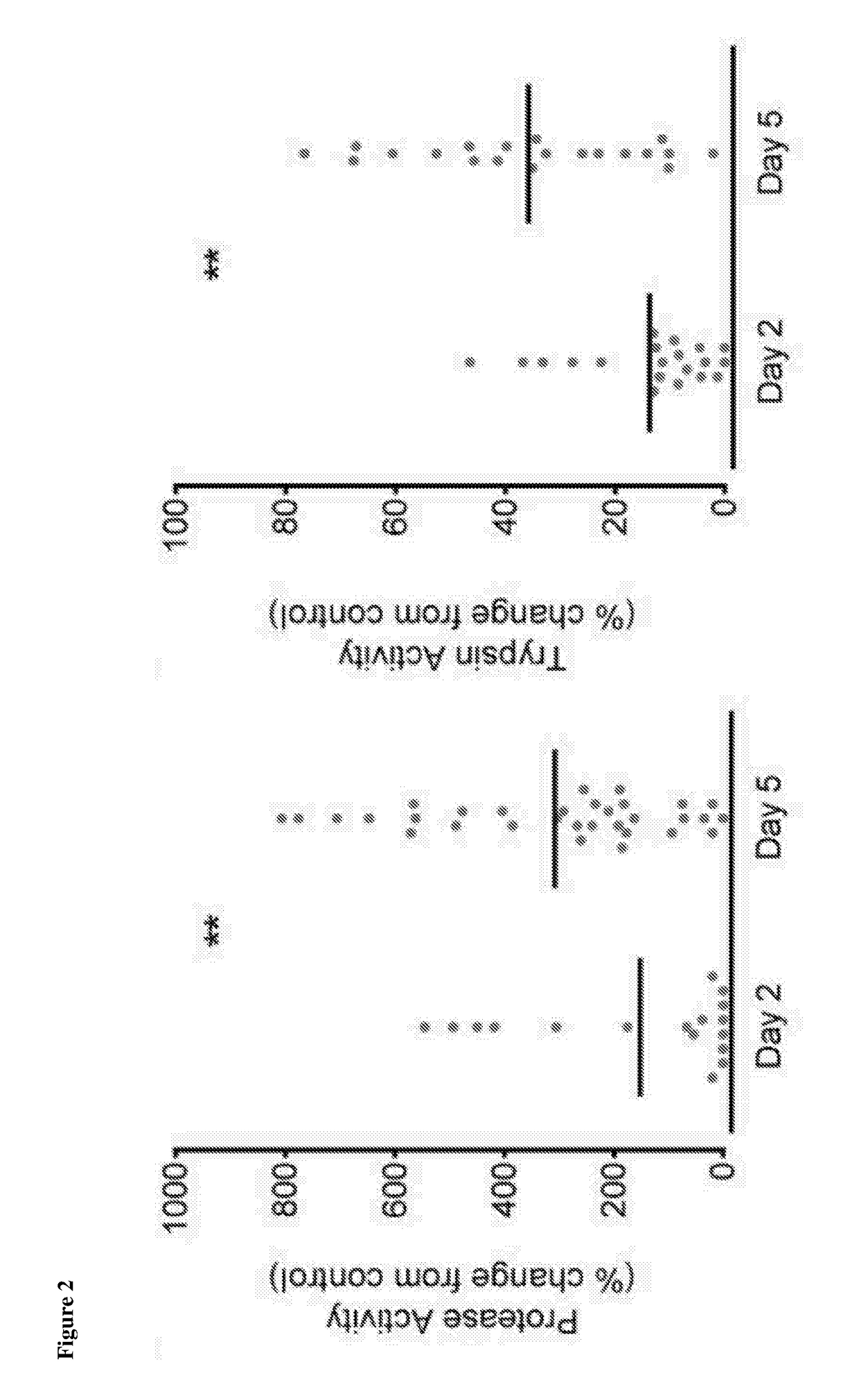
[0085]Experiments were carried out to assess total protease and trypsin activity in samples of conditioned media obtained from the in vitro culture of embryos i.e embryo-conditioned medium.

[0086]Embryo conditioned medium was obtained from two IVF units: Centre for Reproductive Medicine (CRG), Universitair Ziekenhuis Brussel, Laarbeeklaan 101, 1090 Brussels, Belgium; Centre for Reproductive Medicine (CRM), University Hospitals Coventry & Warwickshire, Clifford Bridge Road, Coventry CV2 2DX, UK.

[0087]Embryos produced were cultured in individual 20 μl drops of ISM1™ (Origio) and BlastAssist™ (Origio). During this study the CRG changed their culture method to include the ORIGIO® Sequential Series™ (Fert™, Cleav™, Blast™).

[0088]Embryos from CRG Brussels were cultured individually in 25 μl drops of Q1 (Quinn's Protein Plus Cleavage Medium) and Q2 (Quinn's Advantage™ Protein Plus Blastocyst Medium) (Sage In Vitr...

example 3

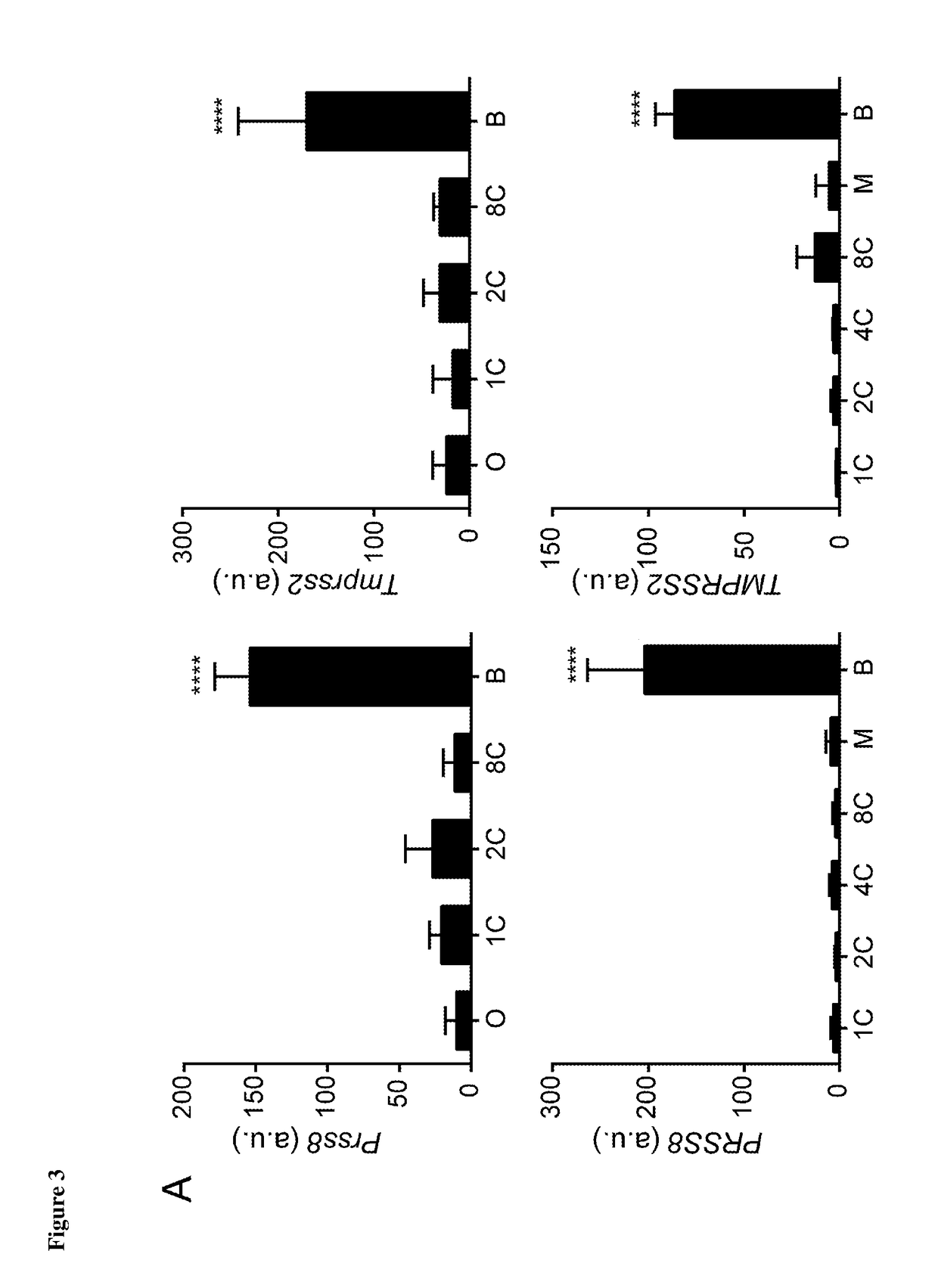
Immunofluorescent Labelling of TMPRSS2 and PRSS8 in both Human and Mouse Blastocyts

[0095]Human and mouse embryos were collected at various stages of development, fixed in 4% Para-formaldehyde / PBS for 1 hour at room temperature then stored at 2-8° C.

[0096]Embryos selected for use were washed in PBS, permeabilised for 1 hour in 0.01% Triton X-100 / PBS at room temperature and incubated in BSA for 1 hour at 4° C. to prevent non-specific binding. Embryos were then incubated in selected primary antibodies (diluted 1:100 in PBS) or BSA in the case of the negative control, overnight at 4° C. The antibodies used in this study are shown below in Table 1.

TABLE 1NameCompanyCat. NumberAlpha-1-MicroglobulinNovus BiologicalsH00000259-M01Enterokinase*Novus BiologicalsNBP1-55616NANOG*Fisher Scientific12907229PRSS8 (Protease. serine, 8)*Antibodies OnlineABIN761891Anti-SPINT1*SigmaSAB1409704-50UGTMPRSS2 / PRSS10*Antibodies OnlineABIN716876*No commercially available blocking peptides

AMBP blocking peptide ...


PUM


Abstract

The present invention relates to a method of assessing the viability of an embryo, wherein the method comprises a step of: measuring the level and/or activity of a protein marker selected from either TMPRSS2 or PRSS8 in a sample of culture medium, wherein the sample has been obtained from the in vitro culture medium of an embryo produced by in vitro fertilization, wherein the level and/or activity of the protein marker is indicative of the viability of the embryo.

Description

CROSS REFERENCE TO RELATED APPLICATIONS[0001]This application is a U.S. National Phase application of International Appl. No. PCT / GB2016 / 053535, filed Nov. 11, 2016, which claims priority to United Kingdom Appl. No. GB 1519944.1, filed Nov. 12, 2015, each of which is incorporated herein by reference.FIELD OF THE INVENTION[0002]The present invention relates to methods of assessing the viability of embryos following in vitro fertilization (IVF). The embryos are assessed for the purposes of assessing their suitability for embryo transfer to a female recipient. The invention also relates to methods of selecting embryos suitable for implantation based on a comparison of embryos produced by IVF carried out with oocytes from the same female donor. The methods of the invention are based on measuring the levels and / or activity of a protein marker, selected from either TMPRSS2 or PRSS8, in samples of embryo-conditioned culture media. The levels of these two markers in embryo-conditioned media...

Claims


Application Information

Patent Timeline
IPC IPC(8): C12Q1/37G01N33/68
CPCC12Q1/37G01N33/689C12M41/46
Inventor SALTER, SCARLETTBROSENS, JAN
Owner UNIVERSITY OF WARWICK
Why Eureka
Try Eureka