Deep learning-based diagnosis and referral of ophthalmic diseases and disorders

a deep learning and diagnosis technology, applied in the field of deep learning-based diagnosis and referral of ophthalmic diseases and disorders, can solve the problems of inability to adequately perform image analysis without significant human intervention, creation and refinement of multiple classifiers, and inability to adequately perform so as to achieve the effect of effective image analysis and/or diagnosis, less computational power, and improved speed, efficiency and computational power

Active Publication Date: 2019-04-18
AITECH +1
View PDF1 Cites 80 Cited by
  • Summary
  • Abstract
  • Description
  • Claims
  • Application Information

AI Technical Summary

Benefits of technology

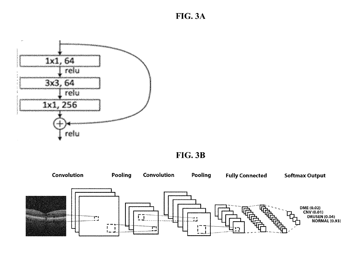
[0004]The present disclosure solves these technical problems with existing computer systems carrying out image analysis by providing improved systems and techniques that do not require substantial intervention by an expert to generate the classifiers. These include, for example, convolutional neural network layers that provide multiple processing layers to which image analysis filters or convolutions are applied. The abstracted representation of images within each layer is constructed by systematically convolving multiple filters across the image to produce a feature map used as input for the following layer. This overall architecture enables images to be processed into pixels as input and to generate the desired classification as output. Accordingly, the multiple resource-intensive steps used in traditional image analysis techniques such as handcrafted object segmentation, identification of the segmented objects using a shallow classifier, and classification of the image is no longer required.
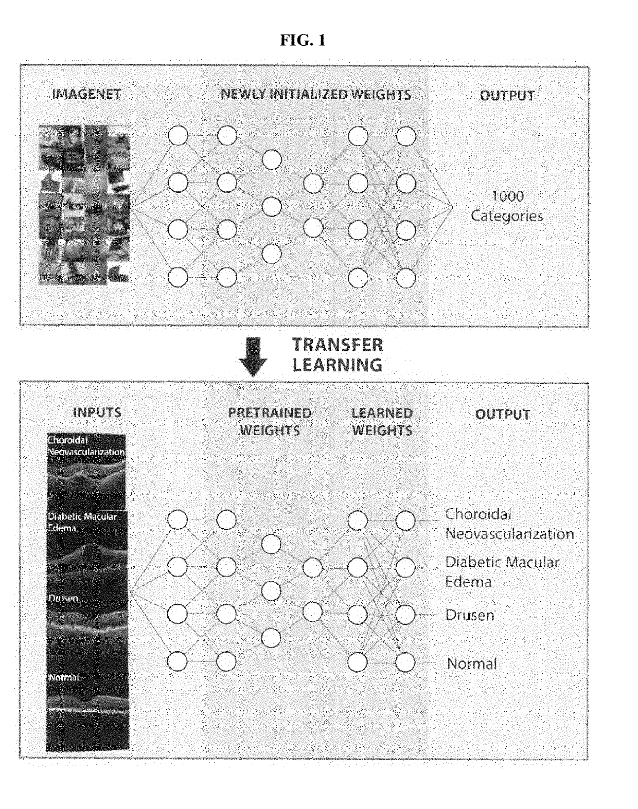
[0005]In addition, the present disclosure solves the technical problem of insufficient images in the relevant domain (e.g. medical images for a specific ophthalmic disease) for training algorithms to effectively perform image analysis and / or diagnosis. Certain embodiments of the present disclosure include systems and techniques applying a transfer learning algorithm to train an initial machine learning algorithm such as a convolutional neural network on images outside of the specific domain of interest to optimize the weights in the lower layer(s) for recognizing the structures found in the images. The weights for the lower layer(s) are then frozen, while the weights of the upper layer(s) are retrained using images from the relevant domain to identify output according to the desired diagnosis (e.g. identification or prediction of specific ophthalmic diseases or conditions). This approach allows the classifier to recognize distinguishing features of specific categories of images (e.g. images of the eye) far more quickly using significantly fewer training images and while requiring substantially less computational power. The use of non-domain images to partially train or pre-train the classifier allows optimization of the weights of one or more of the neural network layers using a deep reservoir of available images corresponding to thousands of categories. The result is a classifier having a sensitivity, specificity, and accuracy that is unexpected and surprising compared to the traditional approach, especially in view of the improvements in speed, efficiency, and computational power required. Indeed, certain embodiments of the classifier outperform human experts in correctly diagnosing medical images according to sensitivity, specificity, accuracy, or a combination thereof.
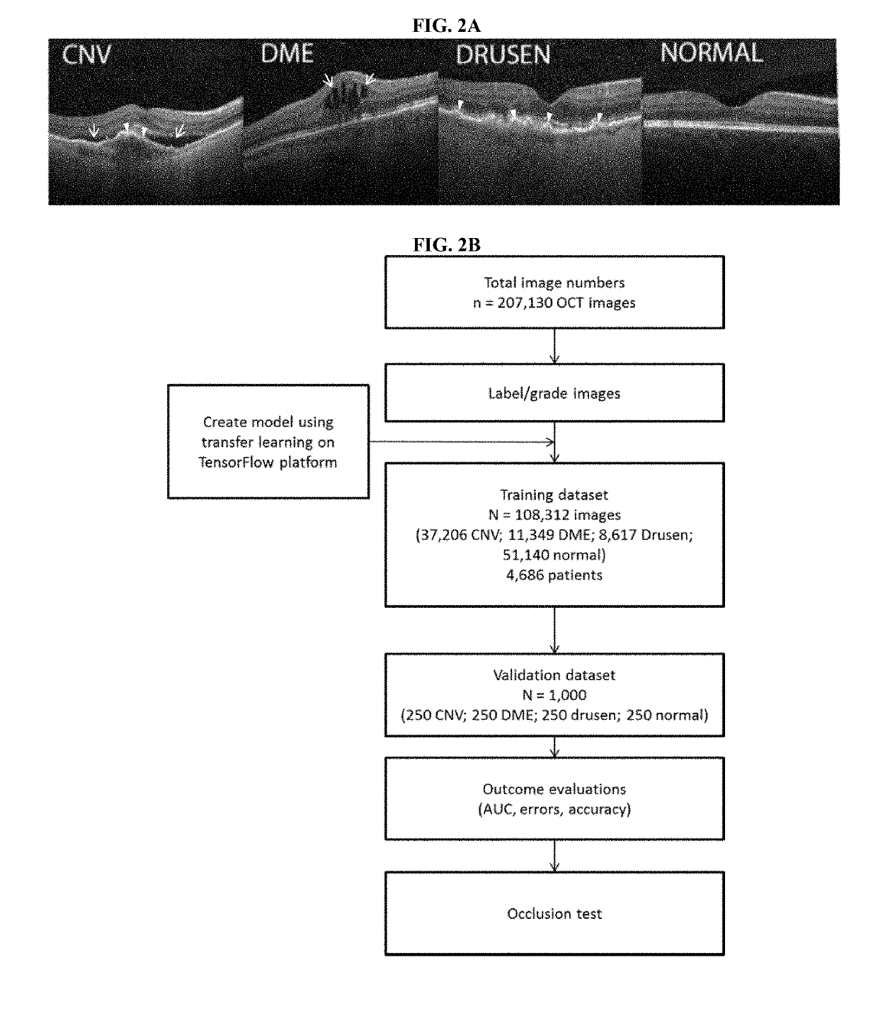
[0006]The present disclosure also addresses the black box nature of machine learning by allowing identification of the critical areas contributing most to the classifier's predicted diagnosis. Certain embodiments of the present disclosure utilize occlusion testing on test images to identify the regions of interest that contribute the highest importance to the classifier's ability to generate accurate diagnoses. These regions can be verified by experts to validate the system, which creates greater transparent and increases trust in the diagnosis.

Problems solved by technology

Traditional algorithmic approaches to medical image analysis suffer from numerous technical deficiencies related to an inability to adequately perform the analysis without significant human intervention and / or guidance, which belies the supposed promise of artificial intelligence and machine learning to revolutionize disease diagnosis and management.
As a result, the creation and refinement of multiple classifiers required considerable expertise and time, and was computationally expensive.
In addition, the training of machine learning classifiers is often deficient due to a lack of sufficient medical images in the training set.
This problem is exacerbated in the case of diseases or conditions that are relatively rare or lack adequate access to the medical images.
Moreover, because machine learning often behaves like a black box, acceptance of diagnoses generated through such methods can be hindered due to the lack of transparency on how the classifier evaluates a medical image to generate a prediction.
Indeed, certain embodiments of the classifier outperform human experts in correctly diagnosing medical images according to sensitivity, specificity, accuracy, or a combination thereof.

Method used


Image

  • Deep learning-based diagnosis and referral of ophthalmic diseases and disorders
  • Deep learning-based diagnosis and referral of ophthalmic diseases and disorders
  • Deep learning-based diagnosis and referral of ophthalmic diseases and disorders

Examples


example 1

[0144]A total of 207,130 OCT images were initially obtained of which 108,312 images (37,206 with CNV, 11,349 with DME, 8617 with drusen, and 51,140 normal) from 4,686 patients were used to train the AI system. The trained model (e.g. trained machine learning algorithm or classifier) was then tested with 1,000 images (250 from each category—CNV, DME, drusen, and normal) from 633 patients.

[0145]After 100 epochs (iterations through the entire dataset), the training was stopped due to the absence of further improvement in both accuracy (FIG. 4A) and cross-entropy loss (FIG. 4B).

[0146]In a multi-class comparison between CNV, DME, drusen, and normal, an accuracy of 96.6% (FIG. 4), with a sensitivity of 97.8%, a specificity of 97.4%, and a weighted error of 6.6% were achieved. ROC curves were generated to evaluate the model's ability to distinguish “urgent referrals” (defined as CNV or DME) from normal and drusen exams. The area under the ROC curve was 99.9% (FIG. 4).

[0147]Another “limited...

example 2

[0151]An independent test set of 1000 images from 633 patients was used to compare the AI network's referral decisions (e.g. using the AI from Example 1) with the decisions made by human experts. Six experts with significant clinical experience in an academic ophthalmology center were instructed to make a referral decision on each test patient using only the patient's OCT images. Performance on the clinically most important decision of distinguishing patients needing “urgent referral” (those with CNV or DME) compared to normal patients is displayed as a ROC curve, and this performance was comparable between the AI system and the human experts (FIG. 4A).

[0152]Having established a standard expert performance evaluation system, the potential impact of patient referral decisions between the network and human experts was compared. The sensitivities and specificities of the experts were plotted on the ROC curve of the trained model, and the differences in diagnostic performance, measured ...

example 3

Summary

[0168]Despite the overall effectiveness of cataract surgery in improving vision, a significant number of patients still have decreased vision after cataract extraction. Understanding the underlying etiologies of persistent post-operative vision impairment is of great importance. In China, myopia is the number one cause of visual impairment, with nearly half of population being affected. Despite its prevalence, its impact on the visual outcomes after cataract surgery remains unclear. In this study, a prospective trial was conducted on the effects of high myopia on visual outcomes after cataract surgery in 1,005 patients with high myopia patients (axial length ≥26 mm). Multi-variable analysis was conducted to identify factors influencing visual outcomes. Macular sensitivity images were then used to train a deep learning model for predicting post-operative visual acuity. This model demonstrated an accuracy of over 90% in the validation set, suggesting the high utility of artific...


PUM


Abstract

Disclosed herein are systems, methods, devices, and media for carrying out medical diagnosis of ophthalmic diseases and conditions. Deep learning algorithms enable the automated analysis of ophthalmic images to generate predictions of comparable accuracy to clinical experts.

Description

CROSS-REFERENCE[0001]This application claims the benefit of U.S. Provisional Application No. 62 / 572,384, filed Oct. 13, 2017, U.S. Provisional Application No. 62 / 668,698, filed May 8, 2018, and U.S. Provisional Application No. 62 / 694,939, filed Jul. 6, 2018, which each of the applications is incorporated herein by reference in its entirety.BACKGROUND OF THE DISCLOSURE[0002]Many ophthalmic diseases and disorders are diagnosed based on medical imaging, such as for example retinal imaging. Medical imaging has traditionally relied upon human experts to analyze images individually. As the number of medical imaging procedures increase, demand for efficient and accurate image analysis is outstripping the supply of experts capable of performing this function.SUMMARY OF THE DISCLOSURE[0003]Traditional algorithmic approaches to medical image analysis suffer from numerous technical deficiencies related to an inability to adequately perform the analysis without significant human intervention an...

Claims


Application Information

Patent Timeline
Patent Type & Authority Applications(United States)
IPC IPC(8): A61B5/00A61B3/00A61B3/14A61B3/12A61B3/10G16H30/40G06N20/00
CPCA61B5/7267A61B3/0025A61B3/14A61B3/12A61B3/102A61B5/6898A61B5/0013A61B5/0022A61B5/7282A61B5/486G16H30/40G06N20/00A61B2576/02A61B5/021A61B5/024A61B5/0531A61B5/4872A61B5/4875G16H50/20G16H40/63G06N3/084G06N3/045
Inventor ZHANG, KANGHOU, RUIZHENG, LIANGHONG
Owner AITECH
Why Eureka
Try Eureka