Methods and compositions for the treatment of intervertebral disc herniation
Inactive Publication Date: 2019-04-18
GU VENTURES AB
0 Cites 1 Cited by
AI-Extracted Technical Summary
Problems solved by technology
This can cause pressure on an adjacent nerve root which, in combination with chemical factors as further discussed below, causes radiating pain.
For a long time, sciatic pain in disc herniation was believed to be caused solely by mechanical pressure on a nerve root, but during the last three decades this has been proven wrong.
However, these different histological characteristics have not been found useful in predicting clinical outcome [26].
Other studies have failed to correlate morphological development with clinical outcome, and as such this correlation remains a controversial subject.
TNF then activates and increases the expression of MMP's, leading to degradation of the extracellular matrix and thus spontaneous resorption of the hernia.
Interestingly, dis...
Abstract
The invention relates to methods and compositions for use in the treatment or prophylaxis of intervertebral disc herniation in a mammal. The composition according to the invention comprises an inhibitor of T-cell activation, said inhibitor being capable of inhibiting CD28-mediated co-stimulation of T-cells. The said inhibitor of T-cell activation is preferably a protein comprising either an exact or a modified version of the extracellular domain of CTLA-4, such as abatacept, belatacept, XPro9523 and/or ASP2408.
Application Domain
Cell receptors/surface-antigens/surface-determinantsPeptide/protein ingredients +5
Technology Topic
CTLA-4Intervertebral disc herniation +6
Image
Examples
- Experimental program(4)
Example
Example 1: Pilot Study, Treatment with Abatacept
[0062]20 female Sprague-Dawley rats weighing approximately 225 g were evenly divided into two groups: disc puncture (DP, n=10) and disc puncture+abatacept (DP+abatacept, n=10). The rats were housed with free access to food and water in environmental-enriched cages. All rats underwent the same surgical procedure as described below.
[0063]Anesthesia was induced and maintained by the inhalation of Isoflurane. A single dose of 0.05 mg/kg buprenorphine (Temgesic®) was administered subcutaneously before the procedure for per- and postoperative analgesia.
[0064]Following induction of anesthesia, a skin incision of approximately 6 cm was made in the midline over the spinous processes of the lumbar and caudal spine. The spinal muscles on the left side of the lumbar spine were dissected to expose the left L4/L5 facet joint. The left L4/L5 facet joint was removed to expose the underlying dural sac, L4 nerve root and L4/L5 intervertebral disc. The disc was then punctured using a 23 g needle. A small amount of air (0.2-0.3 ml) was injected into the disc to facilitate leakage of nucleus pulposus. The spinal muscles and the thoracolumbar fascia were then sutured and the skin closed with metal clips.
[0065]For the DP group, no additional treatment was given. For the DP+abatacept group, 10 mg/kg abatacept was administered intraperitoneally immediately before surgery. This was repeated 7 and 14 days after surgery.
[0066]To evaluate the effects on the morphology of the hernia-like nodule a macroscopic analysis was performed. 21 days after surgery anesthesia was again induced by inhalation of isoflurane followed by euthanization by incision of the heart, causing the rat to quickly bleed to death. The lumbar spine was then dissected by removing the spinal muscles and the vertebral arches of L4 and L5, exposing the L4/L5 intervertebral disc. A macroscopic analysis was performed through the surgical microscope by an experienced researcher. A description of the different types of data collected can be found in Table 1.
[0067]The data from the macroscopic analysis was considered ordinal. To test for statistical significance between the DP and DP+abatacept groups, Mann-Whitney U-tests were performed. Significance was defined as p≤0.05.
[0068]All rats survived the surgery and displayed good general condition throughout the study. In the DP group, 8 out of 10 rats displayed clear or pronounced disc nodules (Table 2). In the DP+abatacept group, no clear or pronounced nodules were found. The difference in nodule size was significant between the two groups (p=0.000). There was also significantly less inflammation observed in the DP+abatacept group (p=0.005). Tendencies of less osteophytes were also noted, but this difference was not significant (p=0.143).
Example
Example 2: Longitudinal Assessment of Nodule Size, Treatment with Abatacept
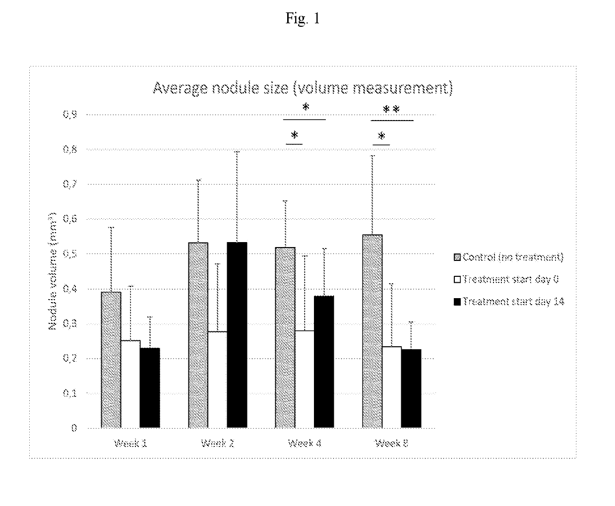
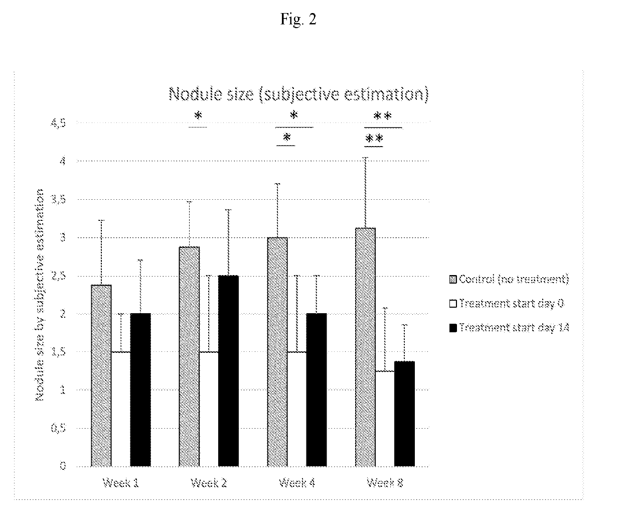
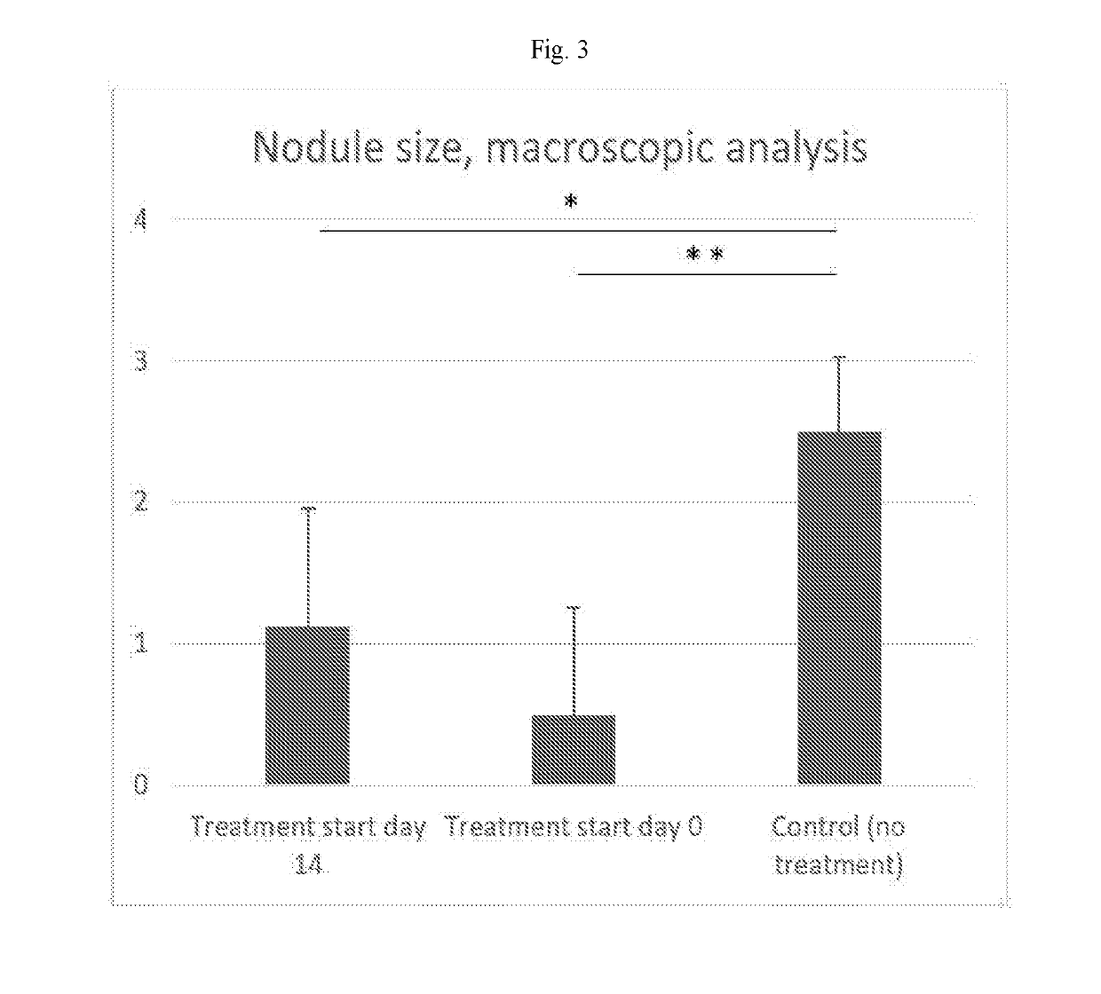
[0069]The purpose of this study was to assess 1) how the nodule formed after disc puncture in the rat varies in size over time, 2) if the formation of the nodule can be inhibited by administration of the T-cell inhibitor abatacept, and 3) if the size of existing nodules can be reduced by administration of the T-cell inhibitor abatacept.
[0070]24 female Sprague-Dawley rats (n=24) weighing approximately 225 g were evenly divided into three groups: Control group (no treatment, n=8); Treatment start day 0 (n=8); and Treatment start day 14 (n=8). The rats were housed with free access to food and water in environmental-enriched cages. All rats underwent the same surgical procedure as described below.
[0071]Anesthesia was induced and maintained by the inhalation of Isoflurane. A single dose of 0.05 mg/kg buprenorphine (Temgesic®) was administered subcutaneously before the procedure for per- and postoperative analgesia.
[0072]Following induction of anesthesia, a skin incision of approximately 6 cm was made in the midline over the spinous processes of the lumbar and caudal spine. The spinal muscles on the left side of the lumbar spine were dissected to expose the left L5/L6 facet joint. The left L5/L6 facet joint was removed to expose the underlying dural sac, L5 nerve root and L5/L6 intervertebral disc. The disc was punctured using a 23 g needle. A small amount of air (0.2-0.3 ml) was injected into the disc to facilitate leakage of nucleus pulposus. The spinal muscles and the thoracolumbar fascia were then sutured and the skin closed with metal clips. The researcher performing the surgery was blinded to treatment groups.
[0073]All rats received an intraperitoneal injection immediately before surgery and subsequently once a week throughout the study period. For the different groups, the compound administered at the different time periods were as shown in Table 3.
[0074]For dosage calculations, a body weight of 225 g was assumed for all rats throughout the study period. When abatacept was not administered, saline was administered for blinding purposes. The rats were shortly anesthetized by inhalation of isoflurane before each injection to allow for a safe administration of the compound and to minimize pain and stress to the animal.
[0075]To assess the size of the disc nodules following disc puncture, MRI images were obtained with a 7T MRI system (Bruker Bio Spec®) week 1, 2, 4 and 8 after surgery. The rats were anesthetized by inhalation of isoflurane throughout the MRI measurements. During the measurements, breathing frequency was monitored and the body temperature maintained by placing the rat on a heated pad.
[0076]A sagittal T2-weighted 3D RARE sequence with isotropic voxels of 150 μm was used. The volume of the nodule was measured by manually defining a region of interest (ROI) of the disc aggrandizement dorsal to a longitudinal line between the most dorsal aspects of the adjacent vertebrae on each image where the disc could be identified. The number of voxels within the ROI was assessed three times for each image. The average number of voxels for each sagittal section of the disc were added and then multiplied by the voxel volume (0.003375 mm3), resulting in the total nodule size expressed in mm3. In addition to this, a subjective assessment of the nodule size was also performed by the observer for each MM. The researcher collecting the data was blinded to treatment groups.
[0077]Upon termination of the study, a macroscopic analysis was performed in addition to the MRI measurements. After the final MRI measurement 8 weeks after surgery the rats were euthanized by incision of the heart while still anesthetized by inhalation of isoflurane, causing the rat to quickly bleed to death. The lumbar spine was dissected by removing the spinal muscles and the vertebral arches of L5 and L6, exposing the L5/L6 intervertebral disc. A macroscopic assessment of the nodule size was performed through the surgical microscope. The assessments of the nodule size were performed according to the description in Table 1. The observer was not blinded to treatment groups for logistic reasons.
[0078]All rats survived the surgery and displayed good general condition throughout the study. The data from the macroscopic analysis and the subjective assessment from the MRI were considered ordinal. The data from the volume measurement on the MM was considered continuous Mann-Whitney U-tests were performed, and statistical significance defined as p≤0.05.
[0079]The results from the MM measurements can be found in FIG. 1 and FIG. 2. The results from the macroscopic analysis can be found in FIG. 3. The nodule was found to be significantly smaller in both treatment groups as compared to control by all types of assessments on week 4 and 8. In addition, a significant difference in nodule size as determined by subjective assessment was noted between “Control (no treatment)” and “Treatment start day 0” on week 2.
[0080]To conclude, the results of this study confirmed that administration of abatacept inhibited the formation of the hernia-like nodule when treatment started immediately before disc puncture. Also, when treatment with abatacept was initiated two weeks after disc puncture, the hernia-like nodules that had formed during the two weeks became significantly smaller compared to the control group, indicating that abatacept can also cause shrinkage of the hernia-like nodule after it has formed. These results contradict the generally accepted hypothesis that autoimmunity and inflammation are beneficial for the resorption of hernias, but rather suggest that specific inhibition of the immune system, more specifically T-cells, can cause resorption and thus shrinkage of hernias.
Example
Example 3: Longitudinal Assessment of Nodule Size and Immunohistochemistry, Treatment with Specific Inhibitor of CD28-Mediated Co-Stimulation of T-Cells
[0081]The purpose of this study is 1) to reproduce previous results indicating that treatment with a specific inhibitor of CD28-mediated co-stimulation of T-cells can be used to induce and/or expedite resorption of disc hernias in the modified animal model described in Example 3, and 2) to make a more detailed assessment of specific morphological changes in the hernia-like nodules caused by treatment with a specific inhibitor of CD28-mediated co-stimulation of T-cells.
[0082]48 female Sprague-Dawley rats weighing approximately 225 g are evenly divided into three groups: Control group (Control, n=16); low dose treatment (T-L n=16); high dose treatment (T-H, n=16). The rats are housed with free access to food and water in environmental-enriched cages. All rats undergo the same surgical procedure as described below. The researcher performing the surgery is blinded to treatment groups.
[0083]Anesthesia is induced and maintained by the inhalation of Isoflurane. A single dose of 0.05 mg/kg buprenorphine (Temgesic®) is administered subcutaneously before the procedure for per- and postoperative analgesia.
[0084]Following induction of anesthesia, a skin incision of approximately 5 cm is made in the midline over the spinous processes of the lumbar and caudal spine. An incision is made in the thoracolumbar fascia from level L3-L6. The spinal muscles on both sides on level L4/5 are dissected as well as the supra- and interspinous ligaments to expose the laminae and the interlaminar space at L4/5. A small laminotomy is performed on the caudal aspect of L4 using a surgical micro drill. A 23 g needle is then used to carefully incise the exposed dural sac. The needle is introduced into the cauda equina and the underlying L4/5 disc identified by probing with the needle. The disc is punctured and a small amount of air (0.2 ml) is injected into the disc to facilitate leakage of nucleus pulposus. The needle is then removed, the thoracolumbar fascia sutured and the skin closed with metal clips.
[0085]Therapeutic compounds are administered by intraperitoneal injections once a week.
[0086]To assess the size of the disc nodules following disc puncture, MRI images are obtained with a 7T MRI system (Bruker BioSpec®). Measurements are performed week 24 and 8 after surgery. The rats are anesthetized by inhalation of isoflurane throughout the MRI measurements. During the measurements, breathing frequency is monitored and the body temperature maintained by placing the rat on a heated pad.
[0087]A sagittal T2-weighted 3D RARE sequence with isotropic voxels of 150 μm is used. The volume of the nodule is measured by manually defining a region of interest (ROI) of the disc aggrandizement dorsal to a longitudinal line between the most dorsal aspects of the adjacent vertebrae on each image where the disc can be identified. The number of voxels within the ROI is assessed three times for each image. The average number of voxels for each sagittal section of the disc are added and then multiplied by the voxel volume (0.003375 mm3), resulting in the total nodule size expressed in mm3. In addition to this, a subjective assessment of the nodule size is also performed by the observer for each MM. The researcher collecting the data is blinded to treatment groups.
[0088]Upon termination of the study, a macroscopic analysis is performed in addition to the MRI measurements. After the final MRI measurement 8 weeks after surgery the rats are euthanized by incision of the heart while still anesthetized by inhalation of isoflurane, causing the rat to quickly bleed to death. The lumbar spine is dissected by removing the spinal muscles and the vertebral arches of L3 through L6, exposing the underlying intervertebral disc. A macroscopic assessment of the nodule size is performed through the surgical microscope. The assessments of the nodule size were performed according to the description in Table 1.
[0089]Some of the animals from all groups are terminated at various time points previous to week 8 to allow for harvest of the punctured intervertebral discs for assessments with histology and immunohistochemistry.
[0090]The results from this study support that targeted inhibition of T cell activation, more specifically inhibition of CD28-mediated co-stimulation, can be used to treat disc herniation by inducing and/or expediting resorption of the hernia as well as reducing inflammation in the hernia.
PUM
Property | Measurement | Unit |
Fraction | 0.9 | fraction |
Mass | 0.125 | g |
Dimensionless property | 1.0E-7 ~ 2.0E-5 | dimensionless |
Description & Claims & Application Information
We can also present the details of the Description, Claims and Application information to help users get a comprehensive understanding of the technical details of the patent, such as background art, summary of invention, brief description of drawings, description of embodiments, and other original content. On the other hand, users can also determine the specific scope of protection of the technology through the list of claims; as well as understand the changes in the life cycle of the technology with the presentation of the patent timeline. Login to view more.