Label-free contrast enhancement for translucent cell imaging by purposefully displacing the detector

a technology of translucent cells and label-free enhancement, applied in the field of retinal imaging, can solve the problems of limited basic science and clinical investigation, lack of cellular contrast in ophthalmic imaging, and inability to fully understand the source of contrast, so as to achieve the effect of optimizing contrast and improving non-invasive imaging

Active Publication Date: 2019-06-06
UNIVERSITY OF ROCHESTER
View PDF0 Cites 1 Cited by
  • Summary
  • Abstract
  • Description
  • Claims
  • Application Information

AI Technical Summary

Problems solved by technology

Most of the inner retinal neurons are translucent, providing a benefit for vision but also a formidable challenge to image these cells employing non-invasive microscopic retinal imaging techniques.
This lack of cellular contrast in ophthalmic imaging has thus limited basic science and clinical investigation.
While some inner retinal cells have been successfully imaged employing offset imaging techniques, the source of the contrast has not been completely understood.

Method used


Image

  • Label-free contrast enhancement for translucent cell imaging by purposefully displacing the detector
  • Label-free contrast enhancement for translucent cell imaging by purposefully displacing the detector
  • Label-free contrast enhancement for translucent cell imaging by purposefully displacing the detector

Examples


Embodiment Construction

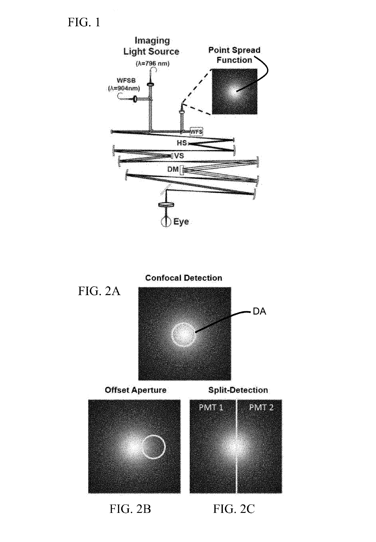
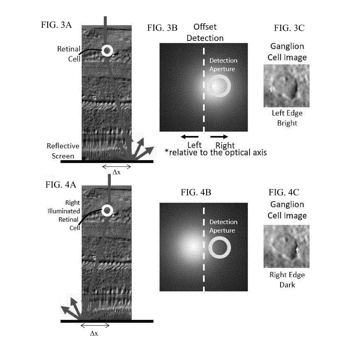
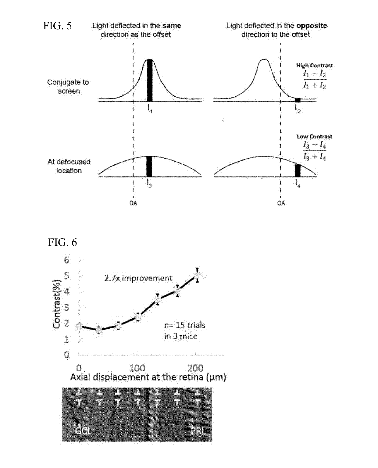
[0016]With non-confocal, off-axis detection imaging methods such as offset aperture and split detector techniques, several studies have demonstrated new classes of cells that can be identified using safe-levels of near infrared light. Among the new capabilities provided by these studies are imaging blood vessel wall, individual red blood cells, photoreceptor inner segments, photoreceptor somas, horizontal cells and ganglion cells. Previous models of light scatter provide a partial description of contrast mechanisms. In work by Elsner et al. and further refined by Chui and colleagues referenced above, authors provide a schematic of how light is scattered in offset aperture detection suggesting that light is forward scattered and then reflected by a deeper screen in what they call multiply scattered light. While this model provides an understanding that light interactions in the retina are complex, there is no developed optical model that describes the nature of the asymmetry in the c...


PUM


Abstract

A method for imaging vertebrate translucent retinal structures includes imaging a translucent retinal structure at a first imaging plane in the retina with a light source focused at such first imaging plane, and detecting reflected light with a non-confocal off-axis detector, wherein the detector is axially displaced from a plane conjugate to the first imaging plane to a plane conjugate to a reflective layer deeper in the retina along a path of illumination from the light source. The method may employ scanning ophthalmic instruments, and enables an enhancement in contrast and collected intensity by axially displacing the image detector away from the object of interest to positions closer to conjugate to layers of strong retinal reflection, like the photoreceptors or the interface between the choroid and the sclera, that act as a diffusive screen. This improvement was found using a model that describes the source of the contrast in offset aperture and split-detector ophthalmoscopy by emphasizing the role of cellular refractive index change within the plane of illumination.

Description

CROSS-REFERENCE TO RELATED APPLICATION[0001]This application claims the benefit of U.S. Provisional Application No. 62 / 593,471, filed Dec. 1, 2017, the disclosure of which is incorporated by reference herein in its entirety.STATEMENT REGARDING FEDERALLY SPONSORED RESEARCH OR DEVELOPMENT[0002]This invention was made with government support under grant number P30 EY001319 awarded by National Eye Institute. The government has certain rights in the invention.FIELD OF THE DISCLOSURE[0003]This disclosure relates to the field of retinal imaging, and more particularly to improvements in non-invasive, label-free imaging of translucent retinal cells.BACKGROUND[0004]The retina is the light sensitive tissue at the back of the eye where the sensation of vision begins. In the vertebrate retina, light must travel through the entirety of retinal neurons before being detected by the photoreceptors. Most of the inner retinal neurons are translucent, providing a benefit for vision but also a formidabl...

Claims


Application Information

Patent Timeline
Patent Type & Authority Applications(United States)
IPC IPC(8): A61B3/10A61B3/12A61B3/14
CPCA61B3/1025A61B3/1241A61B3/14A61B2503/40A61B3/12
Inventor GUEVARA-TORRES, RAUL ANDRESSCHALLEK, JESSE
Owner UNIVERSITY OF ROCHESTER
Why Eureka
Try Eureka