Methods and kits for assessing neurological function and localizing neurological lesions

a neurological function and kit technology, applied in the field of methods and kits for assessing neurological function and localizing neurological lesions, can solve the problems of permanent neurologic impairment or death, chemical dilatation of the pupil, loss of vision, etc., and achieve the effect of impaired integration of the central nervous system

Active Publication Date: 2019-06-27
NEW YORK UNIV
View PDF1 Cites 3 Cited by
  • Summary
  • Abstract
  • Description
  • Claims
  • Application Information

AI Technical Summary

Benefits of technology

[0088]The integrity of the central nervous system may be impaired by, for instance, trauma, stroke, a neurodegenerative disease, inflammation, an infectious process, a neoplastic process, vascular disease, an autoimmune disease, a genetic disorder

Problems solved by technology

If untreated, acute elevations in intracranial pressure (ICP) due to hydrocephalus, brain injury, stroke, or mass lesions can result in permanent neurologic impairment or death.
Chemical dilatation of the pupil to assess for papilledema may be unpleasant for the examinee, relies on the experience of the examiner and obfuscates further examination of the pupillary reflex.
Misalignment results in loss of this vision.
However, if the image from the weaker eye is suppressed by higher cortical centers, there is only one image with loss of visual acuity (or a blurred image).
Despite this capability, these tests are not used routinely in the emergency setting due to the need for a trained practitioner to administer them, the requirement for sophisticated equipm

Method used


Image

  • Methods and kits for assessing neurological function and localizing neurological lesions
  • Methods and kits for assessing neurological function and localizing neurological lesions
  • Methods and kits for assessing neurological function and localizing neurological lesions

Examples


example 1

Background

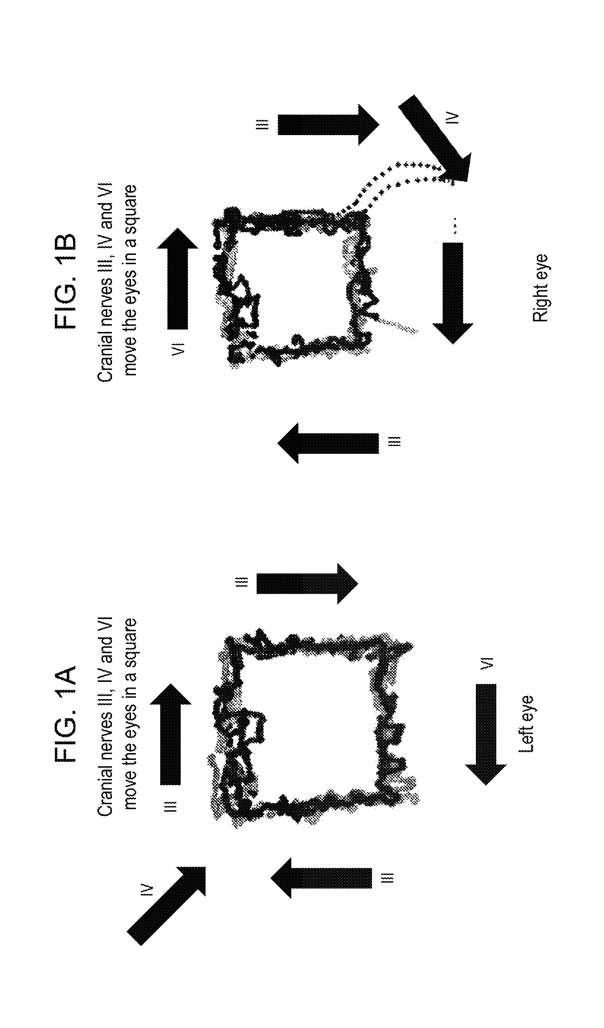
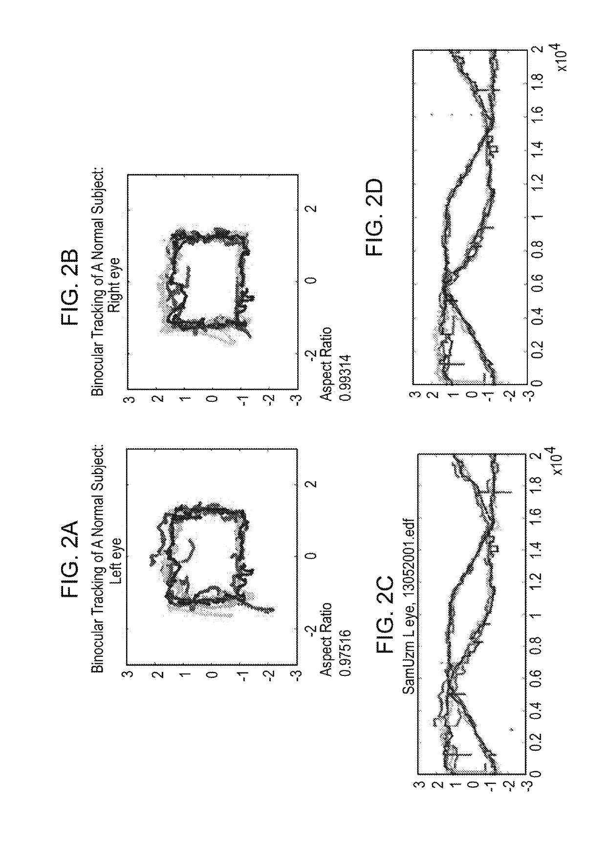
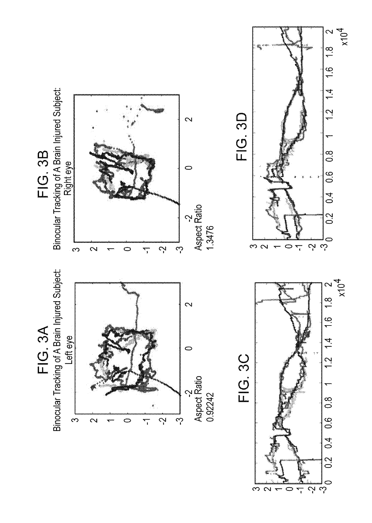
[0330]Eye movements contain clinically important information about neurological integrity. Clinical devices may take advantage of the relative ease of automated eye-movement tracking, for applications such as assessing recovery following clinical intervention. A technique was designed that can reliably measure eye movements with precision, without initial spatial calibration. We tracked eye movements without spatial calibration in neurologically intact adults and in neurosurgical patients as they watched a short music video move around the perimeter of a screen for 220 s. Temporal features of the data were measured, rather than traditional spatial measures such as accuracy or speed.

[0331]The methods reliably discriminated between the presence and absence of neurological impairment using these uncalibrated measurements. The results indicate that this technique may be extended to assess neurologic integrity and quantify deficits, simply by having patients watch TV.

[0332]Thes...

example 2

Materials and Methods

[0366]Subjects.

[0367]Healthy subjects were recruited in a university setting in accordance with IRB approved protocols. All other subjects were recruited directly from our neurosurgical practice. Informed consent from the subject or their legal proxy was obtained for prospective data collection in all cases in accordance with IRB guidelines.

[0368]Eye Movement Tracking.

[0369]The subjects' eye movements were recorded using an Eyelink 1000 binocular eye tracker (500 Hz sampling, SR Research). Healthy volunteers were seated 55 cm from the screen with their head stabilized using a chinrest. Stimulus was presented on average 55 cm from patient eyes, with the presentation monitor adjusted to match gaze direction. Subjects used a chinrest.

[0370]Innovations for Tracking Patients.

[0371]Two innovations were provided to measure ocular motility in a patient population. The first was a paradigm, consisting of a stimulus and an analysis stream that allows interpreting raw eye ...

example 3

Materials and Methods

[0393]Patient Selection.

[0394]Control subjects were employees, volunteers, visitors and patients at the Bellevue Hospital Center recruited in accordance with Institutional Review Board policy. Inclusion criteria for normal control subjects were: age 7 to 100 years, vision correctable to within 20 / 500 bilaterally, intact ocular motility, and ability to provide a complete ophthalmologic, medical and neurologic history as well as medications / drugs / alcohol consumed within the 24 hours prior to tracking. Parents were asked to corroborate details of the above for children aged 7-17. Exclusion criteria were history of: strabismus, diplopia, palsy of cranial nerves III, IV or VI, papilledema, optic neuritis or other known disorder affecting cranial nerve II, macular edema, retinal degeneration, dementia or cognitive impairment, hydrocephalus, sarcoidosis, myasthenia gravis, multiple sclerosis or other demyelinating disease, and active or acute epilepsy, stroke / hemorrhag...


PUM


Abstract

The invention provides methods and kits for detecting, screening, quantifying or localizing the etiology for reduced or impaired cranial nerve function or conduction; localizing a central nervous system lesion; detecting, diagnosing or screening for increased intracranial pressure, pressure or disruption of central nervous system physiology as seen with concussion; or detecting, diagnosing, monitoring progression of or screening for a disease or condition featuring increased intracranial pressure or concussion by tracking eye movement of the subject. The invention also provides methods and kits useful for detecting, screening for or quantitating disconjugate gaze or strabismus, useful for diagnosing a disease characterized by disconjugate gaze or strabismus in a subject, useful for detecting, monitoring progression of or screening for a disease or condition characterized by disconjugate gaze or strabismus in a subject or useful for quantitating the extent of disconjugate gaze or strabismus. Further, the invention provides methods for assessing or quantifying structural and non-structural traumatic brain injury or diagnosing a disease characterized by or featuring structural and non-structural traumatic brain injury.

Description

FIELD OF THE INVENTION[0001]The present invention relates to methods and kits for assessing physiologic function of the cranial nerves II, III, IV and VI, screening for, diagnosing, and quantitating the extent of elevated intracranial pressure, transtentorial herniation, concussion, normal pressure hydrocephalus, posterior fossa mass effect, optic neuropathy, neurodegenerative diseases, and diagnosing, localizing and monitoring progression of intracranial lesions and disease processes. The present invention also relates to methods and kits for assessing or quantitating conjugacy or disconjugacy of gaze or strabismus and for screening for, diagnosing, and assessing neurological diseases characterized by or featuring the same. In addition, the present invention relates to methods and kits for assessing or quantitating structural and non-structural traumatic brain injury and for screening for, diagnosing, and assessing the same.BACKGROUND OF THE INVENTIONEye Movement Tracking[0002]Auto...

Claims


Application Information

Patent Timeline
IPC IPC(8): A61B5/00A61B3/113A61B3/08A61B3/00
CPCA61B5/4064A61B3/113A61B3/085A61B5/7278A61B5/7282A61B3/0091A61B3/103A61B3/107A61B3/12A61B3/14A61B3/16A61B5/031A61B5/1114
Inventor SAMADANI, UZMA
Owner NEW YORK UNIV
Why Eureka
Try Eureka