Method for examining liver cancer

a liver cancer and method technology, applied in the field of liver cancer methods, can solve the problems of poor effectiveness of therapeutic methods, no reports so far regarding the relationship between non-bound and bound, and no reports on a method that can discriminate hcc, etc., and achieve the effect of superior sensitivity and specificity

Inactive Publication Date: 2019-10-17
SEKISUI MEDICAL CO LTD +2
View PDF5 Cites 0 Cited by
  • Summary
  • Abstract
  • Description
  • Claims
  • Application Information

AI Technical Summary

Benefits of technology

[0034]According to the present invention, there is provided a method for detecting liver cancer or a method for assisting diagnosis of liver cancer that is superior in sensitivity and specificity, as well as a kit that can be used therefor.

Problems solved by technology

Liver cancer (hepatocellular carcinoma; HCC) is a disease that is poor in effective therapeutic methods, and is one of the top causes of death among carcinomas.
However, there have been no reports thus far regarding the relationship between non-bound AIM that has not formed a complex with other binding partners, in particular IgM, and liver cancer.
There have also been no reports on a method that can discriminate HCC and other liver diseases including NAFL or NASH with high precision.

Method used


Image

  • Method for examining liver cancer
  • Method for examining liver cancer
  • Method for examining liver cancer

Examples


example 1

on of Mouse Anti-Human AIM Monoclonal Antibody

[0089]As the antigen, full length human rAIM (2 mg / ml) was mixed with an equal amount of TiterMax Gold (G-1, Funakoshi) to prepare an emulsion. Two 8 week old female Balb / c mice (from Charles River) were employed as immune animals, and 50 μL of the antigen solution was administered to the posterior plantar region. A similar administration was performed after 2 weeks, and 50 μg of the antigen solution was administered to the posterior plantar region after another 2 weeks or more in order to prepare for cell fusion 3 days later.

[0090]Mouse P3U1 was employed as the myeloma cell, and a medium which was RPMI1640 (11875-119 GIBCO) supplemented with glutamine and pyruvic acid and having FBS (S1560 from BWT) added to be 10% was employed as the proliferation culture. As antibiotics, penicillin and streptomycin were added at appropriate amounts.

[0091]From a mouse from which cardiac blood was collected under anesthesia, popliteal lymph nodes were a...

example 2

of Free AIM in Serum Specific for NASH Liver Cancer (HCC)

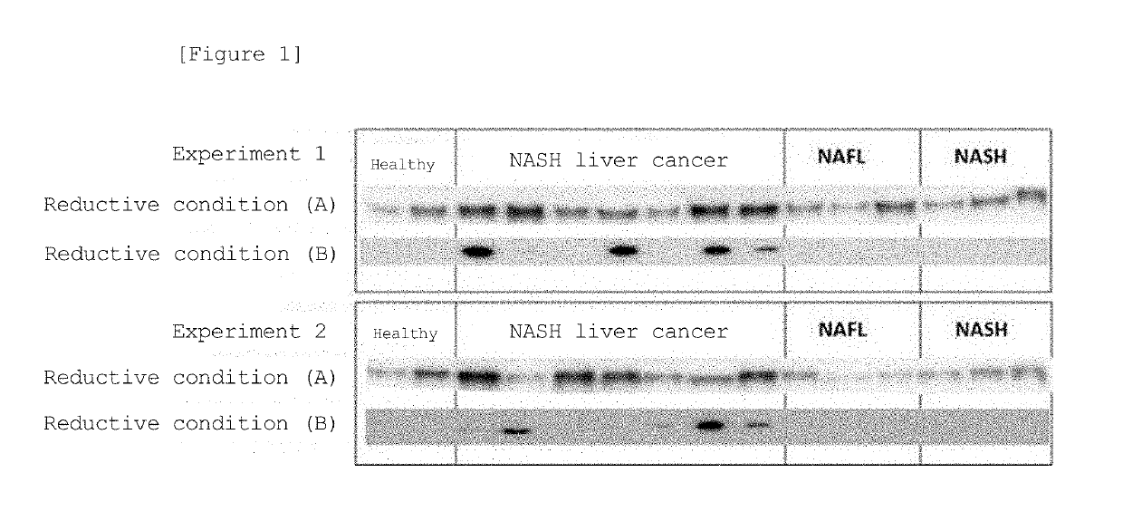
[0093]The serum of 4 healthy individual cases, 6 NAFL patient cases, 6 NASH patient cases, and 14 NASH liver cancer patient cases (HCC) were subjected to agarose gel electrophoresis under reductive or non-reductive condition, and Western blot was performed with an anti-AIM antibody.

[0094]In a reductive condition, the patient serum was mixed with a sample buffer comprising SDS and mercaptoethanol, allowed to be reduced at 99° C. for 5 minutes, and then added to e-PAGEL (from ATTO) gel and separated by SDS-PAGE method. In a non-reductive condition, the patient serum was mixed with a sample buffer that does not comprise SDS and mercaptoethanol, and separated by Native-PAGE method. After electrophoresis, the protein was transferred onto a PVDF membrane (Immobilon, from Millipore), and primary antibody reaction was performed with the mouse anti-human AIM monoclonal antibody prepared in Example 1 at 4° C. overnight. HRP-bound anti-m...

example 3

ation of Free AIM in Serum

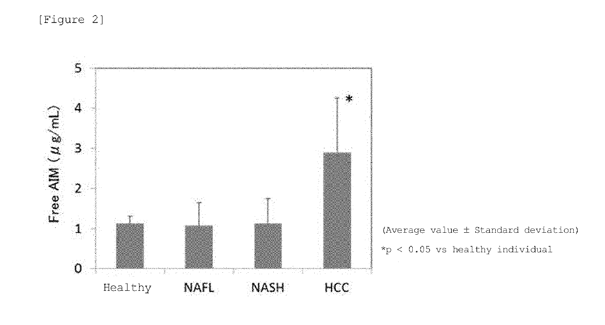
[0096]Human AIM ELISA kit (CY-8080, from CircuLex) that quantifies free AIM was employed to measure the concentration of free AIM in the serum of 10 healthy individual cases, 42 NAFL patient cases, 135 NASH patient cases, and 26 NASH liver cancer patient cases (HCC). Measurement was carried out according to the instruction of the kit. Man-Whittney U test was employed for statistical analysis, and p<0.05 was considered to be significantly different.

[0097]As a result, the concentration of free AIM in NAFL and NASH patients was 1.07±0.568 μg / mL and 1.12±0.632 μg / mL, respectively, and did not show difference to healthy individuals (1.12±0.187 μg / mL), but NASH liver cancer patients (HCC) showed significantly high free AIM at 2.90±1.375 μg / mL (FIG. 2).


PUM

PropertyMeasurementUnit
molecular weightaaaaaaaaaa
concentrationsaaaaaaaaaa
adhesiveaaaaaaaaaa

Abstract

A method for examining liver cancer or a method for assisting diagnosis of liver cancer that is superior in sensitivity and specificity, as well as a kit that can be used therefor is provided. The present invention provides a method for detecting liver cancer or a method for assisting diagnosis of liver cancer comprising a step of detecting or quantifying free AIM in a biological sample derived from a test subject, as well as a kit for examining or assisting diagnosis of liver cancer comprising an antibody that binds to free AIM.

Description

TECHNICAL FIELD[0001]The present invention typically relates to a method for examining liver cancer or a method for assisting diagnosis of liver cancer. Specifically, the present invention relates to a method for examining liver cancer or a method for assisting diagnosis of liver cancer, comprising detecting free AIM in a biological sample of a test subject. The present invention further relates to a kit for examining or assisting diagnosis of liver cancer.BACKGROUND ART[0002]AIM (apoptosis inhibitor of macrophage; also referred to as CD5L, api6, or Spα) is a secretory protein having a molecular weight of approximately 50 kDa that is specifically produced by tissue macrophage (Non-Patent Literature 1). AIM has a structure of tandemly connected three scavenger receptor cysteine-rich (SRCR) domains which are specific sequences containing a large number of cysteine residues, and it is thought to have a compact spherical conformation due to each cysteine residue being bound to one anoth...

Claims


Application Information

Patent Timeline
Patent Type & Authority Applications(United States)
IPC IPC(8): G01N33/574
CPCG01N33/57438C12Q1/02
Inventor MIYAZAKI, TORUOKANOUE, TAKESHIKOYAMA, NORIYUKI
Owner SEKISUI MEDICAL CO LTD
Why Eureka
Try Eureka