Methods and kits for treating pain
- Summary
- Abstract
- Description
- Claims
- Application Information
AI Technical Summary
Benefits of technology
Problems solved by technology
Method used
Image
Examples
example 1
ibits Acute Inflammatory Pain and Increases Pain Threshold in Naïve Animals
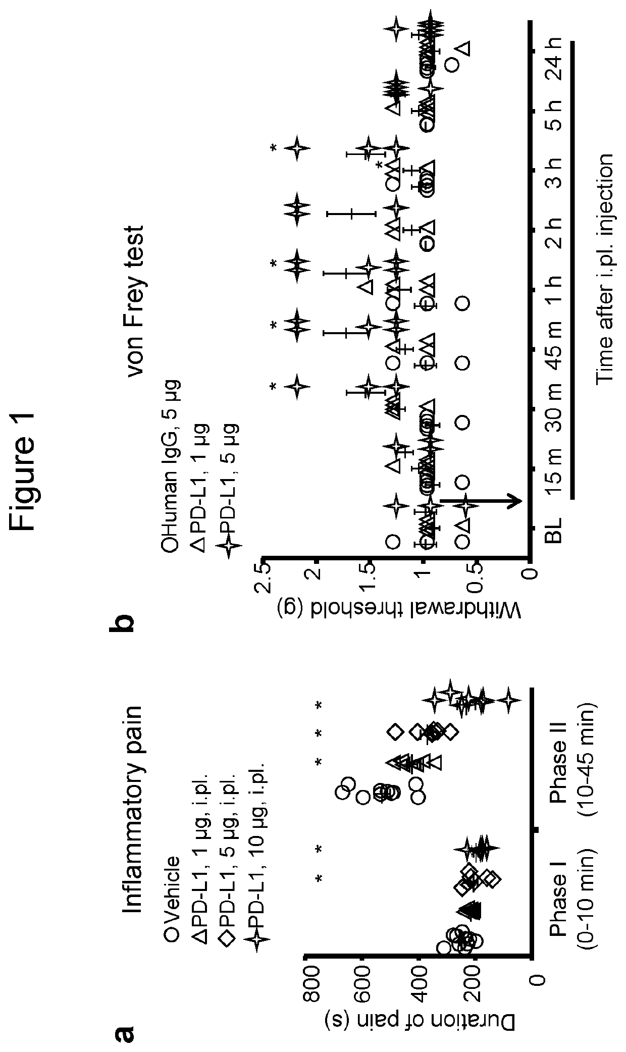
[0131]As a first step to address a role of PD-L1 in acute pain modulation, we examined the effects of PD-L1 in an acute inflammatory pain model. Intraplantar (i.pl) injection of formalin (5%) induced typical bi-phasic inflammatory pain as previously reported (Berta, T, et al. 2014), but the 2nd-phase pain (10-45 min) was substantially inhibited by PD-L1 pre-treatment (i.pl., 1-10 μg, Pst-phase pain (FIG. 1a).
[0132]Next, we tested if PD-L1 would also alter pain threshold in naïve mice. Von Frey test revealed a significant increase in paw withdrawal threshold after i.pl. injection of PD-L1 (5 μg=0.1 nmol, P<0.05, Two-Way ANOVA). The threshold increase was rapid and evident at 30 min and maintained for 3 h after the injection (FIG. 1b). Since PD-L1 (CD274) is a chimera protein fused with human IgG at the C-terminal, we used human IgG as an inactive control (http: / / www.abcam.com / recombinant-mouse-pd-l1-protein-fc...
example 2
an Endogenous Pain Inhibitor and Alters Basal Pain Thresholds Via PD-1
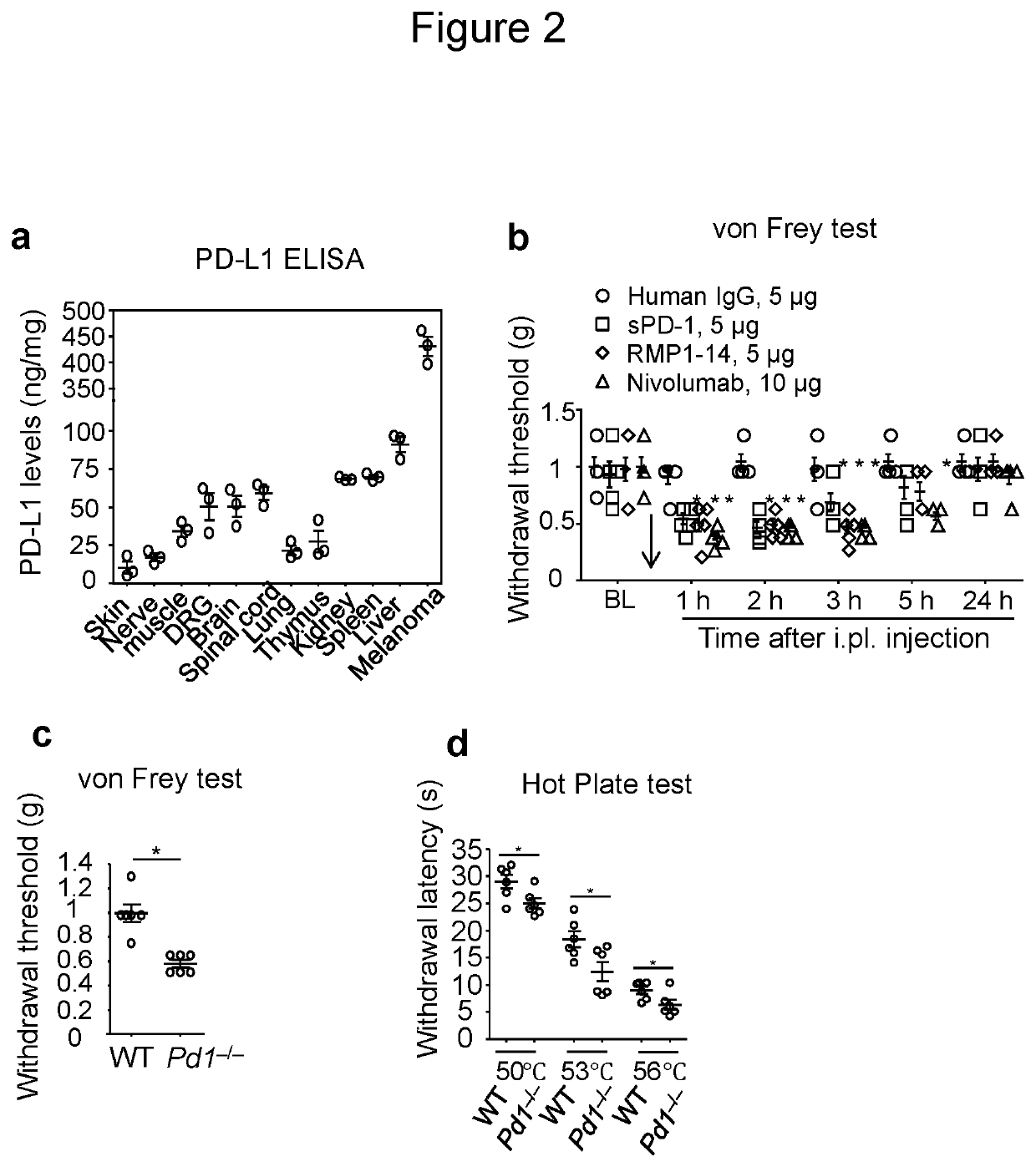
[0133]PD-L1 is produced by malignant tissues and serves as a predictive biomarker in cancer immunotherapy (Patel, S P & Kurzrock, R. Mol Cancer Ther, 2015, 14:847-856). As expected, mouse B16 melanoma tissue has high expression levels of PD-L1 (≈450 ng / mg tissue, FIG. 2a), as evaluated by ELISA analysis. PD-L1 was also secreted in cultured medium of melanoma cells (FIG. 9a). To determine if normal tissues also produce PD-L1, we compared PD-L1 contents in non-neural and neural tissues. Non-neural tissues, such as liver, spleen, and kidney have high levels of PD-L1 (≈70-90 ng / mg tissue, FIG. 2a). Interestingly, neural tissues, including brain, spinal cord, and dorsal root ganglia (DRG) contact PD-L1 at levels around 50 ng / mg tissue (FIG. 2a). Furthermore, PD-L1 was detected in the sciatic nerve and hindpaw skin tissues (FIG. 2a), which contain pain-sensing nerve fibers. These results suggest that PD-L1 is broadly sy...
example 3
ptor is Expressed by Primary Sensory Neurons in Mouse DRGs
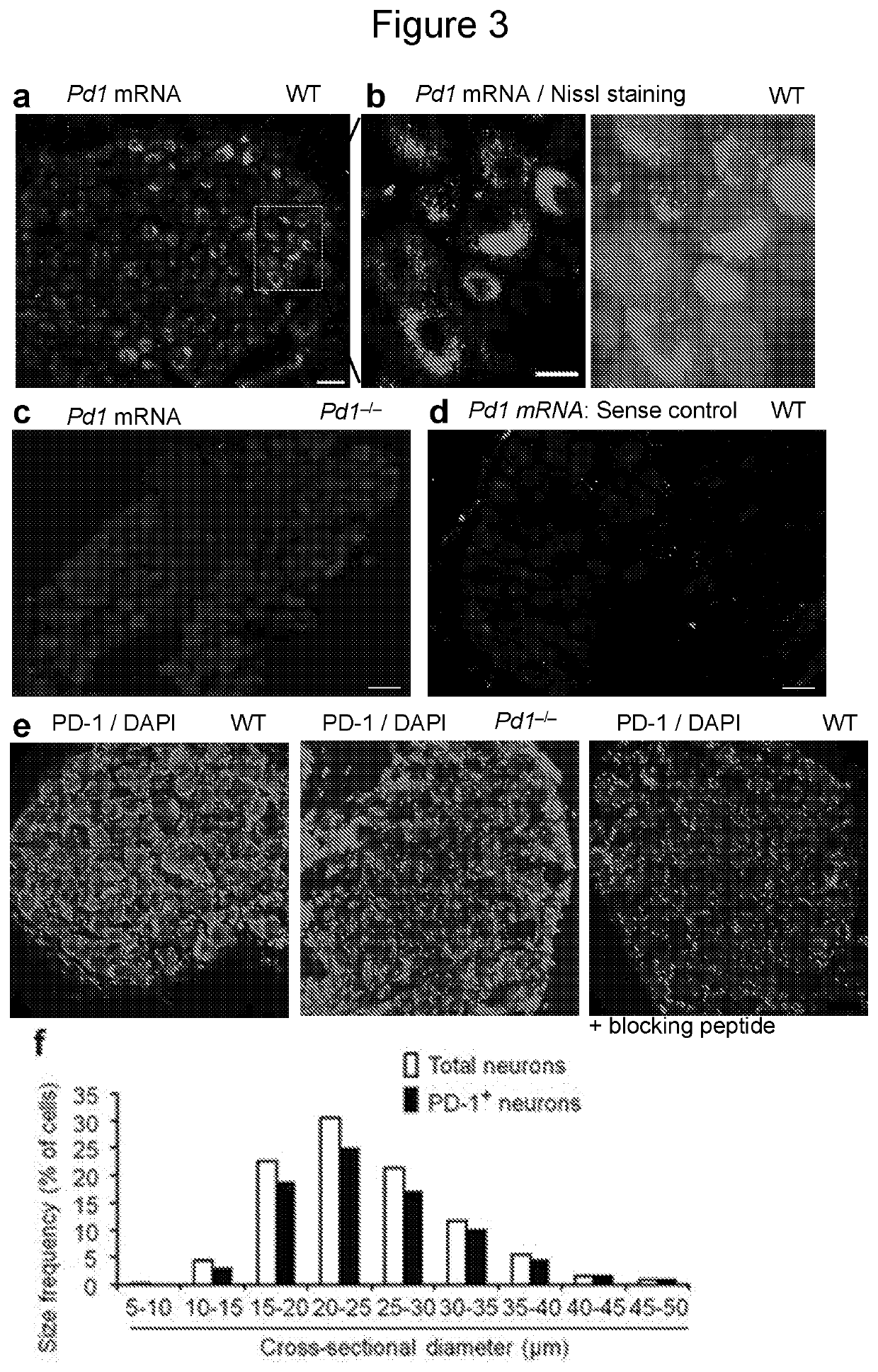
[0137]To determine peripheral mechanisms by which PD-L1 modulates pain, we examined Pd1 mRNA and PD-1 protein expression in mouse DRG neurons. In situ hybridization showed Pd1 mRNA expression in majority of DRG neurons with various sizes (FIG. 3a,b). This expression was lost in Pd1− / − mice (FIG. 3c) and in DRG sections treated with sense control probe (FIG. 3d), confirming the specificity of Pd1 mRNA expression. Immunohistochemistry reveled PD-1 immunoreactivity (IR) in majority of DRG neurons (FIG. 3e). The specificity of the PD-1 antibody was validated by loss of PD-1 immunostaining in DRG neurons of Pd1− / − mice (FIG. 3e) and further confirmed by absence of staining in WT DRG after co-incubation of the antibody with a blocking peptide (FIG. 3e). Size frequency analysis showed a broad expression of PD-1 by DRG neurons with small, medium, and large sizes (FIG. 3f). Double staining confirmed PD-1 expression in both large-diame...
PUM
| Property | Measurement | Unit |
|---|---|---|
| Time | aaaaa | aaaaa |
| Time | aaaaa | aaaaa |
| Time | aaaaa | aaaaa |
Abstract
Description
Claims
Application Information
 - R&D
- Intellectual Property
- Life Sciences
- Materials
- Tech Scout
- Unparalleled Data Quality
- Higher Quality Content
- 60% Fewer Hallucinations
Browse by: Latest US Patents, China's latest patents, Technical Efficacy Thesaurus, Application Domain, Technology Topic, Popular Technical Reports.
© 2025 PatSnap. All rights reserved.Legal|Privacy policy|Modern Slavery Act Transparency Statement|Sitemap|About US| Contact US: help@patsnap.com


