


Quick Research
Generate reliable direction feasibility study reports for your R&D in just a few steps.

Technical Q&A
Discover and master advanced knowledge NOW. Basics, ideas, possibilities, all at once.

Find Solutions
As an expert in R&D theories, this can generate solutions to your technical problems instantly.

Evaluate Feasibility
Analyze your overall solution with one click, know your potential R&D risks in advance.

Monitor Landscape
Get weekly tech updates, stay abreast of the latest tech innovations and key insights.
Antibody conjugate for treating and detecting bladder cancer
- Summary
- Abstract
- Description
- Claims
- Application Information
AI Technical Summary
Benefits of technology
Problems solved by technology
Method used
Image

Examples
example i
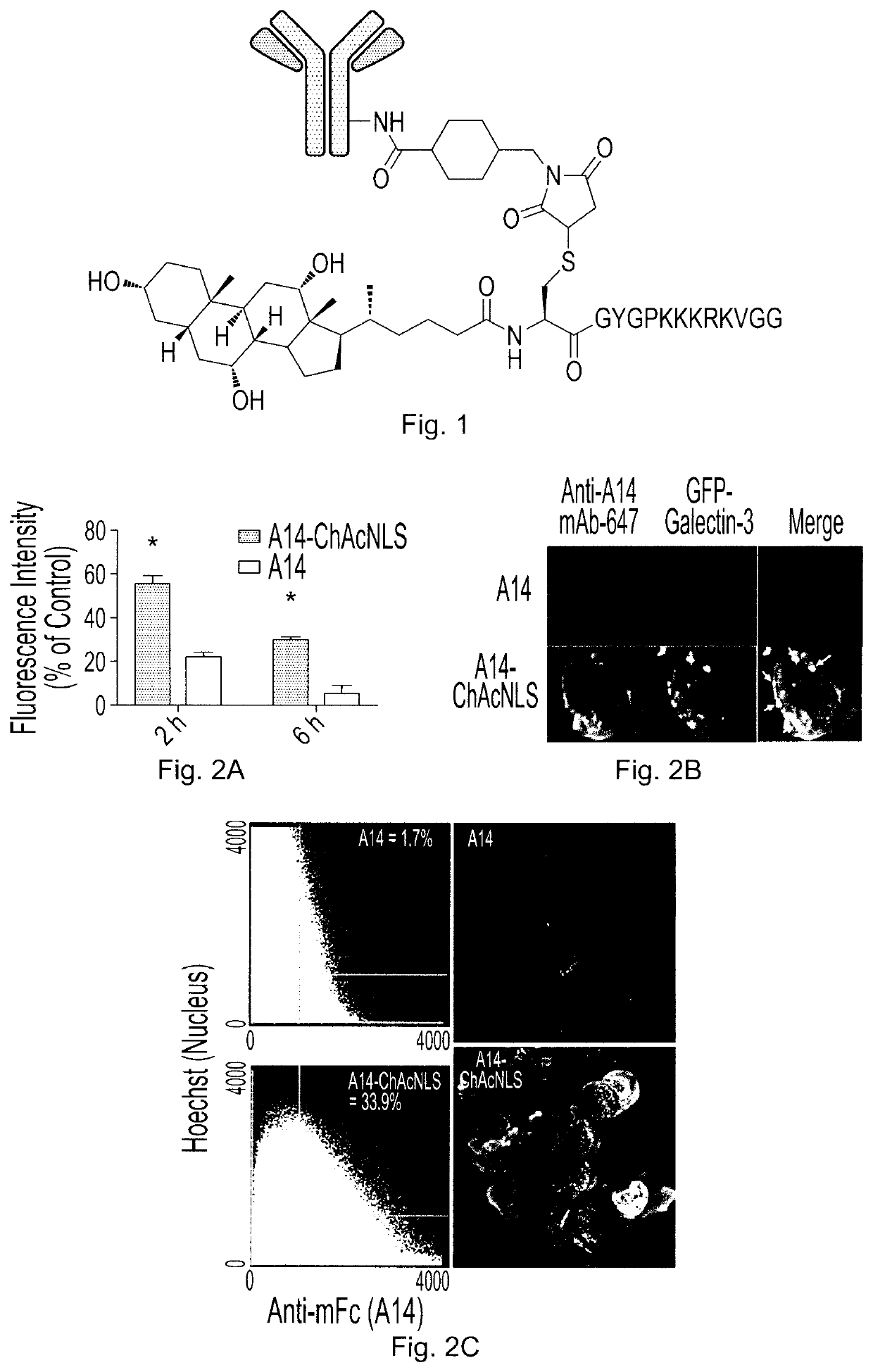
ChAcNLS Attachment and Preparation of Conjugates
[0092]A14 was obtained and purified as previously described (Sun et al., 1996, Blood, 87: 83-92.
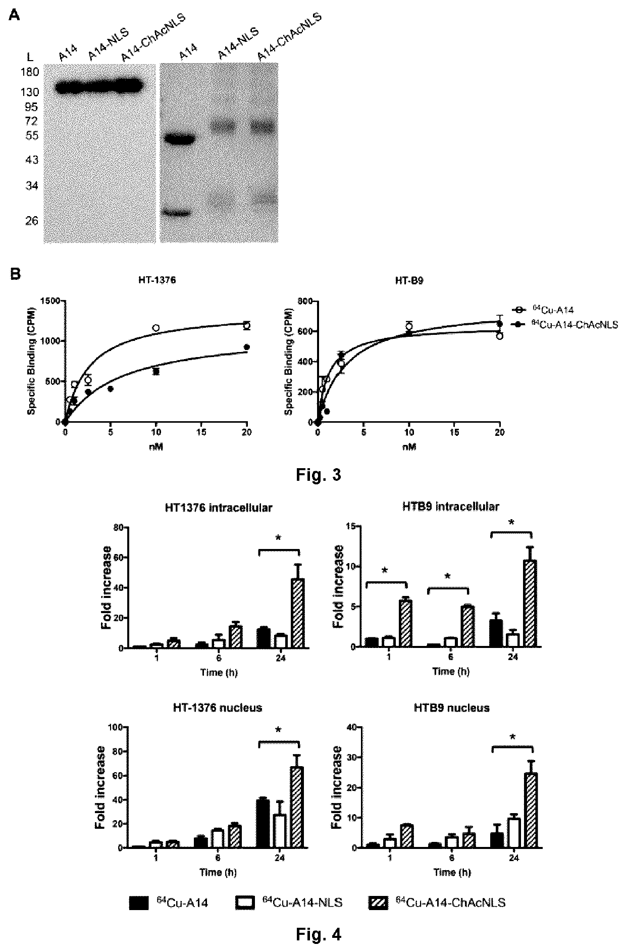
[0093]To evaluate conjugation, a reducing 12% polyacrylamide gel was loaded with A14, A14-NLS, A14-ChAcNLS, and protein standards (BioRad, Ontario, Canada). The gel was stained with Coomassie and protein standard retention factor (Rf) values obtained. The size of the A14-NLS and A14-ChAcNLS heavy and light chains were extrapolated by plotting the distance of migration against the Rf values. The numbers of ChAcNLS or NLS compounds per A14 were calculated by dividing the difference in molecular weight (MW) between the modified A14 conjugates and unmodified A14 by 1768.5 g / mol and 1418.8 g / mol, respectively.
[0094]A14 was first reacted with NOTA-NHS in a 5-to-1 NOTA-to-A14 ratio, purified, and then conjugated to NLS or ChAcNLS followed by purification. Radiolabeling efficiency was determined by autoradiography on both instant thin-layer chromato...
example ii
Mechanistic Studies
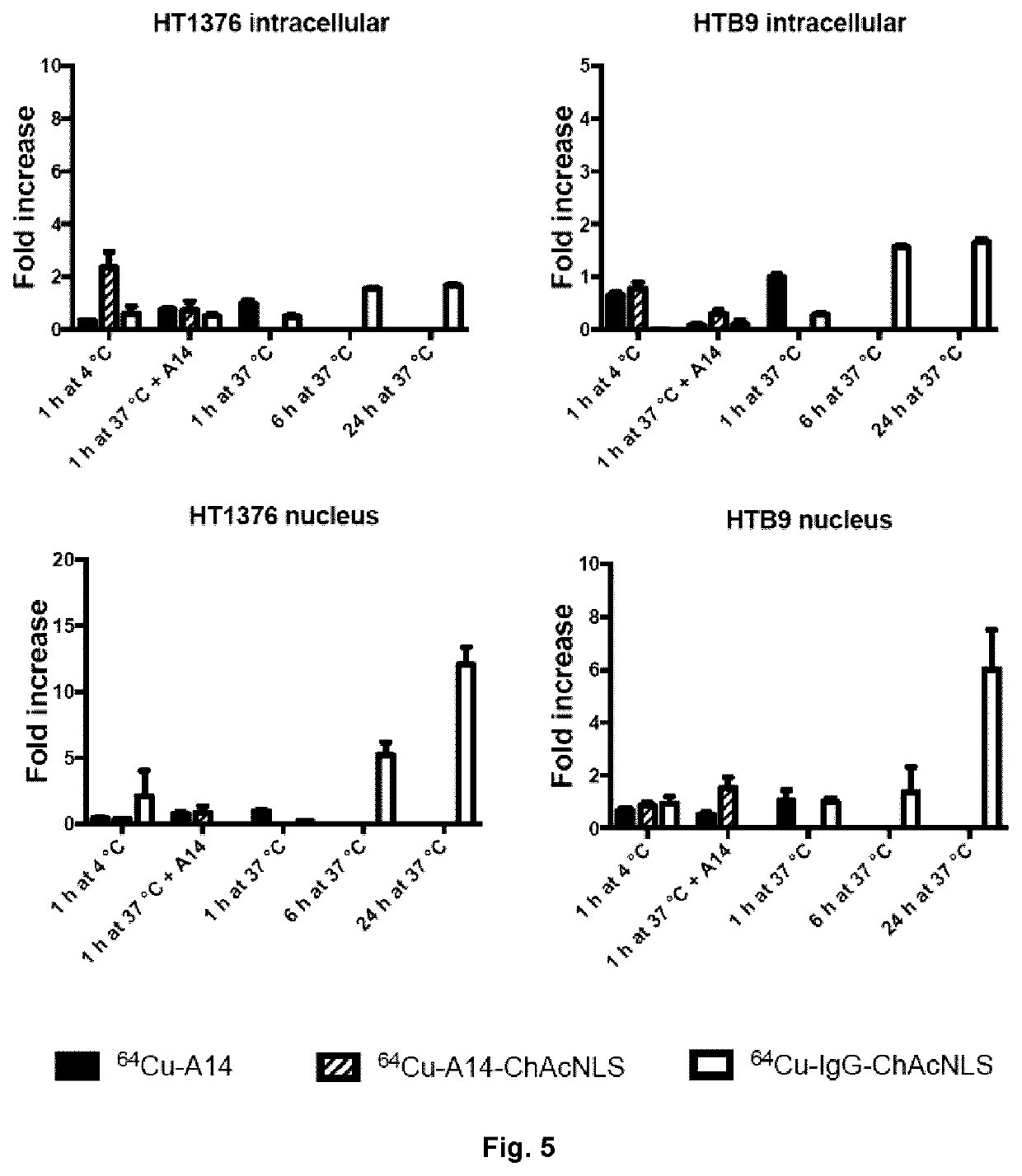
[0095]HT-1376 cells were treated with 200 nmol / L of A14 or A14-ChAcNLS for 1 h at 37° C. followed by washing in ice cold PBS. Cells were then replenished with fresh antibody-free media and placed back at 37° C. for 1 h. Cells were then prepped for nuclear and antibody staining for evaluation by confocal microscopy as previously described (Beaudoin et al., 2016, Mol Pharm, 13: 1915-1926). For determination of ceramide levels, HT-1376 cells were treated with A14 or A14-ChAcNLS for increasing time points at 37° C. Cells were fixed and permeabilized. Cells were incubated with the anti-ceramide antibody conjugated to the fluorophore AlexaFluor 488 (Cedarlane, Ontario, Canada). Flow cytometric analysis measured the mean fluorescence intensity (MFI).
[0096]To explore endosome escape, HT-1376 cells were transfected with cDNA encoding for GFP-Galectin-3. Cells were then treated with 200 nM of A14 or A14-ChAcNLS for 1 h at 37° C. Cells were fixed and permeabilized as describ...
example iii
In Vivo IL-5Rα-Positive Invasive Bladder Cancer Tumor Targeting
[0097]When tumors were 65-100 mm3, mice (n=5) were intravenously injected with 64Cu-A14, 64Cu-A14-NLS, or 64Cu-A14-ChAcNLS (˜25 μg; ˜7 MBq; radiochemical purity ≥98%). Nicking of the saphenous vein and collection of blood was performed daily. Mice anesthetized under 2.5% isoflurane were then euthanized by CO2 inhalation at 48 h and 96 h post-injection. Major organs and tumors were excised, rinsed in saline, blotted dry, and placed in pre-weighted tubes and gamma counted. Radioactivity accumulation was corrected for decay and expressed as the injected dose per gram of tissue (% ID / g).
[0098]PET imaging studies were performed on 5 mice per group on a PET / CT Triumph™ scanner (Trifoil, Calif., USA) at 24 h and 48 h post-injection. PET scans were acquired for 30 and 45 min at 24 h and 48 h, respectively, with tumors near the center of the field of view, in double axial sampling mode to improve spatial resolution. The images we...
PUM
| Property | Measurement | Unit |
|---|---|---|
| Length | aaaaa | aaaaa |
| Length | aaaaa | aaaaa |
| Length | aaaaa | aaaaa |
Abstract
Description
Claims
Application Information
 - R&D Engineer
- R&D Manager
- IP Professional
- Industry Leading Data Capabilities
- Powerful AI technology
- Patent DNA Extraction
Browse by: Latest US Patents, China's latest patents, Technical Efficacy Thesaurus, Application Domain, Technology Topic, Popular Technical Reports.
© 2024 PatSnap. All rights reserved.Legal|Privacy policy|Modern Slavery Act Transparency Statement|Sitemap|About US| Contact US: help@patsnap.com