Methods for identifying free thiols in proteins

a technology of free thiol and protein, applied in the field of systems and methods of characterizing free thiol group in proteins, can solve the problems of affecting the structural integrity, stability, biological functions, and the thermal stability of mab products, so as to reduce the sample, reduce the thermal stability, and reduce the effect of degradation

Active Publication Date: 2020-07-16
REGENERON PHARM INC
View PDF0 Cites 2 Cited by
  • Summary
  • Abstract
  • Description
  • Claims
  • Application Information

AI Technical Summary

Benefits of technology

[0008]Compositions and methods for identifying free thiols (also referred to as free sulfhydryls) are provided. Free thiols can exist in proteins, for example mAbs, as a result of incomplete disulfide bond formation or disulfide bond breakage. The presence of free thiols can lower the thermal stability of mAb products and affect their structural integrity, stability, and biological functions. It is therefore important to characterize free thiols in mAb production. Methods for identifying site specific free thiols in protein drug products are disclosed herein. An exemplary method includes labeling peptides with a tag to identify free thiols and a tag to identify native disulfide bonds and analyzing the tags using targeted MS2. In one embodiment, the method provides complete coverage of all 32 cysteine residues in an IgG molecule. In other embodiments the method covers the 16 cysteine residues on the heavy and light chains in an IgG molecule. In another embodiment, the method covers the 5 cysteine residues on each light chain of an IgG molecule. In another embodiment, the method covers the 11 cysteine residues on each heavy chain of an IgG molecule.
[0009]One embodiment provides a method for identifying the presence of free thiols in a protein, for example an antibody or fragment thereof. An exemplary method for identifying free thiols in a protein drug product includes the steps of labeling a sample containing the protein drug product with a first label containing a sulfhydryl-reactive iodoacetyl group, an MS-neutral spacer arm, and a first MS / MS reporter having a unique reporter ion mass. Excess label can be removed and the sample is denatured and reduced. The sample is then labeled with a second label having a sulfhydryl-reactive iodoacetyl group, an MS-neutral spacer arm, and a second MS / MS reporter having a unique reporter ion mass. The method includes enzymatically digesting the sample and analyzing the sample using mass spectrometry, for example ultra-performance liquid chromatography tandem mass spectrometry system (UPLC-MS2 system) including a charged surface hybrid column and a formic acid buffer mobile phase. Next, the method includes quantifying the first MS / MS reporter and the second MS / MS reporter, wherein the amount of the first MS / MS reporter correlates to the amount of free thiols in the protein drug product and the amount of the second MS / MS reporter correlates to the amount of bound thiols in the protein drug product. In one embodiment the first MS / MS reporter has a mass of 128 and the second MS / MS reporter has a mass of 131. The protein drug product is typically an antibody, for example a monoclonal or chimeric antibody. The presence of free thiols is likely a result of incomplete disulfide bond formation or disulfide bond degradation. In one embodiment, the method provides complete coverage of all 32 cysteine residues in an IgG molecule. In other embodiments the method covers the 16 cysteine residues on the heavy and light chains in an IgG molecule. In another embodiment, the method covers the 5 cysteine residues on each light chain of an IgG molecule. In another embodiment, the method covers the 11 cysteine residues on each heavy chain of an IgG molecule.
[0010]Another embodiment provides a method of identifying disulfide heterogeneity in a protein drug product including the steps of labeling a sample containing the protein drug product with a first label containing a sulfhydryl-reactive iodoacetyl group, an MS-neutral spacer arm, and a first MS / MS reporter having a unique reporter ion mass. Excess label can be removed and the sample is denatured and reduced. The sample is then labeled with a second label having a sulfhydryl-reactive iodoacetyl group, an MS-neutral spacer arm, and a second MS / MS reporter having a unique reporter ion mass. The method includes enzymatically digesting the sample and analyzing the sample using mass spectrometry, for example an ultra-performance liquid chromatography tandem mass spectrometry system (UPLC-MS2 system) including a charged surface hybrid column and a formic acid buffer mobile phase. Next, the method includes quantifying the first MS / MS reporter and the second MS / MS reporter, wherein the amount of the first MS / MS reporter correlates to the amount of free thiols in the protein drug product and the amount of the second MS / MS reporter correlates to the amount of bound thiols in the protein drug product. If the assay detects free thiols in the protein drug product, the protein drug product contains disulfide heterogeneity. The presence of these free thiols is likely a result of incomplete disulfide bond formation or disulfide bond degradation. In one embodiment the first MS / MS reporter has a mass of 128 and the second MS / MS reporter has a mass of 131. The protein drug product is typically an antibody, for example a monoclonal, chimeric antibody, bispecific antibody, or antigen binding fragments thereof. In one embodiment, the method provides complete coverage of all 32 cysteine residues in an IgG molecule. In other embodiments the method covers the 16 cysteine residues on the heavy and light chains in an IgG molecule. In another embodiment, the method covers the 5 cysteine residues on each light chain of an IgG molecule. In another embodiment, the method covers the 11 cysteine residues on each heavy chain of an IgG molecule.
[0011]A method for selecting a protein drug product including the steps of labeling a sample containing the protein drug product with a first label containing a sulfhydryl-reactive iodoacetyl group, an MS-neutral spacer arm, and a first MS / MS reporter having a unique reporter ion mass. Excess label can be removed and the sample is denatured and reduced. The sample is then labeled with a second label having a sulfhydryl-reactive iodoacetyl group, an MS-neutral spacer arm, and a second MS / MS reporter having a unique reporter ion mass. The method includes enzymatically digesting the sample and analyzing the sample using an ultra-performance liquid chromatography tandem mass spectrometry system (UPLC-MS2 system) including a charged surface hybrid column and a formic acid buffer mobile phase. Next, the method includes quantifying the first MS / MS reporter and the second MS / MS reporter, wherein the amount of the first MS / MS reporter correlates to the amount of free thiols in the protein drug product and the amount of the second MS / MS reporter correlates to the amount of bound thiols in the protein drug product. If a free thiol is detected, the protein drug product has disulfide heterogeneity. The method includes selecting the protein drug product that does not display disulfide heterogeneity. In one embodiment the first MS / MS reporter has a mass of 128 and the second MS / MS reporter has a mass of 131. The protein drug product is typically an antibody, for example a monoclonal, chimeric antibody, or a bispecific antibody, or antigen binding fragments thereof. Another embodiment provides a pharmaceutical composition containing the protein drug product selected using the method described above. In one embodiment, the method provides complete coverage of all 32 cysteine residues in an IgG molecule. In other embodiments the method covers the 16 cysteine residues on the heavy and light chains in an IgG molecule. In another embodiment, the method covers the 5 cysteine residues on each light chain of an IgG molecule. In another embodiment, the method covers the 11 cysteine residues on each heavy chain of an IgG molecule.

Problems solved by technology

The presence of free thiols can lower the thermal stability of mAb products and affect their structural integrity, stability, and biological functions.
The presence of these free thiols is likely a result of incomplete disulfide bond formation or disulfide bond degradation.

Method used


Image

  • Methods for identifying free thiols in proteins
  • Methods for identifying free thiols in proteins
  • Methods for identifying free thiols in proteins

Examples


example 1

of Site-Specific Free Thiols

[0054]Methods

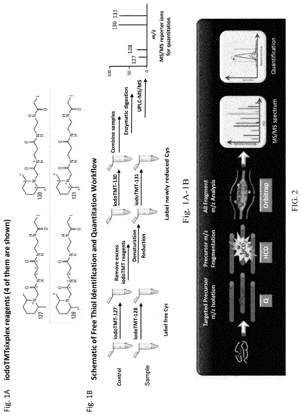
[0055]Cysteine Labeling

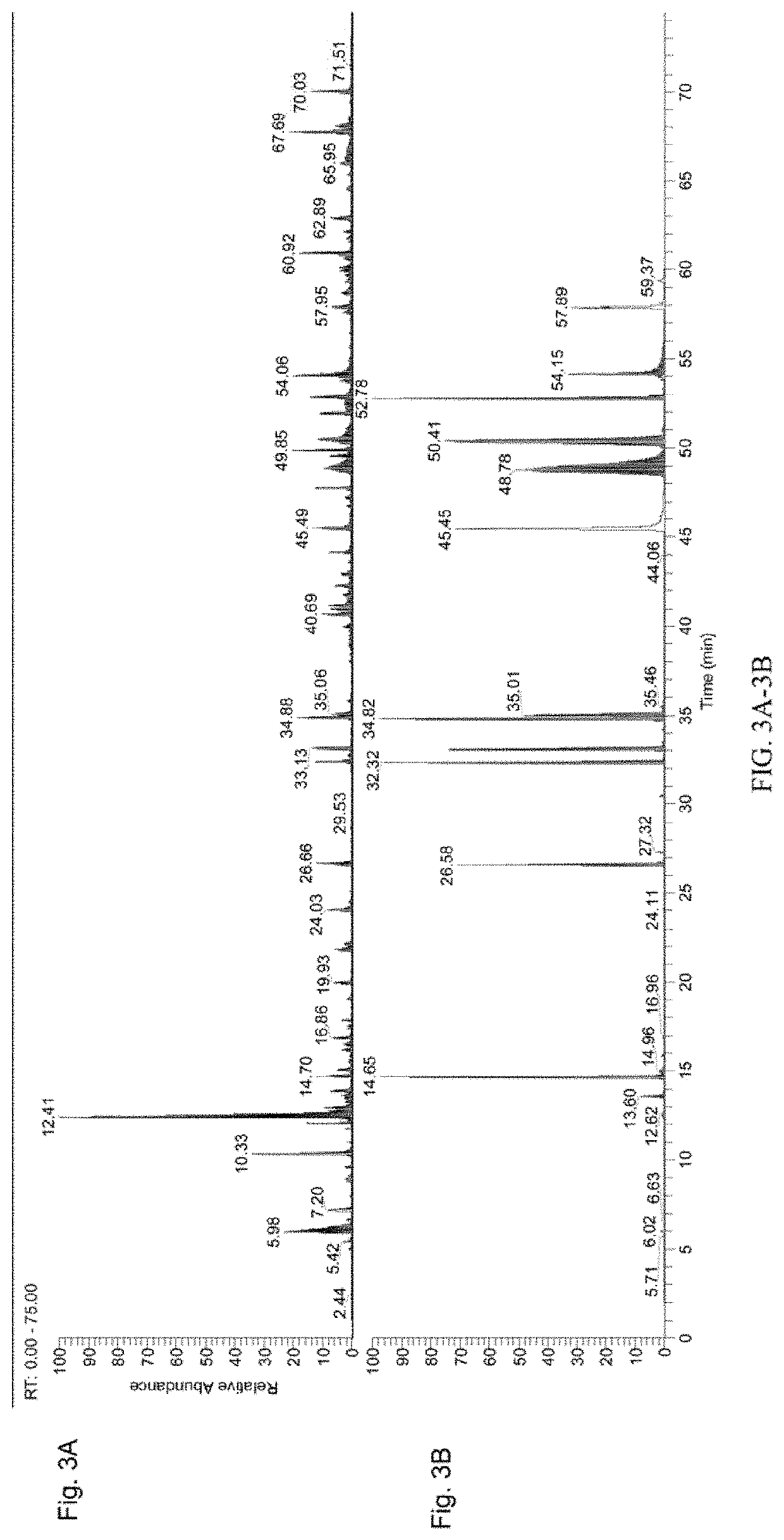
[0056]The workflow from FIG. 1B was followed. In brief, a control sample and an antibody sample were labeled with iodo-TMT reagents (FIG. 1A). The excess reagents were removed and the samples were denatured and reduced using 0.5M TCEP and incubated for 1 hour. After denaturation and reduction, a second label was added to the samples. The samples were then combined, enzymatically digested, and analyzed using UPLC-MS / MS. UPLC conditions were as follows: Samples were run on a 150 mm CSH column for a 150 minutes run, including a 40 minute re-equilibration step. FA buffers were used.

[0057]Spike-in Study

[0058]For the spike-in study, aliquots of two different samples of the same protein were prepared. The proteins were denatured using GuanHCl and reduced. The samples were labeled with two different IodoTMT tags. A spike-in concentration gradient was then prepared. 10%, 5%, 1%, 0.5%, or 0.1% of protein sample 1 was added to ali...


PUM

PropertyMeasurementUnit
massaaaaaaaaaa
massaaaaaaaaaa
massaaaaaaaaaa

Abstract

Compositions and methods for identifying free thiols in protein are provided. An exemplary method labeling peptides with a tag to identify free thiols and a tag to identify native disulfide bonds and analyzing the tags using targeted MS2. In one embodiment, the method provides complete coverage of all 32 cysteine residues in an IgG molecule. In other embodiments the method covers the 16 cysteine residues on the heavy and light chains in an IgG molecule. In another embodiment, the method covers the 5 cysteine residues on each light chain of an IgG molecule. In another embodiment, the method covers the 11 cysteine residues on each heavy chain of an IgG molecule.

Description

CROSS-REFERENCE TO RELATED APPLICATIONS[0001]This application claims benefit of and priority to U.S. Provisional Patent Application No. 62 / 792,994 filed Jan. 16, 2019, incorporated herein by reference in its entirety.TECHNICAL FIELD OF THE INVENTION[0002]The invention is generally related to systems and methods of characterizing free thiol group in proteins, particularly in antibodies.BACKGROUND OF THE INVENTION[0003]During the development of monoclonal antibodies (mAbs) from drug candidate to marketed product, issues with stability, post-translational modifications, or other changes to the antibody can occur. Alterations in antibody structure and function can cause problems such as poor shelf-life or even immunogenicity in the patient. It is therefore important to properly characterize antibody structure and monitor it throughout production. Antibody quality control and quality assurance are critical to the purity and safety of mAb products.[0004]Disulfide bonds are important for s...

Claims


Application Information

Patent Timeline
Patent Type & Authority Applications(United States)
IPC IPC(8): G01N33/68G01N1/28
CPCG01N1/28G01N33/6854G01N33/6848G01N33/6815G01N30/7233G01N2030/027G01N2030/8831G01N2458/15A61K2039/505A61K39/395
Inventor E, SOOK YENBRAMHALL, DAVIDQIU, HAIBO
Owner REGENERON PHARM INC
Why Eureka
Try Eureka