Method for Motion Correction of 3D Contrast Enhanced Ultrasound Without the Availability of Bmode Data

a technology of contrast enhancement and ultrasound, applied in the field of motion correction of contrast fluid in medical imaging, can solve the problems of affecting the quality of quantification, prone to sampling errors, and biased quantitative results of imaging using conventional 2d dce-us, and achieves the effects of improving image similarity, reducing the risk of motion artifacts, and improving the volume of lesion overlap

Inactive Publication Date: 2020-09-10
THE BOARD OF TRUSTEES OF THE LELAND STANFORD JUNIOR UNIV
View PDF0 Cites 2 Cited by
  • Summary
  • Abstract
  • Description
  • Claims
  • Application Information

AI Technical Summary

Benefits of technology

[0009]The present invention provides a validated method for motion correction with great potential for mitigating motion artifacts in 3D DCE-US. In an exemplary embodiment, the inventors demonstrated that the method consistently generates improvement across a variety of different liver metastasis imaging acquisitions qualitatively, and highlights its robustness quantitatively through improvement in lesion volume overlap, increa...

Problems solved by technology

In contrast, imaging tumors via conventional 2D DCE-US is prone to sampling errors and can produce biased quantitative results because of plane-to-plane perfusion variation and tumor heterogeneity.
However, the quality of quantification is affected by several imaging and clinical limitations.
In particular, ultrasound-based dynamic imaging is highly susceptible to motion artifacts arising from patient movement, respiration, and peristalsis, as well as operator hand stability.
Motion artifacts can be especially hampering in lengthy acquisitions and in the context of quantification by introducing significant errors to the quantified contrast signal.
In ultrasound imaging of focal liver lesions technical failures can be encountered due to motion artifacts from patient movement.
While MC algorithms exist for 2D-based DCE...

Method used


Image

  • Method for Motion Correction of 3D Contrast Enhanced Ultrasound Without the Availability of Bmode Data
  • Method for Motion Correction of 3D Contrast Enhanced Ultrasound Without the Availability of Bmode Data
  • Method for Motion Correction of 3D Contrast Enhanced Ultrasound Without the Availability of Bmode Data

Examples


Embodiment Construction

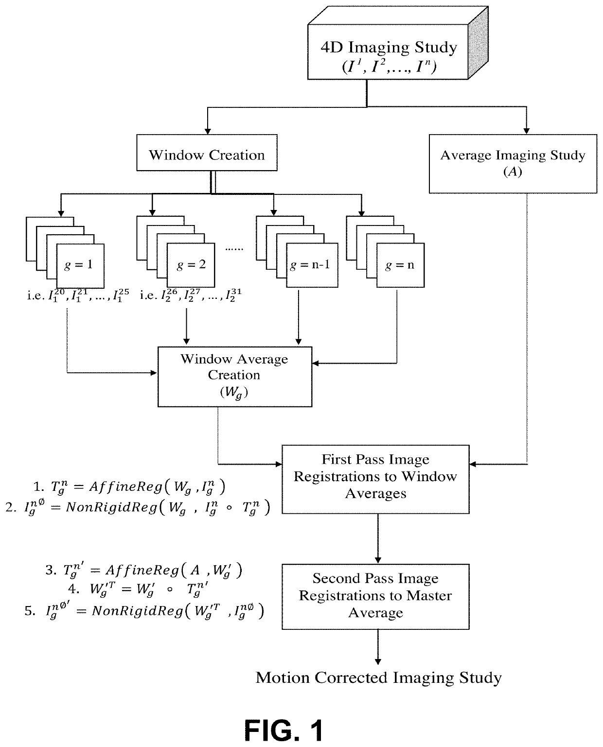
[0019]This invention provides a motion correction technology for 3D DCE-US data unaccompanied by Bmode anatomical imaging that improves the reliability of bolus time-intensity analysis for perfusion parameters. The following is an example of the method and should not be regarded as limiting to the invention.

[0020]Materials and Methods

[0021]Clinical Case Analysis

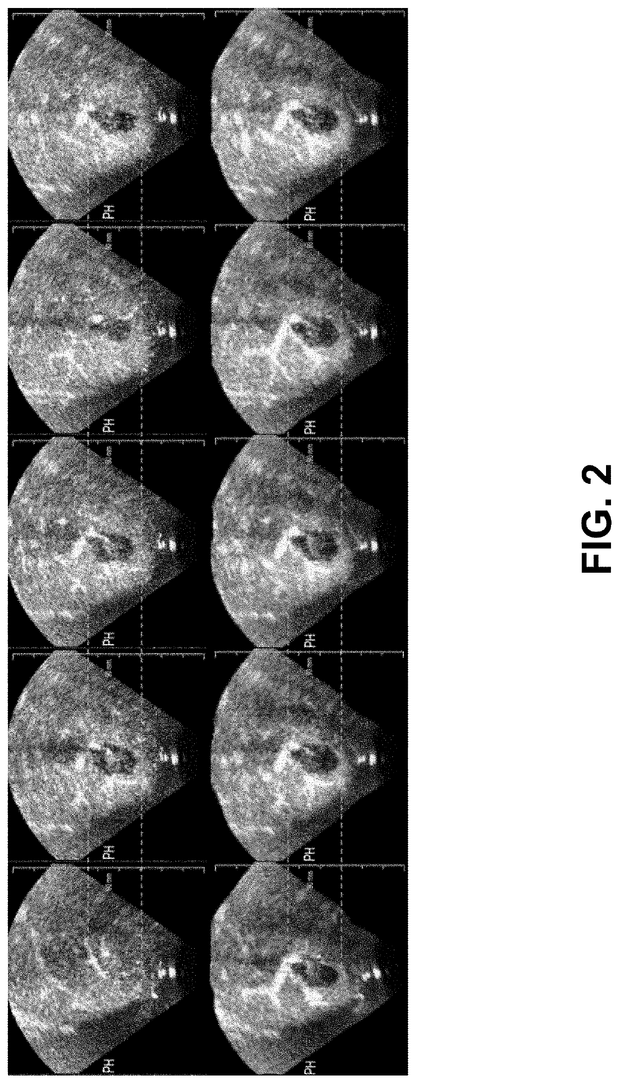
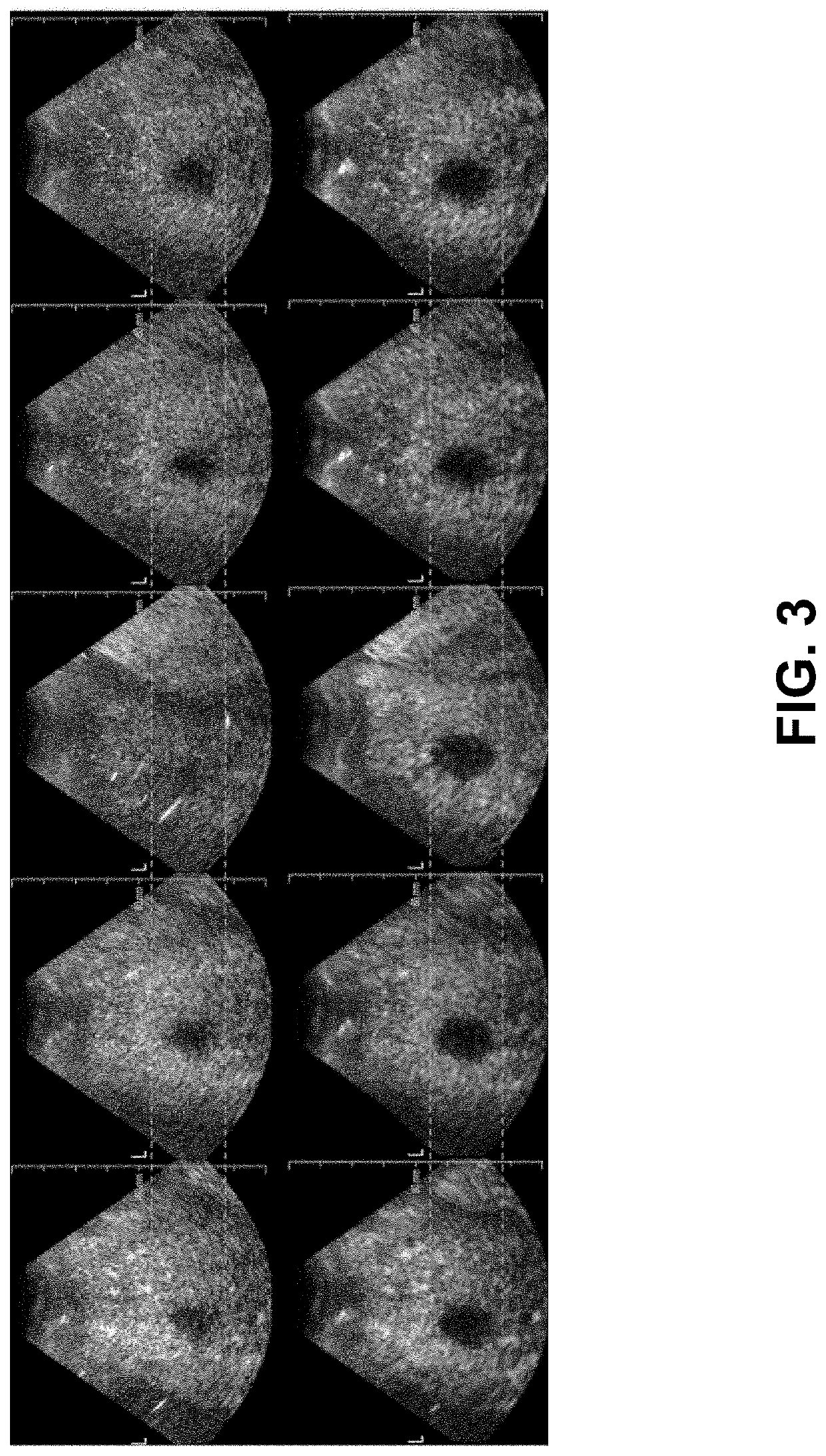
[0022]The MC method was evaluated using eight human 3D-DCE US imaging studies of liver metastasis. The imaging studies were acquired from patients who provided written consent, were 18 years of age or older, and presented with at least one liver metastasis that was within the range of 1-14 cm in diameter and confirmed using MRI / CT imaging studies. The 3D-DCE US scanning was performed by an experienced sonographer using the commercial Philips X6-1 MHz xMATRIX array transducer at a setting of 1-3 Hz. Each original imaging study (pre-MC) was processed using the method to produce a motion corrected imaging study (post-MC).

[0023]P...


PUM


Abstract

The present invention provides a validated method for motion correction with great potential for mitigating motion artifacts in 3D DCE-US. The method is described as a method for motion correction of three-dimensional contrast enhanced ultrasound without the availability of Bmode data. Four-dimensional cine data including three-dimensional contrast enhanced ultrasound image frames are acquired. The acquired three-dimensional contrast enhanced ultrasound image frames are subdivided into groups of similar images referred to as windows. A first pass registration is performed for each of the images in the window to a window representative image. A second pass is performed for each of the registered images from the first pass to a master reference image.

Description

CROSS-REFERENCE TO RELATED APPLICATIONS[0001]This application claims priority from U.S. Provisional Patent Application 62 / 814,054 filed Mar. 5, 2019, which is incorporated herein by reference.FIELD OF THE INVENTION[0002]This invention relates to methods, devices and systems for motion correction due to contrast fluid in medical imaging.BACKGROUND OF THE INVENTION[0003]The introduction of contrast-enhanced ultrasound imaging has significantly expanded the diagnostic potential of ultrasound as a non-invasive and inexpensive imaging modality. Imaging tissue perfusion is feasible using dynamic contrast-enhanced ultrasound (DCE-US) methods in contrast-mode to isolate contrast signal from tissue signals in several abdominal indications, as well as other anatomical sites accessible to ultrasound, and has been adopted in global clinics.[0004]While both CT and MRI imaging modalities also offer contrast-enhanced tissue perfusion measurements, ultrasound-based contrast imaging is advantageous ...

Claims


Application Information

Patent Timeline
IPC IPC(8): A61B8/08
CPCA61B8/481A61B8/5276A61B8/483A61B8/06A61B8/085
Inventor HRISTOV, DIMITRE H.KAFFAS, AHMED EL
Owner THE BOARD OF TRUSTEES OF THE LELAND STANFORD JUNIOR UNIV
Why Eureka
Try Eureka