Elastin reduction allowing recellularization of cartilage implants

a cartilage implant and cartilage technology, applied in the field of tissue implants, can solve problems such as not being expected to raise an immune reaction

Pending Publication Date: 2020-10-15
TRAUMA CARE CONSULT TRAUMATOLOGISCHE FORSCHUNG GEMEINNUTZIGE GES MBH
View PDF0 Cites 1 Cited by
  • Summary
  • Abstract
  • Description
  • Claims
  • Application Information

AI Technical Summary

Benefits of technology

The patent describes a way to make a cartilage scaffold that can be used to repair different types of cartilage, including ear, hyaline, and fibrous cartilage. It can also be used to repair bone defects and osteochondral defects. This scaffold can be made from a specific type of protein called elastin, which is found in the connective tissue of the body. Overall, this patent provides a way to create a versatile scaffold that can be used for various types of cartilage repair.

Problems solved by technology

Hence, a scaffold from an allogeneic source or a xenogenic source is not expected to raise an immune reaction after implantation into a human recipient.

Method used


Image

  • Elastin reduction allowing recellularization of cartilage implants
  • Elastin reduction allowing recellularization of cartilage implants
  • Elastin reduction allowing recellularization of cartilage implants

Examples


example 1

rization with BMSCs by Dynamic Seeding

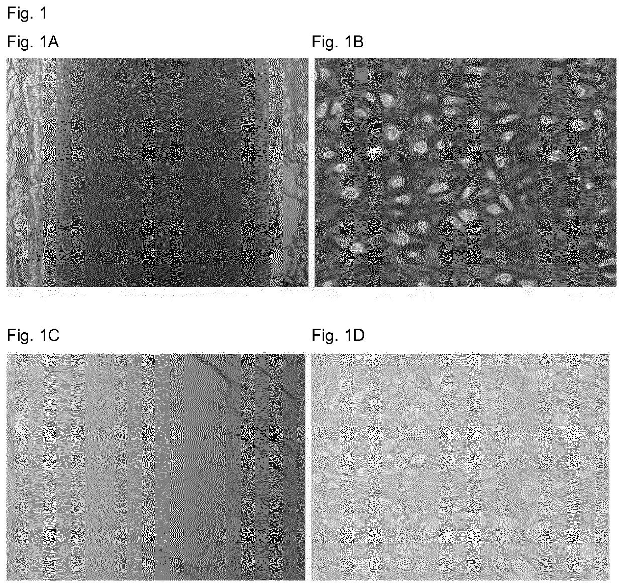
[0073]The outer ears of 12-week-old calves were washed with water and the perichondrium, skin and muscle were removed using a scalpel till only the cartilage remained. Subsequently, biopsies of 6 mm diameter and about 1 mm height were obtained from the auricular cartilage using biopsy punches, hence each sample being about 30 mm3 in volume.

[0074]The following protocol was then employed for removal of the cells and elastin fibers, the volumes given applied to batches of approximately 20 samples unless specified otherwise. First, the samples were rinsed thrice briefly with 40 ml In some embodiments of the invention 3696 Grade I deionized water following referred to as deionized water, to remove debris from the isolation, such as loose pieces of skin. If necessary, samples were frozen in 20 ml of deionized water at −20° C. for storage. Then samples were sterilized with 20 ml of 5% hydrogen peroxide for 60 minutes at 37° C. Subsequently, the samples...

example 2

hondrogenesis after Injection

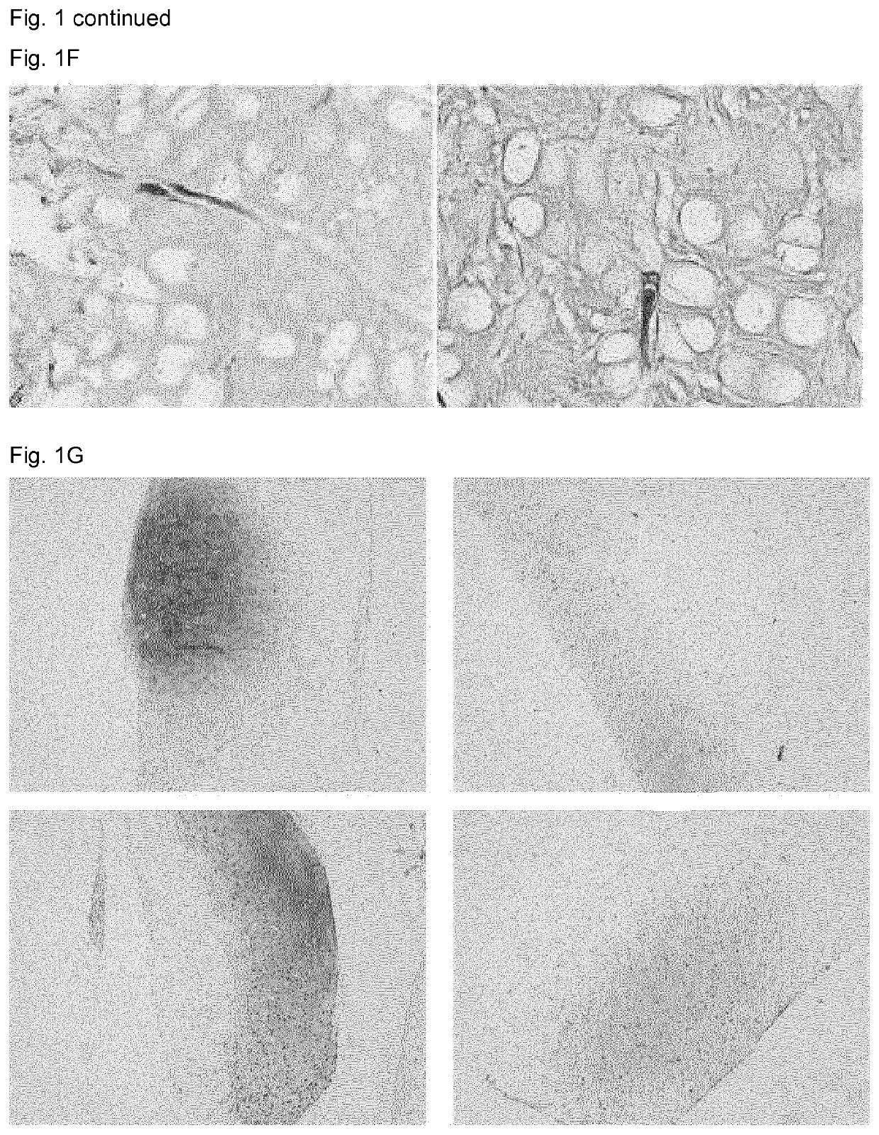
[0078]Some areas of cartilage are difficult to reach for cells by migration, for example the cartilage of the outer ear is surrounded by a dense perichondrium layer. As shown by Utomo et al., 2015 an entire ear can be decellularized using elastase. The perichondrium is tightly attached to the cartilage and removal of it without damaging the cartilage structure is not feasible.

[0079]To test whether recellularization of an entire ear is feasible, bovine auricular cartilage samples without necessitating removal of the perichondrium were recellularized with chondrogenic cells by injection through the perichondrium.

[0080]The outer ears of 12-week-old calves were washed with water and skin and muscle were removed using a scalpel till only the cartilage with its intact perichondrium remained. Subsequently, square biopsies of 15 mm side length were obtained from the auricular cartilage using a scalpel with each sample being about 2-4 mm in height (including peri...

example 3

hondrogenesis in Joint Model

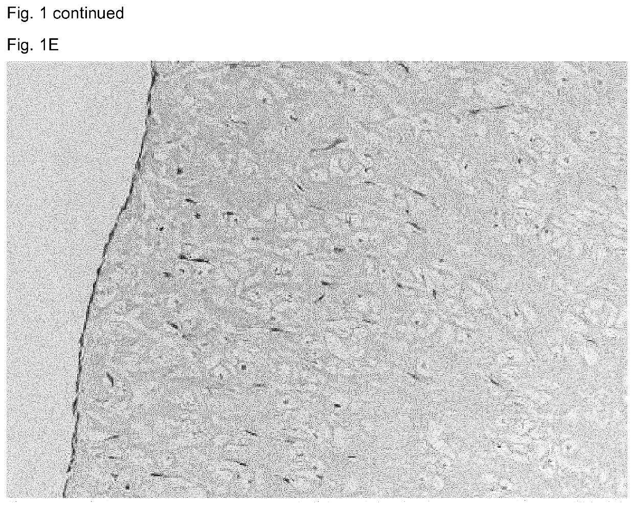
[0085]To determine if elastase-treated elastic cartilage is suitable to repair cartilage defects, it was used to fill defects in a model of articular cartilage damage. First, the outer ears of 12-week-old calves were washed with water and the perichondrium, skin and muscle were removed using a scalpel till only the cartilage remained. Subsequently, biopsies of 6 mm diameter were obtained from the auricular cartilage using biopsy punches and cut to a height of either approximately 0.4 or 1.2 mm.

[0086]The protocol employed for removal of the cells and elastin fibers was identical to that described in example 1. The volumes given applied to batches of about 20 samples unless specified otherwise.

[0087]After removal of the cells and elastin fibers, half the samples were then placed at the bottom of a 0.3 cm2 well (one sample / well, 96 well plate) and covered with 500.000 chondrogenic cells per sample as a solution in 200 μl of chondrocyte expansion medium (DMEM...


PUM

PropertyMeasurementUnit
pHaaaaaaaaaa
temperatureaaaaaaaaaa

Abstract

The invention relates to a method of producing an elastin-reduced cartilage scaffold containing channels and / or lacunae, the method comprising the steps of providing an elastic cartilage sample and reducing elastin from said cartilage sample to produce said channels and / or lacunae. The invention further relates to an elastin-reduced cartilage scaffold. Said elastin-reduced cartilage scaffold can be used in a method of implantation in vivo, for repairment of cartilage, repairment of osteochondral defects and repairment of bone defects.

Description

FIELD OF THE INVENTION[0001]The present invention relates to the field of tissue implants. More specifically the invention is in the field of improvement of cartilage grafts. In particular, the invention relates to methods for reducing elastin of a cartilage scaffold containing channels and / or lacunae that allows for recellularization of the scaffold.BACKGROUND ART[0002]Organs in the body may fail due to congenital disorders, acquired diseases, accidents and old age. Transplantation of organs is rendered difficult by both the scarcity of donor organs and the necessity to suppress the immune system to prevent organ rejection. In the last decades, our knowledge of cells that constitute an organ has rapidly expanded, rendering the creation of organs for transplantation from scratch, so called tissue engineering, increasingly realistic. Organs are a combination of cells and matrices, the latter providing a 3-dimensional structure that holds the cells in place. Cells carry markers unique...

Claims


Application Information

Patent Timeline
Patent Type & Authority Applications(United States)
IPC IPC(8): A61L27/36A61L27/38A61L27/56
CPCA61L2430/40A61L27/56A61L27/3834A61L27/3612A61L2430/06A61L2430/02A61L27/3687A61L27/3817A61L27/3654A61F2/30756A61F2002/30766A61F2002/30762A61L27/3691
Inventor NÜRNBERGER, SYLVIAVAN OSCH, GEERTRUDA JOHANNA VICTORIA MARIALEHMANN, JOHANNES
Owner TRAUMA CARE CONSULT TRAUMATOLOGISCHE FORSCHUNG GEMEINNUTZIGE GES MBH
Why Eureka
Try Eureka