
Ultrasound tracking and visualization

Pending Publication Date: 2020-12-17
KONINKLJIJKE PHILIPS NV
View PDF5 Cites 0 Cited by
  • Summary
  • Abstract
  • Description
  • Claims
  • Application Information

AI Technical Summary

Benefits of technology

The patent describes a system that uses ultrasound imaging to improve the visualization of soft tissue during a medical procedure. The system generates a reference image from live ultrasound images, which helps to avoid the need for separate tracking systems and reduces the risk of complications. The reference image is also more up-to-date than pre-procedural images, as it is generated from live ultrasound images. The system can extend the field of view of the reference image by matching anatomical features in the current image with those in the reference image. This allows for improved navigation and more accurate guidance during the procedure. Additionally, the system can use position data from sensors on the interventional device and the ultrasound imaging probe to more accurately position the device in the reference image. Overall, the system improves the accuracy and efficiency of medical procedures.

Problems solved by technology

Moreover, the extracted reference image is inherently static, and so any movement of the ultrasound probe after the generation of this “map” is not translated into a movement of the map.

Method used


Image

  • Ultrasound tracking and visualization
  • Ultrasound tracking and visualization

Examples


Embodiment Construction

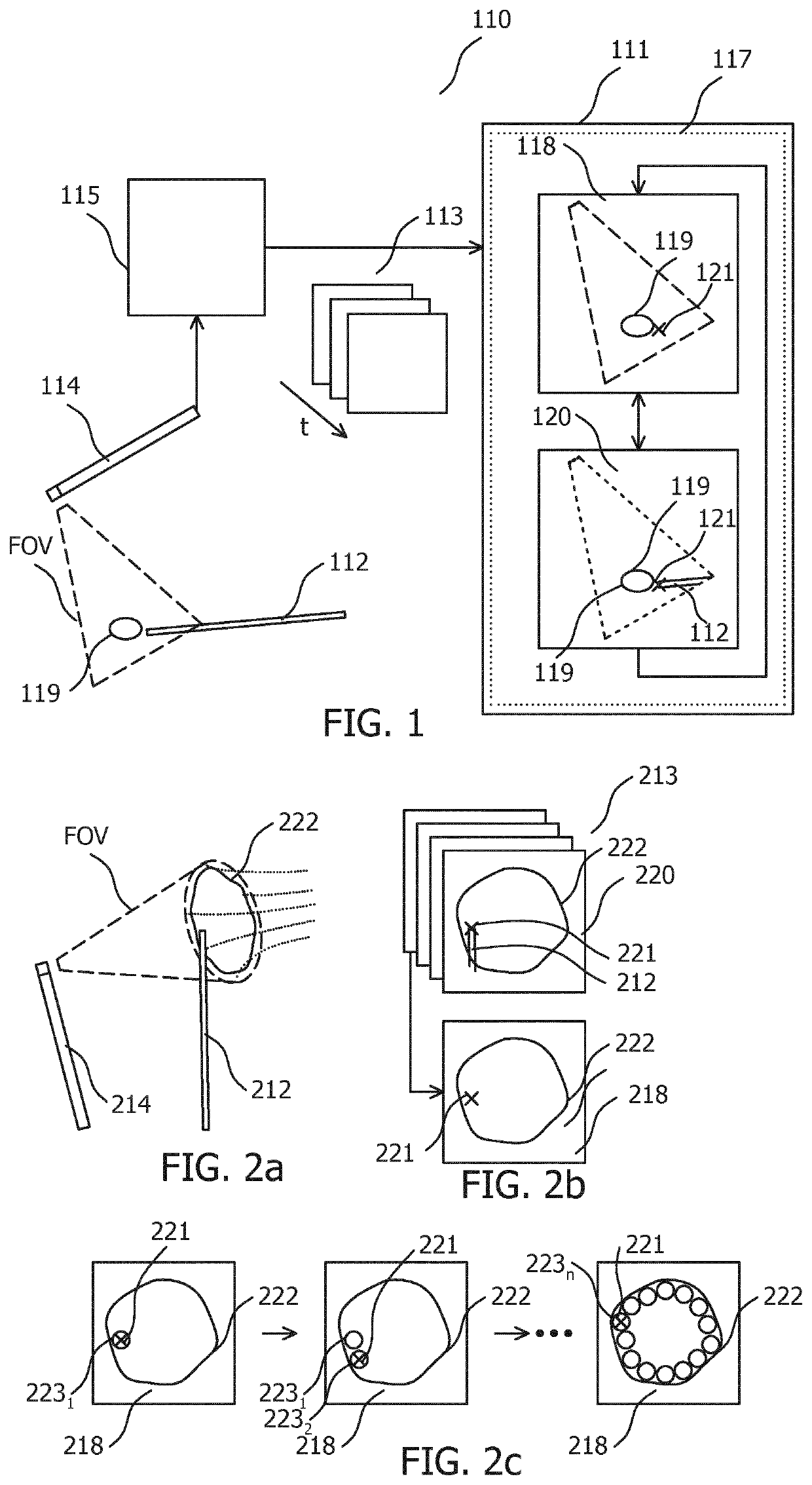
[0015]In order to illustrate the principles of the present invention an ultrasound visualization system is described with particular reference to a cardiac ablation procedure in which an ICE imaging probe serves as the ultrasound imaging probe, and in which an ablation catheter serves as the interventional device. It is however to be appreciated that the invention also finds application in the wider ultrasound imaging field. The ultrasound imaging probe may alternatively be for example a TEE imaging probe, and the interventional device may for example be a catheter, an ablation catheter, an ablation support catheter, a biopsy device, a guidewire, a filter device, a balloon device, a stent, a mitral clip, a left atrial appendage closure device, an aortic valve, a pacemaker lead, an intravenous line, or a surgical tool.

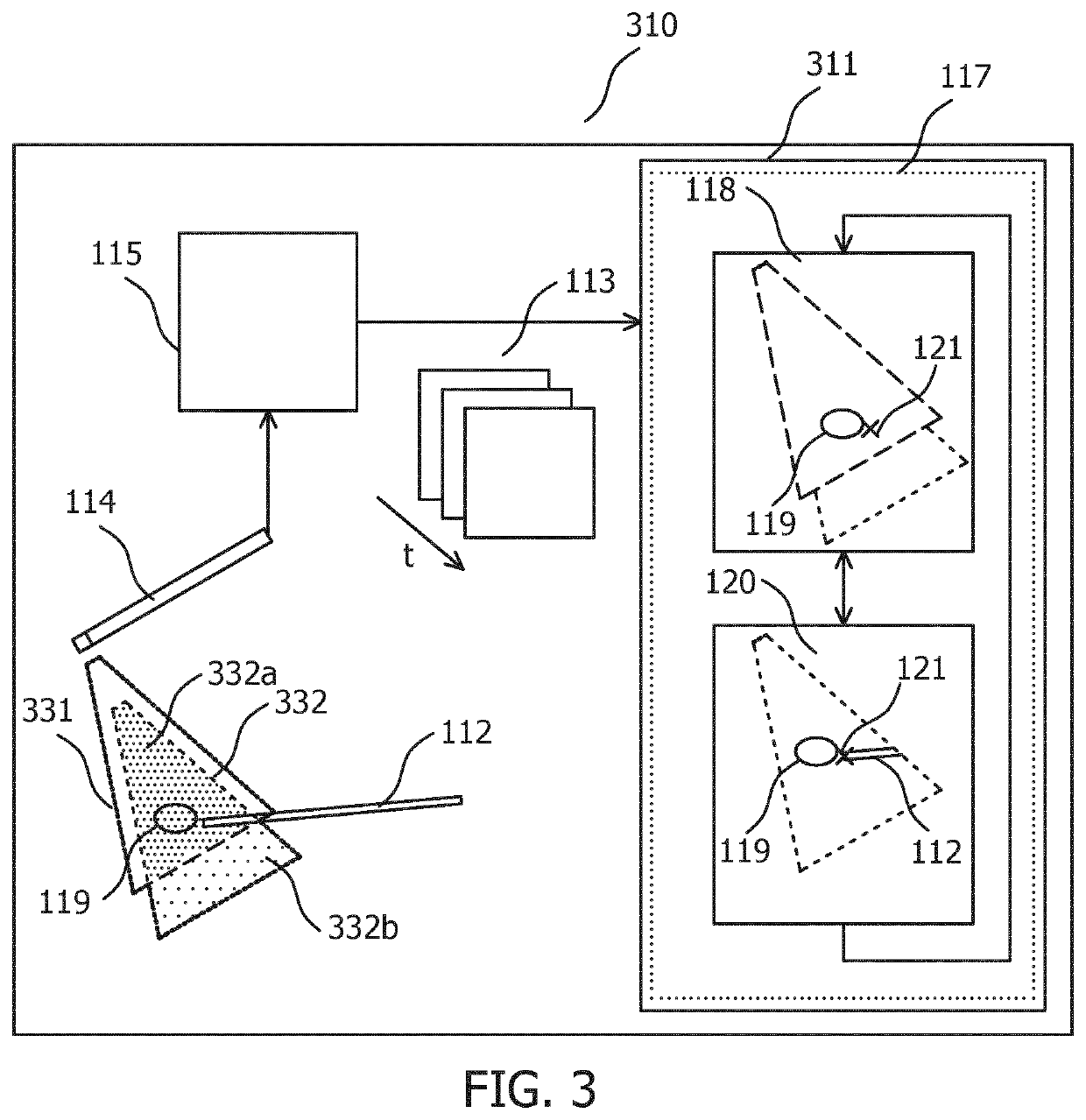
[0016]FIG. 1 illustrates an arrangement 110 that includes an ultrasound visualization system 111. Arrangement 110 may be used to track a position of interventional devi...


PUM


Abstract

The invention relates to an ultrasound visualization system (111, 311) for tracking a position of an interventional device (112) based on a stream of live ultrasound images (113). A processor (117) receives the stream of live ultrasound images (113), extracts a reference image (118) that includes an anatomical feature (119), extracts from the stream of live ultrasound (113) images a current image (120) that includes a portion of the anatomical feature (119) and a portion of the interventional device (112) at a current position (121), matches the portion of the anatomical feature in the current image 120 with the anatomical feature in the reference image (118) to determine a spatial relationship between the current position of the interventional device and the anatomical feature in the reference image (118), and indicates, in the reference image (118), the current position (121) of the interventional device (112), based on the determined spatial relationship.

Description

FIELD OF THE INVENTION[0001]The invention relates to an ultrasound visualization system for tracking a position of an interventional device based on a stream of live ultrasound images of an ultrasound imaging probe. A computer program product is also disclosed. The invention may be used in the medical ultrasound imaging field, and more specifically in ultrasound-based cardiac imaging. The stream of live ultrasound images may exemplarily be provided by a cardiac imaging probe such as an intra cardiac echo, ICE, probe or a trans esophageal echocardiogram, TEE, probe. The interventional device may for example be an ablation catheter.BACKGROUND OF THE INVENTION[0002]Interventional cardiology procedures such as the treatment of cardiac arrhythmias by catheter ablation are increasingly using tracking and mapping techniques to navigate interventional devices within the body. The treatment of atrial fibrillation by catheter ablation is an example of such a procedure. In this, an ablation ca...

Claims


Application Information

Patent Timeline
IPC IPC(8): A61B8/08A61B8/00
CPCA61B8/543A61B2090/364A61B8/5207A61B18/1492A61B2034/2063A61B8/0841A61B2090/378A61B8/0883A61B8/0891A61B8/12A61B8/466A61B8/483A61B8/5246A61N1/00
Inventor GIJSBERS, GERARDUS HENRICUS MARIAKOLEN, ALEXANDER FRANCISCUSBELT, HARM JAN WILLEMHARKS, GODEFRIDUS ANTONIUS
Owner KONINKLJIJKE PHILIPS NV
Learn More