
Nanoengineered surfaces for cancer biomarker capture

a biomarker and nano-engineered technology, applied in the field of surfaces, can solve the problems of difficult isolating and identifying exosome materials, and achieve the effect of reducing non-specific binding and low molecular weigh

Pending Publication Date: 2021-03-11
WISCONSIN ALUMNI RES FOUND
View PDF2 Cites 0 Cited by
  • Summary
  • Abstract
  • Description
  • Claims
  • Application Information

AI Technical Summary

Benefits of technology

This patent describes a method to reduce the stickiness of a surface by adding a layer of polymer brush molecules. These molecules have a lower molecular weight than the bifunctional tethers that stick to the surface. The added layer of polymer brush molecules helps to prevent other molecules from sticking to the surface and reduces the likelihood of unspecific binding. In simple terms, this technology makes the surface less sticky.

Problems solved by technology

Nevertheless, isolating and identifying exosome material is a challenge due to the small size and buoyancy of these vesicles.

Method used


Image

  • Nanoengineered surfaces for cancer biomarker capture
  • Nanoengineered surfaces for cancer biomarker capture
  • Nanoengineered surfaces for cancer biomarker capture

Examples


example 1

on of Capture Surfaces

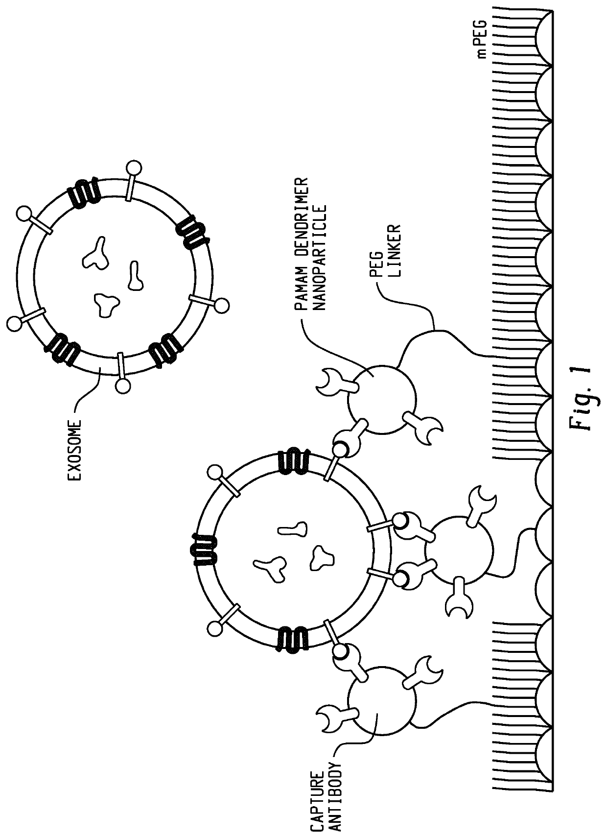
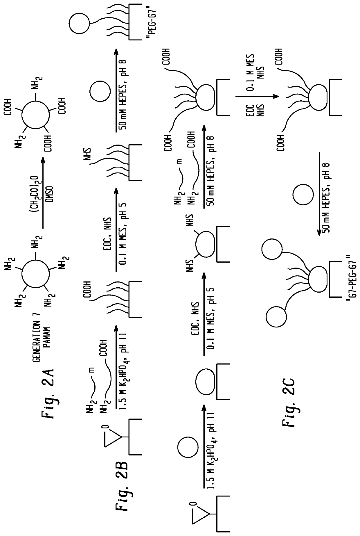
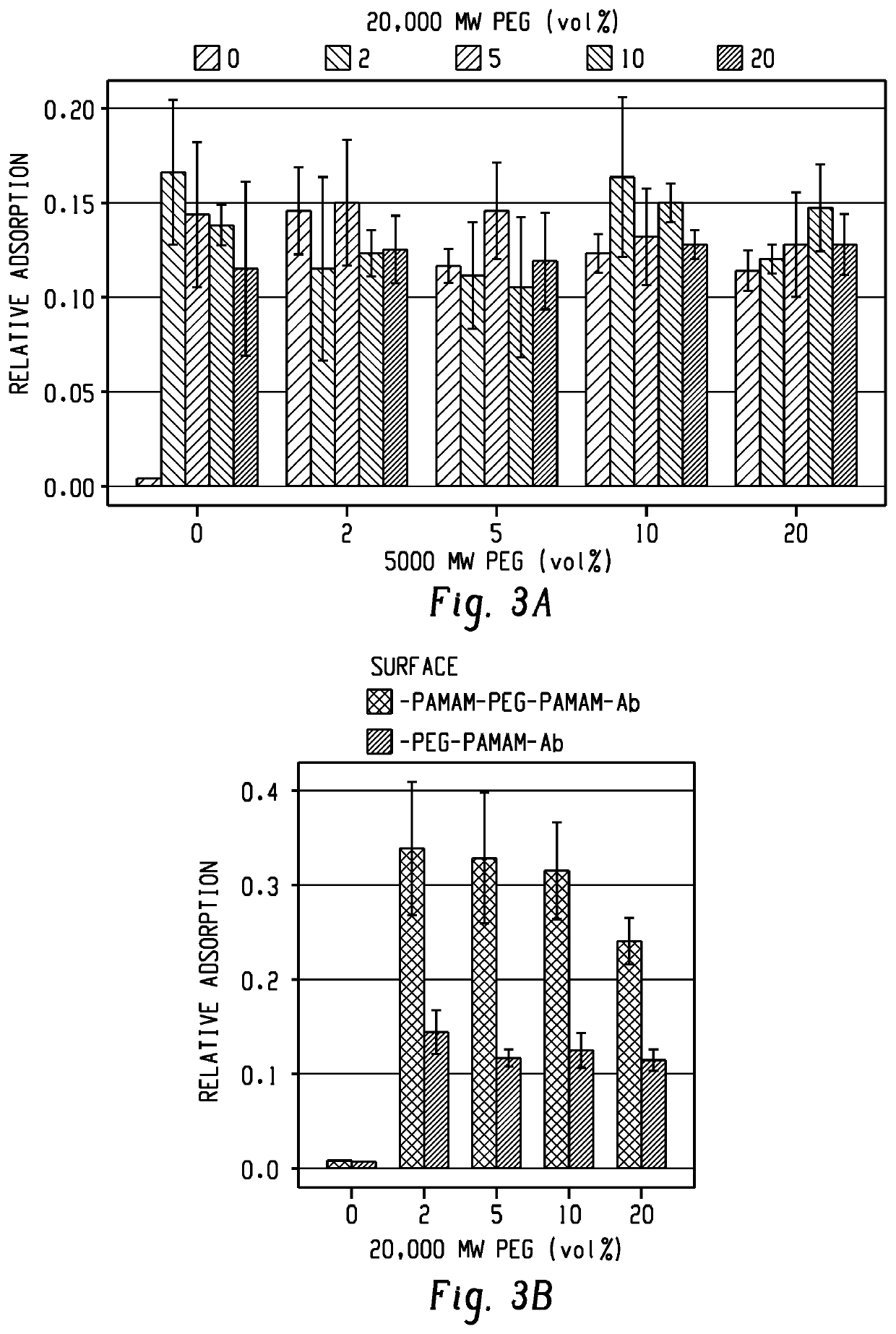
[0109]Capture surfaces for the capture of exosomes included epoxide-functionalized glass coated with partially-carboxylated, generation 7 (G7), poly(amidoamine) dendrimer nanoparticles. The G7 layer was functionalized with a mixture of heterobifunctional polyethylene glycol tethers (PEG) and shorter methoxy-PEG to minimize nonspecific interactions. The PEG tethers were then capped with G7 (FIG. 2). Preliminary experiments with 200 nm functionalized polystyrene beads suggested that a mixture of 0.5 μM 20,000 MW carboxy-PEG-amine, 2 μM 5,000 MW carboxy-PEG-amine, and 240 μM 2,000 MW methoxy-PEG-amine (1:1:48 by mass concentration) resulted in adequate capture rates (FIG. 3). In previous work, antibodies conjugated to PEG-tethered poly(amidoamine) (referred to here as PEG-G7) were shown to be more effective in capturing circulating tumor cells compared with controls with PEG alone (-PEG). In this work, surfaces pre-coated with polyamidoamine are referred to as G7-...

example 2

apture

[0111]The various polymer surface configurations were initially screened using exosome-sized beads. Polystyrene beads nominally 200 nm in diameter (Thermo Fisher FluoSphere™) were coated with recombinant Epithelial-Cell-Adhesion-Molecule (EpCAM, R&D Systems), a commonly-targeted surface antigen for circulating tumor materials. Capture surfaces with various G7 dendrimers and PEG configurations were functionalized with antibodies against EpCAM (aEpCAM, R&D Systems). Our results shown in FIG. 7a revealed significantly higher capture efficiency, as measured by increased fluorescent intensity, on the surfaces with two layers of dendrimers (G7-PEG-G7), compared to those with a single layer of dendrimers (PEG-G7). This enhancement was mostly pronounced when 2% of tethering, longer PEG (20 kDa) was added on the surface with shorter PEG (5 kDa), which shows an agreement with our previous observation using micelles. An increase of the content of the tethering PEG (5-20%) did not result ...


PUM

PropertyMeasurementUnit
molecular weightaaaaaaaaaa

Abstract

Described herein is cancer biomarker, e.g., a vesicle, capture surface that includes a substrate, a first plurality of nanoparticles attached to the substrate; a plurality of bifunctional tethers, wherein a first functionality of the bifunctional tethers is attached to at least a portion of the first plurality of nanoparticles; a second plurality of nanoparticles attached to a second functionality of at least a portion of the plurality of bifunctional tethers; a capture agent attached to at least a portion of the second plurality of nanoparticles; and a plurality of polymer brush molecules attached to the surface, wherein the polymer brush molecules have a lower molecular weight than the bifunctional tethers, and wherein the polymer brush molecules reduce nonspecific binding to the surface. Also described are method of capturing cancer biomarkers such as vesicles from a liquid biopsy sample.

Description

FIELD OF THE DISCLOSURE[0001]The present disclosure is related to surfaces suitable for capture of cancer biomarkers such as vesicles, particularly exosomes, as well as methods of capturing cancer biomarkers.BACKGROUND[0002]Exosomes are nanoscale vesicles that transport functional ribonucleic acid (RNA) and protein between cells. The release rate and composition of exosomes from tumors into circulation has been linked to malignancy and metastasis. Exosomes are therefore a potentially rich source of information about the state of cancer, obtainable from a minimally invasive blood draw (called a “liquid biopsy”). Nevertheless, isolating and identifying exosome material is a challenge due to the small size and buoyancy of these vesicles.[0003]Immunoaffinity separation techniques offer high selectivity, high sensitivity, and a lower likelihood of sample damage compared with the current gold standard of exosome separation by ultracentrifugation. Because exosome membranes are a mixture of...

Claims


Application Information

Patent Timeline
Patent Type & Authority Applications(United States)
IPC IPC(8): G01N33/574G01N33/543
CPCG01N33/57492G01N33/57438G01N33/54346G01N33/57488G01N33/5743
Inventor HONG, SEUNGPYOPOELLMANN, MICHAEL J.
Owner WISCONSIN ALUMNI RES FOUND
Learn More