Cellular image analysis method, cellular image analysis device, and learning model creation method

Pending Publication Date: 2021-05-06
SHIMADZU CORP
View PDF0 Cites 4 Cited by
  • Summary
  • Abstract
  • Description
  • Claims
  • Application Information

AI Technical Summary

Benefits of technology

[0048]According to the present invention, for example in a site where pluripotent stem cells, such as, e.g., iPS cells and ES cells, are cultured, it is possible to quickly and accurately determine noninvasively whether or not the cell that is being cultured maintains the undifferentiated state or is in the deviated state, or whether or not a deviated cell is present in the cell colony, or whether or not a foreign substance such as dust is contaminated. Thereby, the quality control of the cell that is being cultured is facilitated, which in turn can improve the product

Problems solved by technology

However, because the stained cell dies, it is impossible to perform undifferentiated marker-staining to determine pluripotent stem cells for regenerative medicine.
The reason of using a phase-contrast microscope is that a cell is generally transparent and is difficult to be observed under a normal optical microscope.
Therefore, such conventional techniques are not suitable for industrially mass-producing pluripotent stem cells.
Therefore, in order to perform the determination with high accuracy at all times, the feature amounts to be used must be changed depending on the cell species to be observed, the culture condition, and the like, and therefore the processing is very troublesome.
In addition, when determining a cell cultured under a condition different from the previous condition, it is required to examine which

Method used


Image

  • Cellular image analysis method, cellular image analysis device, and learning model creation method
  • Cellular image analysis method, cellular image analysis device, and learning model creation method
  • Cellular image analysis method, cellular image analysis device, and learning model creation method

Examples


Embodiment Construction

"d_n">[0057]Hereinafter, an example of a cell image analysis method and a cell image analysis device according to the present invention will be described with reference to the attached drawings.

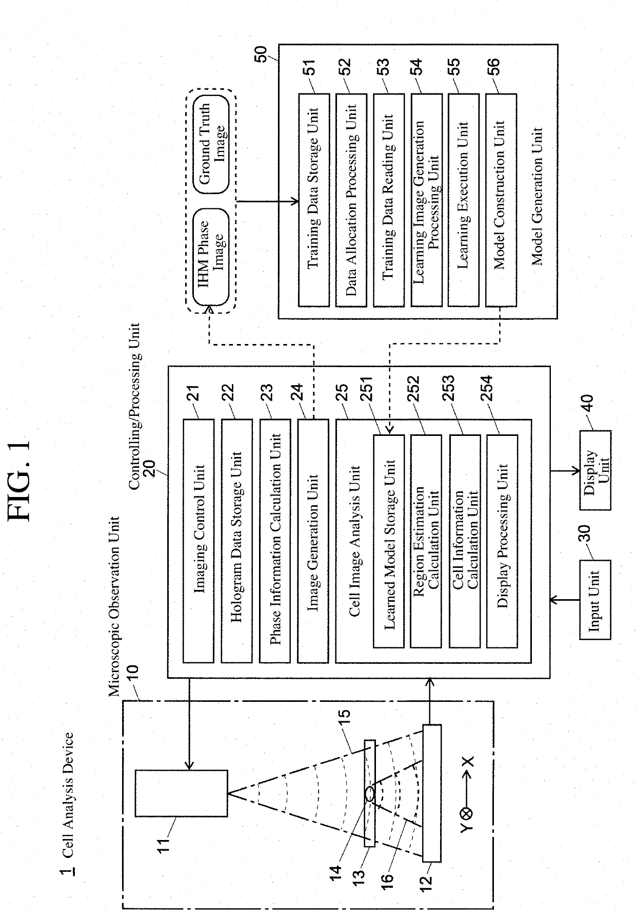
[0058]FIG. 1 is a schematic configuration diagram of a cell analysis device using a cell image analysis device for carrying out a cell image analysis method according to the present invention.

[0059]The cell analysis device 1 of this example is provided with a microscopic observation unit 10, a controlling / processing unit 20, an input unit 30 and a display unit 40 as user interfaces, and a model generation unit 50.

[0060]The microscopic observation unit 10 is an in-line holographic microscopy (IHM), and is provided with a light source unit 11 including a laser diode, etc., and an image sensor 12. A culture plate 13 including a cell colony (or cell alone) 14 is arranged between the light source unit 11 and the image sensor 12.

[0061]The controlling / processing unit 20 controls the operation of the...


PUM


Abstract

A phase image is formed by calculation from a hologram image of a cell, and segmentation is performed for each pixel for the phase image using a fully convolution neural network to identify an undifferentiated cell region, a deviated cell region, a foreign substance region, and the like. When learning, when a learning image included in a mini-batch is read, the image is randomly inverted vertically or horizontally and then is rotated by a random angle. A part that has been lost within the frame by the pre-rotation image is compensated for by a mirror-image inversion with an edge of a post-rotation image as an axis thereof. Learning of a fully convolution neural network is performed using the generated learning image. The same processing is repeated for all mini-batches, and the learning is repeated by a predetermined number of times while shuffling the training data allocated to the mini-batch. The precision of the learning model is thus improved. In addition, since rotationally invariant characteristics can be learned, it is possible to identify cell colonies of various shapes with good precision.

Description

TECHNICAL FIELD[0001]The present invention relates to a method and an apparatus for performing analysis processing of an observation image acquired for a cell and a learning model generation method for the analysis. More particularly, the present invention relates to a cell image analysis method, a cell image analysis device, and a learning model generation method suitable for non-invasively determining the state of a cell or acquiring cell information about the number of cells in the process, etc., of culturing pluripotent stem cells (ES cells or iPS cells).BACKGROUND OF THE INVENTION[0002]In a regenerative medicine field, researches using pluripotent stem cells, such as, e.g., iPS cells and ES cells, have been actively conducted in recent years. In researching and developing regenerative medicine using such pluripotent stem cells, a large number of undifferentiated cells in a state in which the pluripotency is maintained needs to be cultured. Therefore, it is necessary to select a...

Claims


Application Information

Patent Timeline
IPC IPC(8): G06T7/00G06K9/00G06T7/11
CPCG06T7/0012G06K9/0014G06T7/11G06T2207/10056G06T2207/30024G06T2207/20084G06T2207/20081G06T7/62G06T2207/20076G06V20/695G06V10/454G06V10/82
Inventor TAKAHASHI, WATARUAKAZAWA,, AYAKO
Owner SHIMADZU CORP
Why Eureka
Try Eureka