
Examination of a blood vessel based on nuclear resonant absorption

a blood vessel and nuclear resonant technology, applied in the field of nuclear resonant absorption blood vessel examination, can solve the problems of difficult or impossible placement of measurement catheters in certain blood vessels, unnecessary interventions for stenting patients,

Active Publication Date: 2021-10-21
KONINKLJIJKE PHILIPS NV
View PDF0 Cites 2 Cited by
  • Summary
  • Abstract
  • Description
  • Claims
  • Application Information

AI Technical Summary

Benefits of technology

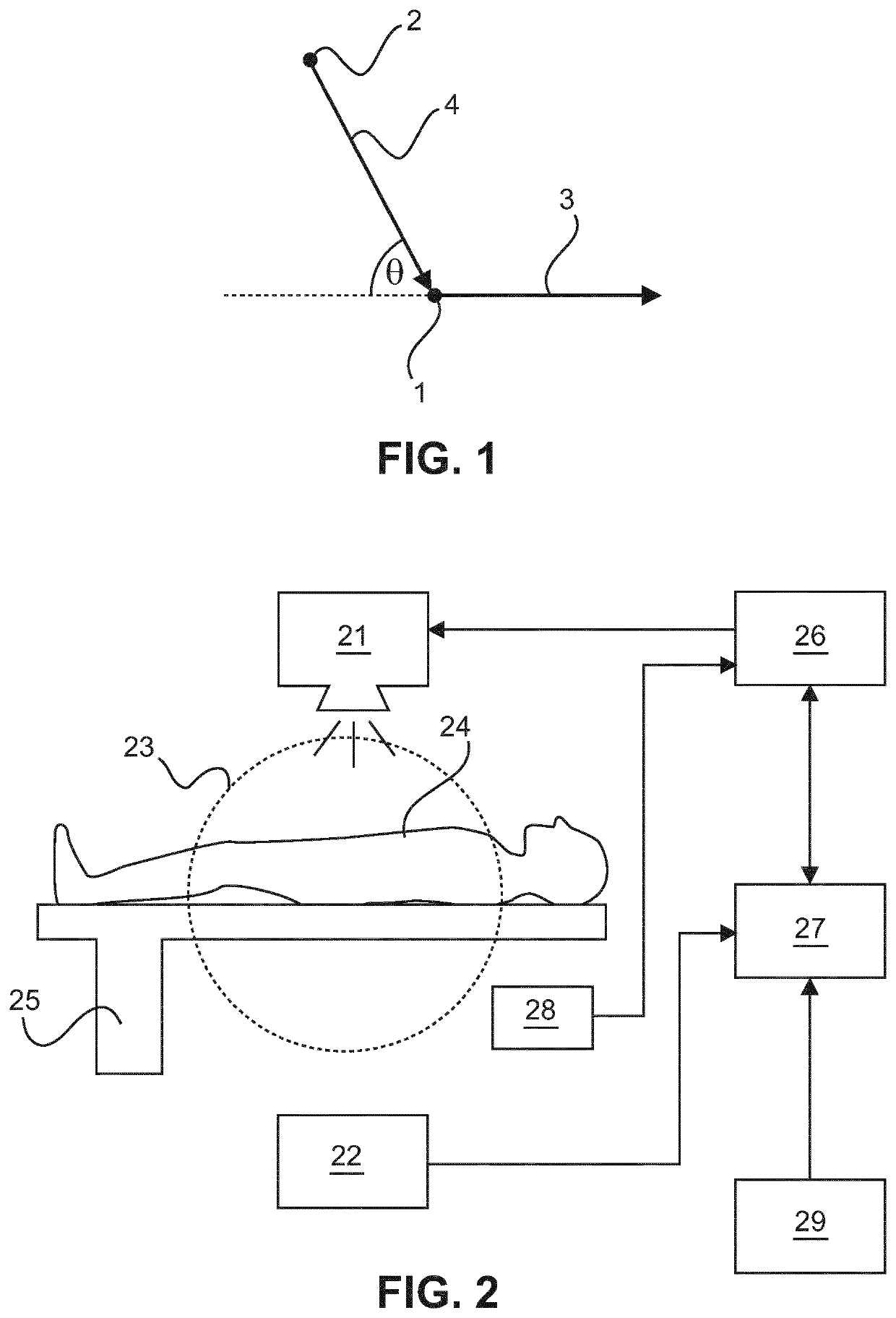
The patent is about using x-rays to determine the velocity of blood flow in blood vessels. By tuning the energy of the x-rays to the energy at which nuclear resonant absorption occurs, the x-rays can be used to create an angiographic image that distinguishes contrast agents from calcium. The patent also explains that in a non-relativistic limit, no Doppler shift of photon energy occurs, so the patient should be positioned with the x-ray source at an angle to the blood vessel to ensure accurate blood velocity measurements.

Problems solved by technology

As a consequence, unnecessary interventions for stenting patients may be performed, if the decision about the intervention is made on the basis of angiography images alone.
However, they are invasive techniques requiring an intervention.
Moreover, it can be difficult or impossible to place the measurement catheter in certain blood vessels, and in such cases a measurement might not be possible.
However, the CT images might be degraded by metal artifacts in case of patients having a pacemaker, an internal defibrillator or a prosthetic heart valve, for example.
For such patients, modelling of the blood flow may not be possible.
However, calcium attenuates x-ray radiation similarly as iodine so that it is not possible to distinguish between calcium and iodine in angiographic images.
As consequence, angiographic images do not allow for determining the spatial distribution of calcified plaque in a blood vessel including a stenosis.

Method used


Image

  • Examination of a blood vessel based on nuclear resonant absorption
  • Examination of a blood vessel based on nuclear resonant absorption
  • Examination of a blood vessel based on nuclear resonant absorption

Examples


Embodiment Construction

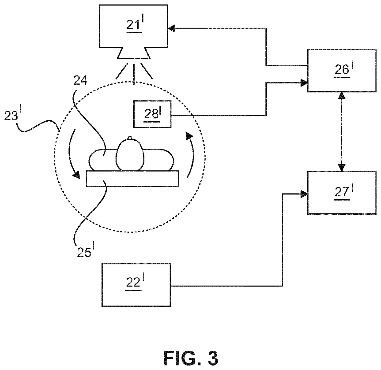
[0031]The invention suggests determining a velocity of blood flowing in a portion of a blood vessel of a body of a patient based on an excitation of atomic nuclei of a contrast agent introduced into the blood vessel. Moreover, an x-ray image showing plaque, including calcified plaque, in the examined portion of the blood vessel can be acquired in the process of measuring the blood velocity. Thus, two characteristics of a portion of a blood vessel—the blood velocity and the distribution of calcified plaque—can be determined in one measurement, if desired. Likewise, it is possible to either determine the blood velocity or to produce an angiographic image showing the distribution of calcified plaque.

[0032]The blood vessel may be a coronary artery in the region of the heart of the patient. However, it is likewise possible to determine the blood velocity in blood vessels in other parts of the body of the patient. The portion of the blood vessel to be examined may have been identified pre...


PUM


Abstract

The invention relates to a system and a method for determining a characteristic of a blood vessel portion, which comprises blood including a contrast agent exhibiting resonant absorption of x-ray photons at a specific energy. The system comprises a tunable monochromatic x-ray source (21) emitting x-ray radiation, an x-ray detector device (22) for detecting the x-ray radiation after it has travelled through the blood vessel portion. A control unit (26) varies a tuning of the x-ray source (21) to vary the energy of the x-ray radiation emitted by the x-ray source (21), and an evaluation unit (27) determines a tuning of the x-ray source (21) at which nuclear resonant absorption of the x-ray radiation incident onto the blood vessel portion occurs and estimates the characteristic on the basis of the determined tuning. The characteristic may particularly be the blood velocity in the blood vessel portion.

Description

FIELD OF THE INVENTION[0001]The invention relates to a system and a method for determining at least one characteristic of a portion of a blood vessel of a patient. The characteristic may correspond to a velocity of blood flowing in the portion of the blood vessel. Likewise, the characteristic may correspond to an anatomy of the portion of the blood vessel and / or to a spatial distribution of calcium included therein.BACKGROUND OF THE INVENTION[0002]A stenosis in a blood vessel can be detected and examined using angiography imaging. In angiography, a contrast agent—typically comprising iodine—is inserted into the blood vessel and the blood vessel is imaged using a suitable imaging modality such as, x-ray imaging, computed tomography (CT) imaging or magnetic resonance (MR) imaging. In the resulting image of the blood flowing through the blood vessel, the stenosis may be detected and its degree may be determined in order to decide about a treatment, such as the insertion of a stent.[000...

Claims


Application Information

Patent Timeline
Patent Type & Authority Applications(United States)
IPC IPC(8): A61B6/00A61B6/03A61K49/04
CPCA61B6/504A61B6/481A61B6/482A61K49/0438A61B6/037A61B6/405A61B6/541A61B6/466G01N23/095G01N33/49G01T1/26G01T1/29A61B6/00A61B6/4064A61B6/4258A61B6/507A61B6/5217A61K49/0004A61K49/04G16H50/30
Inventor DAERR, HEINER
Owner KONINKLJIJKE PHILIPS NV
Learn More