Adenosomes
a technology of adenosomes and adenoviruses, applied in the field of recombinant adenovirus nucleic acid, can solve the problems of high cytotoxicity, strong immune response, purification problems,
- Summary
- Abstract
- Description
- Claims
- Application Information
AI Technical Summary
Benefits of technology
Problems solved by technology
Method used
Image
Examples
example 1
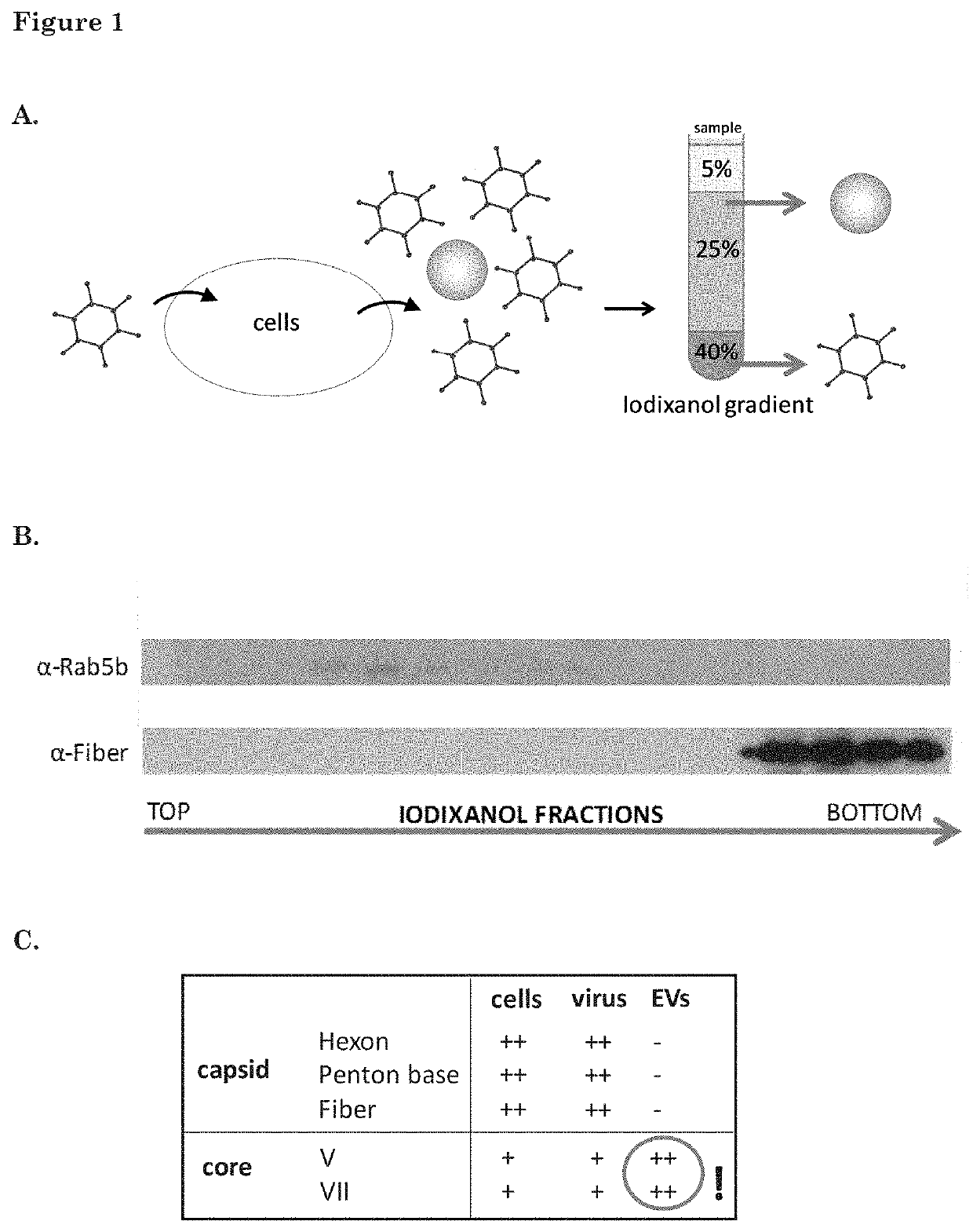
of Adenoviral Core Proteins Inside EVs by Mass-Spectrometry
[0180]GS184 cells (primary glioblastoma cells obtained from the Erasmus MC, Dept. of Neurosurgery) were plated in ten T175 flasks 24 h before infection with wild-type HAdV-5. The cells were cultured as monolayers on a coating of extracellular matrix (ECM 1:100 in dPBS, Cultrex, Sanbio) in NS medium (DMEM-F12+fibroblast growth factor+endothelial growth factor+heparin+B27+1% penicillin-streptomycin). The cells were infected with wild-type human adenovirus type 5 (HAdV-5) with a MOT of 5 infectious particles per cell. Medium was refreshed after 2 h to NS medium. Supernatant and cells were collected at 40 h after infection. EVs were isolated from the cell culture supernatant as described in De Vrij, 2013, Nanomedicine (Lond), 8(9):1443-58, using an iodixanol-based density-gradient (ultracentrifugation at 192,000 g for 4 hr). The iodixanol gradient consisted of three layers: 40% iodixanol as the bottom layer (2 mL), 25% iodixanol...
example 2
Adenovirus DNA is Detected by PCR in EVs and the EVs are Capable to Infect Cells
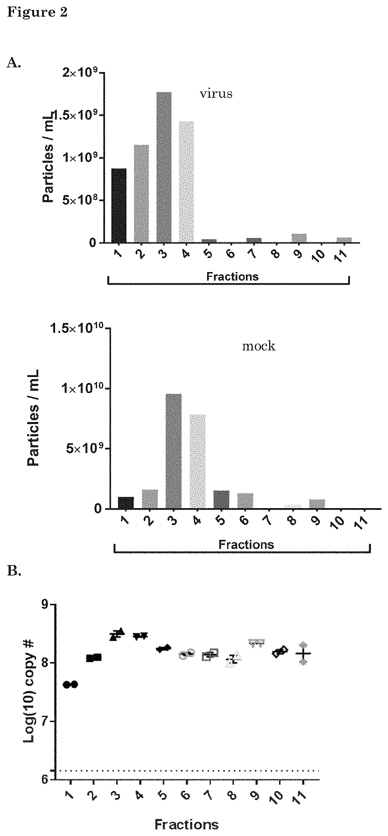
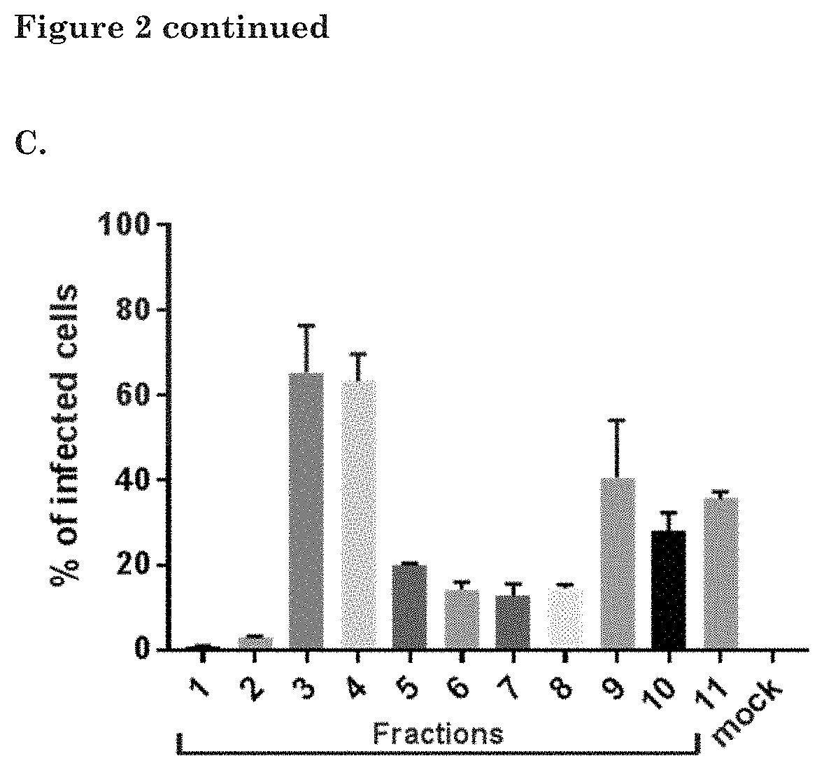
[0181]GS756 cells were plated in two T175 flasks 24 h before infection with the conditionally replicating Adenovirus (CRAd) Ad.5.d24.RGD.GFP (MOI=1 infectious particle per cell). (Virus details including genomic construction are provided in Balvers, 2014, Viruses, 6(8), 3080-3096.) After 72 hours, the supernatant was collected for EV isolation. Low-speed centrifugations were performed to remove cells and cell debris (150×g for 5 minutes plus 3000×g for 20 minutes) followed by ultracentrifugation at 100.000×g for 70 minutes to pellet EVs and viral particles. The pellets were subjected to iodixanol density gradient centrifugation and iodixanol fractions were isolated as described above.
[0182]To determine the amount of EVs within each fraction, samples were prepared for EV-Quant analysis. For this, samples were incubated with a red-fluorescent dye (Rhodamine at a final concentration of 0.33 ng / μl), which la...
example 3
Demonstrates pV. GFP Incorporation in EVs after Infecting Cells with pV. GFP Virus
[0185]Cells (GS184, GS562, A549 and HER911) were plated on 48 well plates 24 h before infection with an adenovirus with green fluorescent protein attached to the pV protein (pV.GFP). The cells were infected with a MOI of 50 viral particles per cell in 100 μL medium per well (NS medium for GS184 and GS562 and serum medium for HER911 / A549). (serum medium: Dulbecco's Modified Eagle Medium+10% Fetal Bovine serum (FBS)+1% penicillin-streptomycin). Medium was refreshed after 2 h to NS medium, DMEM only and serum medium for each cell line. (Serum medium was pre-cleared from bovine EVs by ultracentrifugation for 16 h at 100,000×g). Triplicates were used for each medium condition for all cell lines. Supernatants were collected at 64 h after infection and centrifuged at 500×g for 10 min. to remove cell debris. EV-Quant analysis was performed on 143 μL of all supernatants. With GFP fused to the pV protein it was ...
PUM
| Property | Measurement | Unit |
|---|---|---|
| Stress optical coefficient | aaaaa | aaaaa |
Abstract
Description
Claims
Application Information
 - R&D
- Intellectual Property
- Life Sciences
- Materials
- Tech Scout
- Unparalleled Data Quality
- Higher Quality Content
- 60% Fewer Hallucinations
Browse by: Latest US Patents, China's latest patents, Technical Efficacy Thesaurus, Application Domain, Technology Topic, Popular Technical Reports.
© 2025 PatSnap. All rights reserved.Legal|Privacy policy|Modern Slavery Act Transparency Statement|Sitemap|About US| Contact US: help@patsnap.com


