Automated detection of lung conditions for monitoring thoracic patients undertgoing external beam radiation therapy

a technology for detecting lung conditions and monitoring thoracic patients, which is applied in the field of computerized system for radiation therapy support, can solve the problems of increased risk of secondary cancer, increased risk of developing secondary cancer, and inability to detect medical conditions that may develop in the interval between weekly cbct acquisitions, so as to reduce the total dose, automatic detection of lung conditions, and the effect of reducing the risk of secondary cancer

Pending Publication Date: 2022-03-10
KONINKLJIJKE PHILIPS NV
View PDF0 Cites 2 Cited by
  • Summary
  • Abstract
  • Description
  • Claims
  • Application Information

AI Technical Summary

Benefits of technology

[0035]In embodiments, the proposed system is configured to detect medical condition(s) before the delivery of the treatment fraction, thus allowing medical staff to act before delivering a fraction that might harm or aggravate the detected condition. Specifically, in embodiments, the invention proposes a system to automatically detect lung conditions using daily acquired planar projection X-ray images. The radiation dose from the acquisition of one planar X-ray is much smaller and using daily images would allow timely detection and application of clinical protocols aimed to manage these conditions. Moreover, detection of a lung condition in a planar X-ray image could trigger a warning to acquire a CBCT for further verification and validation.
[0036]Current protocols are based on visual inspection of C

Problems solved by technology

However, visual inspection is error prone and time consuming.
Also, frequent acquisition of CBCTs is associated with increased risk of develo

Method used


Image

  • Automated detection of lung conditions for monitoring thoracic patients undertgoing external beam radiation therapy
  • Automated detection of lung conditions for monitoring thoracic patients undertgoing external beam radiation therapy
  • Automated detection of lung conditions for monitoring thoracic patients undertgoing external beam radiation therapy

Examples


Embodiment Construction

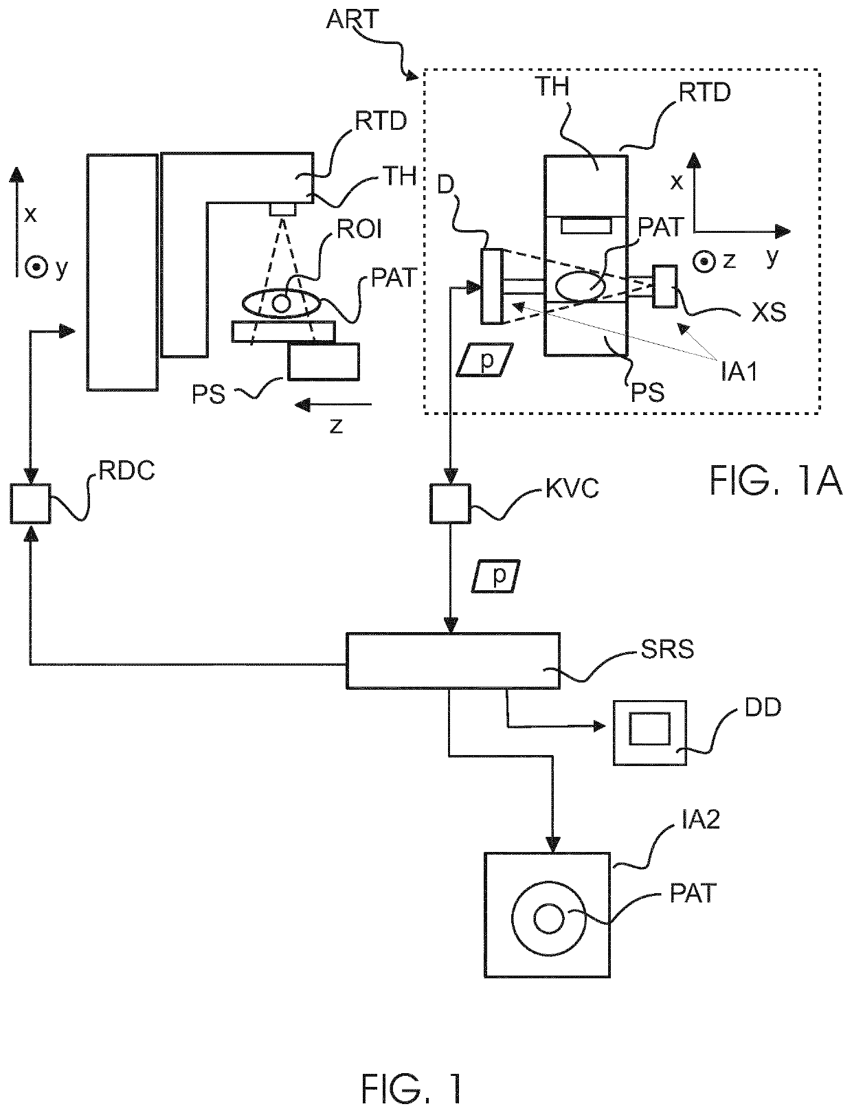
[0054]Radiation therapy (RT), in particular external beam radiation therapy (EBRT), is a mode of treatment of cancer in animal or human patients.

[0055]RT is best thought of as a process in terms of a workflow with certain steps being performed over time. Initially, RT starts with a diagnosis of cancer which usually involves drawing together by clinical professional all available data about the patient, including image data. This (initial) data image data is obtained by using suitable medical imaging devices implementing imaging techniques such as emission imaging (eg, PET / SPEC) or transmission imaging, or other imaging techniques such as MRI (magnetic resonance imaging), or a combination of any of these. Example of transmission imaging envisaged herein includes X-ray based tomography, CT (computed tomography). Like MRI, CT can generate 3D imaging, which is preferred for precise location of a lesion such as a tumor.

[0056]Specifically, based on this initial image data, the area to be ...


PUM


Abstract

A computerized system (SRS) for radiation therapy support. The system comprises an input interface (IN) for receiving an input image acquired by an imaging apparatus (IA1). The input image represents a region of interest (ROI) internal of a patient (PAT) and acquired before delivery of a dose fraction by a radiation therapy delivery apparatus (RTD). A pre-trained machine learning unit (MLU) of the system is configured to process the input image to detect a medical condition. A communication component (RC) of the system is configured to provide, based on the detected medical condition, an indication for one or more clinical actions to be performed in relation to the patient.

Description

FIELD OF THE INVENTION[0001]The invention relates to a computerized system for radiation therapy support, to a computer-implemented method for radiation therapy support, to a method of training a machine learning component for used in the computerized system, to a computer program element, and to a computer readable medium.BACKGROUND OF THE INVENTION[0002]In external beam radiation therapy (EBRT) of, for example, thoracic malignancies, or other anatomical changes and lung medical conditions appearing during the course of the treatment are monitored, measured, and recorded. These records trigger protocols and decisions on the ongoing treatment. Currently, lung tissue changes and lung conditions are monitored and detected using visual inspection of CBCT (cone beam computed tomography) images.[0003]However, visual inspection is error prone and time consuming. Visual interpretation and diagnosis of lung conditions is a specialized skill requiring extensive training. What is more, RTTs (...

Claims


Application Information

Patent Timeline
IPC IPC(8): G16H20/40G16H30/40G16H50/20G16H40/67G16H50/70G16H10/60G06N20/00A61N5/10
CPCG16H20/40G16H30/40G16H50/20G16H40/67G16H40/20G16H10/60G06N20/00A61N5/1039G16H50/70G06N3/082A61B5/7267A61B6/032A61B6/037A61B6/4085A61B6/461A61B6/5217A61N5/1064G06N3/044G06N3/045G16H30/20A61N2005/1074
Inventor BONDAR, MARIA LUIZAKUSTRA, JACEK LUKASZCONSOLI, SERGIO
Owner KONINKLJIJKE PHILIPS NV
Why Eureka
Try Eureka