Endocavity probe and method for processing diagnostic images

a technology of endorectal probe and diagnostic image, which is applied in the field of medical equipment, can solve the problems of disturbance factor, device complex from the programming and controlling viewpoint, and complex bi-functional prob

Pending Publication Date: 2022-06-23
ELESTA SPA
View PDF9 Cites 0 Cited by
  • Summary
  • Abstract
  • Description
  • Claims
  • Application Information

AI Technical Summary

Benefits of technology

The patent is about a set that helps medical professionals accurately merge images from different techniques, like ultrasound and magnetic resonance images. It uses a dummy probe that is inserted into the patient's body to cause the same compression and displacement of the surrounding tissues as the real ultrasound probe. This ensures that the images from the two techniques are similar and can be easily merged together. The use of this set improves the accuracy of the resulting images.

Problems solved by technology

The bi-functional probe is very complex.
This makes the device very complex from the programming and controlling viewpoint.
Moreover, the presence of the endorectal probe in the investigated volume represents a disturbance factor for the operation of the magnetic resonance imaging system.
This results in inaccuracies in the captured images, due to the irregularity of movements that, in turn, affects the quality of the ultrasound data of the investigated volume and, thus, the accuracy of the merged images (ultrasound and magnetic resonance images).
This adversely affects the subsequent use of the captured images during navigation, making the treatment more complex and less safe.
Furthermore, the interventions last more time, due to the slowness of capturing the necessary images, also taking into account the need for repeating the acquisition of an image in case of wrong focusing due to the inaccuracies mentioned above.
This adversely affects the intervention cost and has a greater impact on the patient, to whom larger amounts of anesthetic shall be administered, when necessary.
Similar problems can occur in other applications, where it is necessary to use a substantially cylindrical probe, through which ultrasound images are captured according to multiple scan planes belonging to a bundle of planes and where it is necessary to merge the three-dimensional ultrasound images with a three-dimensional image previously captured through, for example, magnetic resonance or other high-resolution imaging techniques.

Method used


Image

  • Endocavity probe and method for processing diagnostic images
  • Endocavity probe and method for processing diagnostic images
  • Endocavity probe and method for processing diagnostic images

Examples


Embodiment Construction

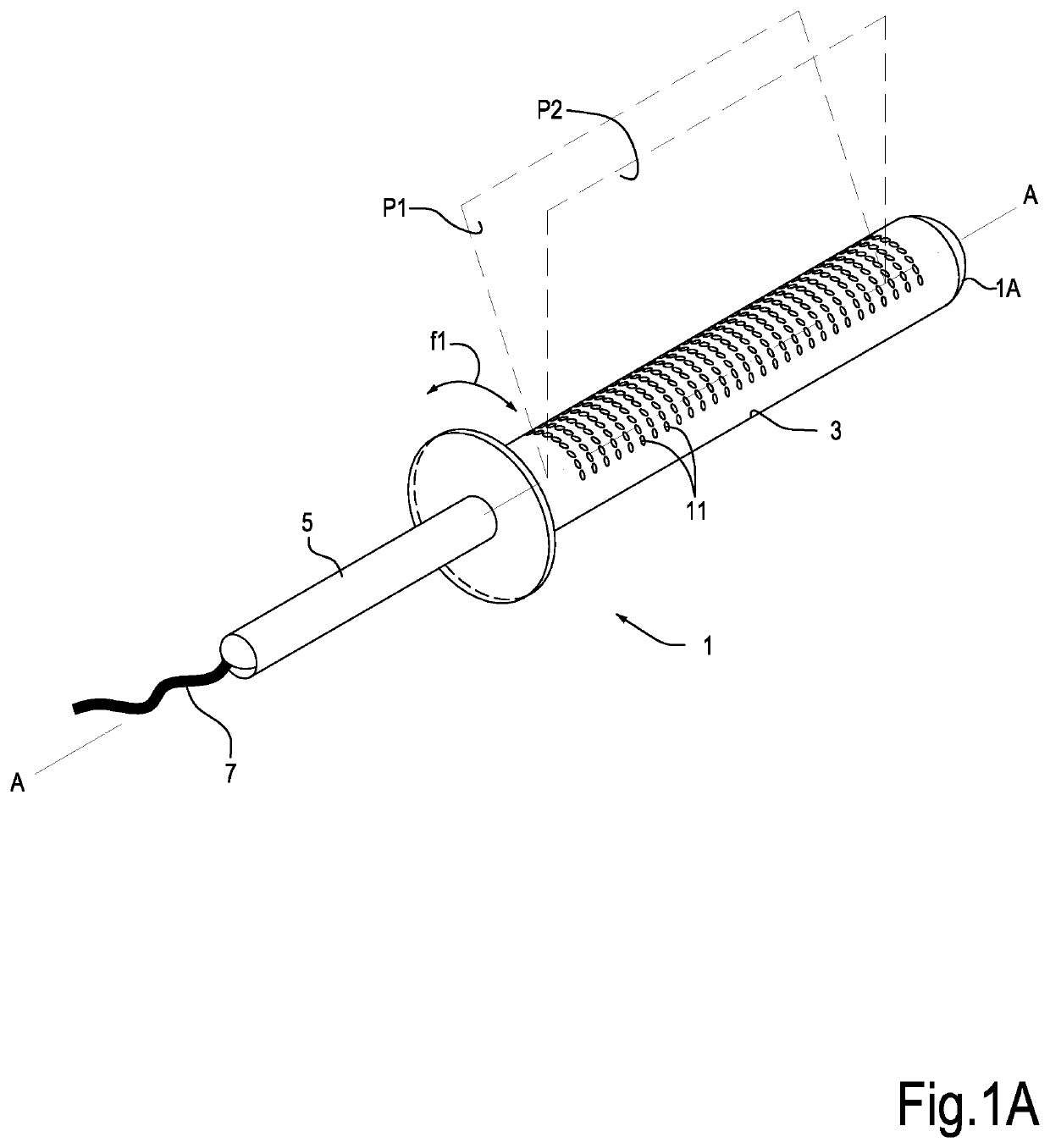
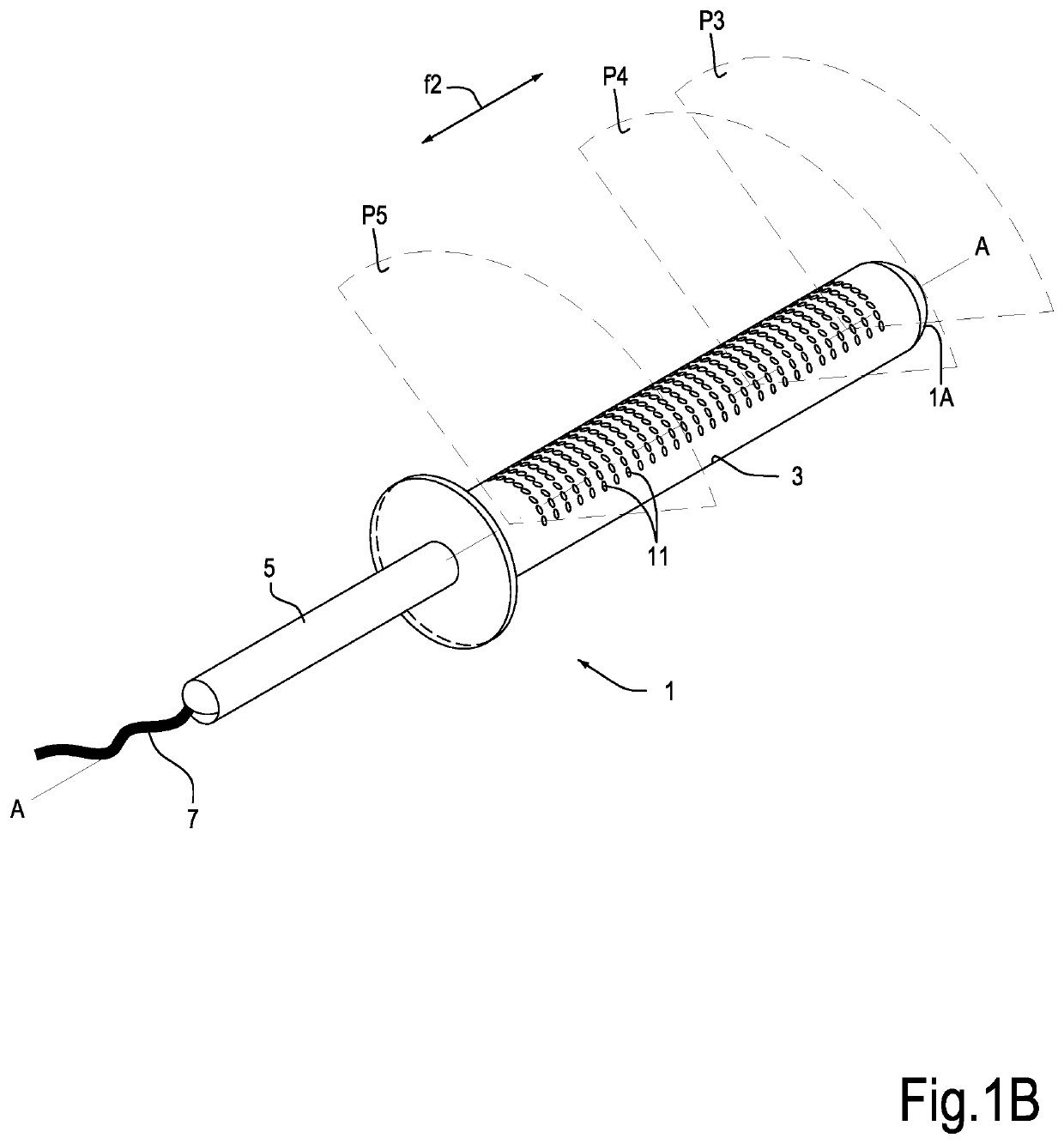
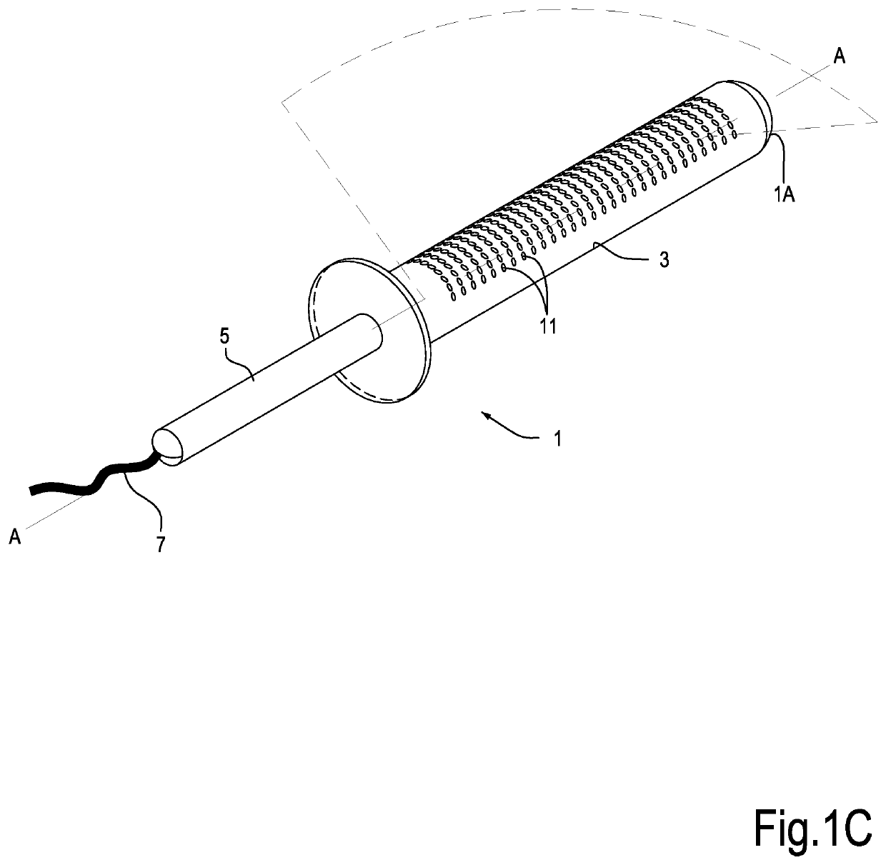
[0036]In the following description, specific reference will be made to endorectal probes to be used in minimally invasive prostate surgery and to methods using the images captured through these probes. However, the novel features disclosed herein with reference to an endorectal probe can be also used in other applications, for producing endocavity probes in general, i.e. probes adapted to be used in cavities of the human or animal body, for constructing three-dimensional images of an organ, a tissue, or a generic portion of the human or animal body, by capturing data on ultrasound signal acquisition planes, in particular a plurality of planes of a bundle of planes containing the probe axis and / or a plurality of parallel planes orthogonal to the probe axis.

[0037]Therefore, what disclosed herein can be conceptually applied also to navigation for performing tumor ablation treatments in all cases when the lesions to be treated are close to the inner wall of a tubular structure of organs...


PUM


Abstract

An innovative method is disclosed for merging images of an organ in vivo captured through a first imaging technique and a second imaging technique, this latter using an endocavity ultrasound probe inserted into a cavity associated with the organ under investigation. For superimposing the images on one another with greater accuracy, the images captured through the first imaging technique, that is different than the ultrasound technique, a dummy probe is inserted into the cavity associated with the organ under investigation. The dummy probe may be applied both using prior art biplane endocavity probes and innovative electronic scanning endocavity probes. Therefore, two types of endorectal probe are described: a biplane probe, that shall be rotated and translated for capturing biplane images; and a new electronic scanning ultrasound probe delivering the two-dimensional images for the merging without the need for moving the endocavity probe.

Description

TECHNICAL FIELD[0001]The present invention refers to medical equipment and the related methods of use. More in particular, the present invention discloses innovations in ultrasound imaging equipment and related methods for image processing, especially, although not exclusively, in the field of endorectal probes for prostate surgery, and, more in general, in the field of endocavity probes for diagnosis and / or interventions on organs adjacent to ducts or cavities of a patient's body.BACKGROUND ART[0002]For many prostate interventions it is useful to have available ultrasound images captured by endorectal probes.[0003]These images are useful, for example, in biopsies and in minimally invasive prostate surgery carried out through the different available technologies, for example local hyperthermia for reducing benign prostatic hyperplasia or for ablation of malignant focal tumors, through hyperthermia techniques, use of radioactive seeds (brachytherapy), or other minimally invasive tran...

Claims


Application Information

Patent Timeline
Patent Type & Authority Applications(United States)
IPC IPC(8): A61B8/08A61B8/12A61B8/00
CPCA61B8/5261A61B8/12A61B8/5246A61B8/54A61B8/085A61B8/4488A61B8/08A61B8/4444A61B8/4494A61B8/40
Inventor MASOTTI, LEONARDOBRESCHI, LUCA
Owner ELESTA SPA
Why Eureka
Try Eureka