
3D Bioprinted Skin Tissue Model

a skin tissue and 3d printing technology, applied in the field of 3d bioprinting of skin tissue models, can solve the problems of labour-intensive methods and inability to control the construction of in vitro models

Pending Publication Date: 2022-08-11
CELLINK AB
View PDF0 Cites 0 Cited by
  • Summary
  • Abstract
  • Description
  • Claims
  • Application Information

AI Technical Summary

Benefits of technology

The present invention provides a method for producing a 3D skin tissue model with controlled deposition of cells, biomaterials, and biomolecules. The model can be produced with high efficiency, ease, and control of construction. It can also be produced in multiple replicates with low variation. The model allows for dispensing at different levels of resolution, and can be adapted to different interests and bioreactors. The model can be bioprinted to form a hypodermal, dermal, and epidermal compartments, and can mimic natural gradients found within native skin tissue. The model is more relevant and natural response of embedded cells.

Problems solved by technology

However, these methods are labour intense and do not allow for controlled construction of the in vitro models.

Method used


Image

  • 3D Bioprinted Skin Tissue Model
  • 3D Bioprinted Skin Tissue Model
  • 3D Bioprinted Skin Tissue Model

Examples


example of

Production Procedure

[0091]Below is an example of producing the skin tissue model with a bioink A, cell type A and an additional cell type A and one factor A:

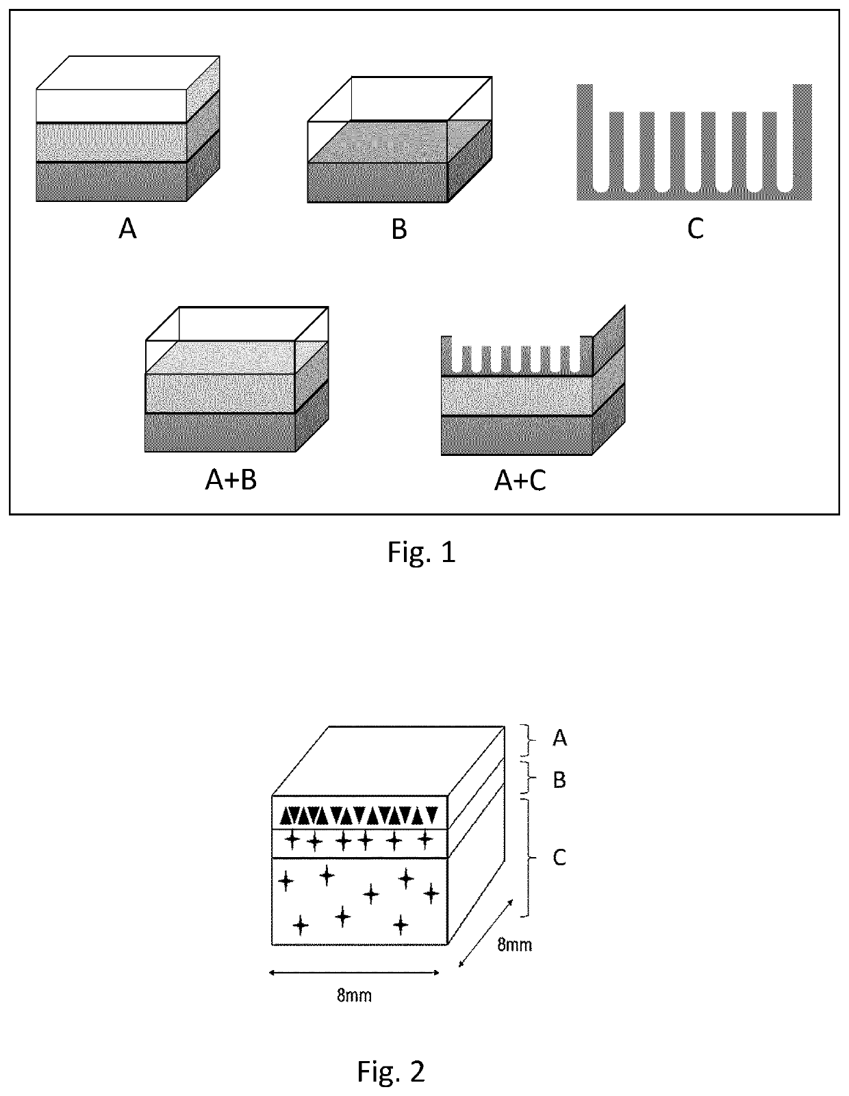
[0092]Design of model (s): To design the model any type of 3D-modeling software can be utilized to either modify a scanned model, such as a CT- or MR-scan, or to create a model from scratch. When creating models from scratch different types of compartments can be used as building blocks. FIG. 1 exemplifies 3 types of compartment types and two examples of how these can be combined to create a model.[0093]A. Block Compartments. These blocks can have different dimensions, and either be stacked and / or placed within each other to form different compartments within the model. Using robust bioink formulations able to retain the shape of the blocks during the print blocks can be utilized to form one or several micro compartments within or throughout a model. They can also be used to form gradients by enabling deposition of different bio...


PUM

PropertyMeasurementUnit
light wavelengthaaaaaaaaaa
temperatureaaaaaaaaaa

Abstract

The present invention relates to a 3D bioprinted skin tissue model, a method for providing said model and the use of said model. The 3D bioprinted skin tissue model comprises at least one bioink A, at least one cell type A, at least one factor A, wherein the bioink A comprises at least one biopolymer, a thickener, at least one extra-cellular matrix or a decellularized matrix, and optionally a photo initiator and / or cellular additions, the at least one cell type A is an epidermal, dermal and / or hypodermal cell or cell line, and the at least one factor A is a growth factor, protein and / or molecule that stimulates altered or abnormal metabolism of cell type A.

Description

TECHNICAL FIELD[0001]The present invention relates to the field of 3D bioprinting of tissue, and in particular to skin tissue models.BACKGROUND ART[0002]In the field of in vitro models for skin, the gold standard for cell culture is cells grown in two-dimensional (2D) culture that are allowed to form confluent layers mimicking the epidermal compartment. This 2D culture is preformed either on tissue culture plastic, potentially covered with a feeder layer of dermal fibroblasts, or on a three-dimensional (3D) construction with dermal fibroblasts. This 3D construction is normally molded, or a scaffold seeded with dermal fibroblasts. Since cells within the human body are organized and distributed in 3D space, these types of cell cultures have enhanced similarities with native tissue environment. However, these methods are labour intense and do not allow for controlled construction of the in vitro models. The development of standardized models that can provide highly relevant information...

Claims


Application Information

Patent Timeline
Patent Type & Authority Applications(United States)
IPC IPC(8): A61L27/20A61L27/38A61L27/44A61L27/60C12N5/00C12N5/071
CPCA61L27/20A61L27/3804A61L27/44A61L27/60C12N5/0062C12N2533/74C12N2533/54C12N2503/02C12N2503/06C12N2513/00C12N2533/52C12N5/0698C12N5/0697C12N2503/04C12N2506/45B33Y70/00G09B23/306B33Y80/00A61L27/22A61F2/105C12M21/08B29C64/10B33Y10/00B33Y30/00B33Y70/10
Inventor BONDESSON, ISABELLANAMRO REDWAN, ADEL ITEDALMARTINEZ, HECTORGATENHOLM, ERIK
Owner CELLINK AB
Learn More