
Methods and devices for detection of coagulation impairment

a technology of impairment and coagulation, applied in the field of methods and devices for detecting impairment, can solve the problems of abnormal or impaired clot degradation, abnormal or impaired clot formation, and type of coagulation management is not possible using traditional factor deficiency and inhibitor

Pending Publication Date: 2022-09-08
FRYDMAN GALIT H
View PDF0 Cites 0 Cited by
  • Summary
  • Abstract
  • Description
  • Claims
  • Application Information

AI Technical Summary

Benefits of technology

The present invention provides methods and devices for evaluating coagulation, which can detect impairments in coagulation factor function caused by factors such as deficiencies or inhibitions, or resistance to natural anticoagulants. The methods and devices can measure coagulation in response to a gradient of coagulation factors and provide a higher level of specificity by better pinpointing where in the entire cascade there may be a deficiency or inhibitor acts. The addition of multiple coagulation factors to the sample allows for the identification of both Factor Xa and Factor IIa anticoagulants and provides a more comprehensive positive control. The clotting curves constructed from the clotting times of the sample can guide patient care and monitor long-term patient treatment. The sample can be any bodily fluid sample in which coagulation may occur, such as blood or cerebral spinal fluid.

Problems solved by technology

An impairment in coagulation factor function may result in abnormal or impaired clot formation (e.g., thrombosis), or in abnormal or impaired clot degradation (e.g., fibrinolysis).
This type of coagulation management is not possible using the traditional factor deficiency and inhibitor assays described above because, in an attempt to be so specific for the factor in question, the contributions of the other endogenous coagulation factors to the patient's clotting phenotype are masked by the plasma adsorption treatment and / or the addition of multiple exogenous coagulation factors in the form of purified factor mixtures or normal plasma.
In other words, a clinician using the coagulation assays currently available may not be able to determine how a patient's other endogenous coagulation factors (factors other than the factor being tested) affect the patient's clotting time, as the assays require the addition of exogenous factors that may overcome any other factor deficiencies and would mask any compensatory changes in factor levels.
There are also situations where currently available tests may result in false positive identification of factor deficiency or inhibitor presence, such as when there is anticoagulant contamination in the sample.
Factor IIa inhibitors may also result in a dose-dependent prolongation of the PT and / or aPTT.
In addition, as discussed above, in the aPTT-based testing approaches the presence of lupus anticoagulant in some circumstances (depending on the particular aPTT reagent used) could also result in a false-positive result for inhibitors or deficiency.

Method used


Image

  • Methods and devices for detection of coagulation impairment
  • Methods and devices for detection of coagulation impairment
  • Methods and devices for detection of coagulation impairment

Examples


example 1

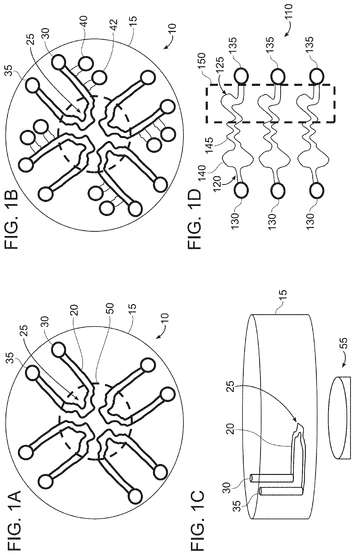
[0176]FIGS. 1A-1D are schematic illustrations of microfluidic device layouts according to example embodiments of the invention.

[0177]FIG. 1A is a top view of a circular layout (it can also be any symmetrical polygon with a center point) of microfluidic device 10 having one or more continuous microfluidic channels (e.g., microchannels) 20 formed in a substrate 15, each channel connected to one inlet (input port) 30 and one outlet (output port) 35. A portion of the channel, e.g., the center of the channel, can have a unique shape, e.g., a clot forming / localizing area 25, in order to result in flow separation or disruption, or stasis of sample flow to promote clot formation. There may be two or more of these microfluidic channels in this single device, dependent on the specific assay being used. This design can allow for multiple samples, such as three or more samples, e.g., up to 10 samples, or more than 10 samples, to be evaluated simultaneously. Typically, each sample (or each aliqu...

example 2

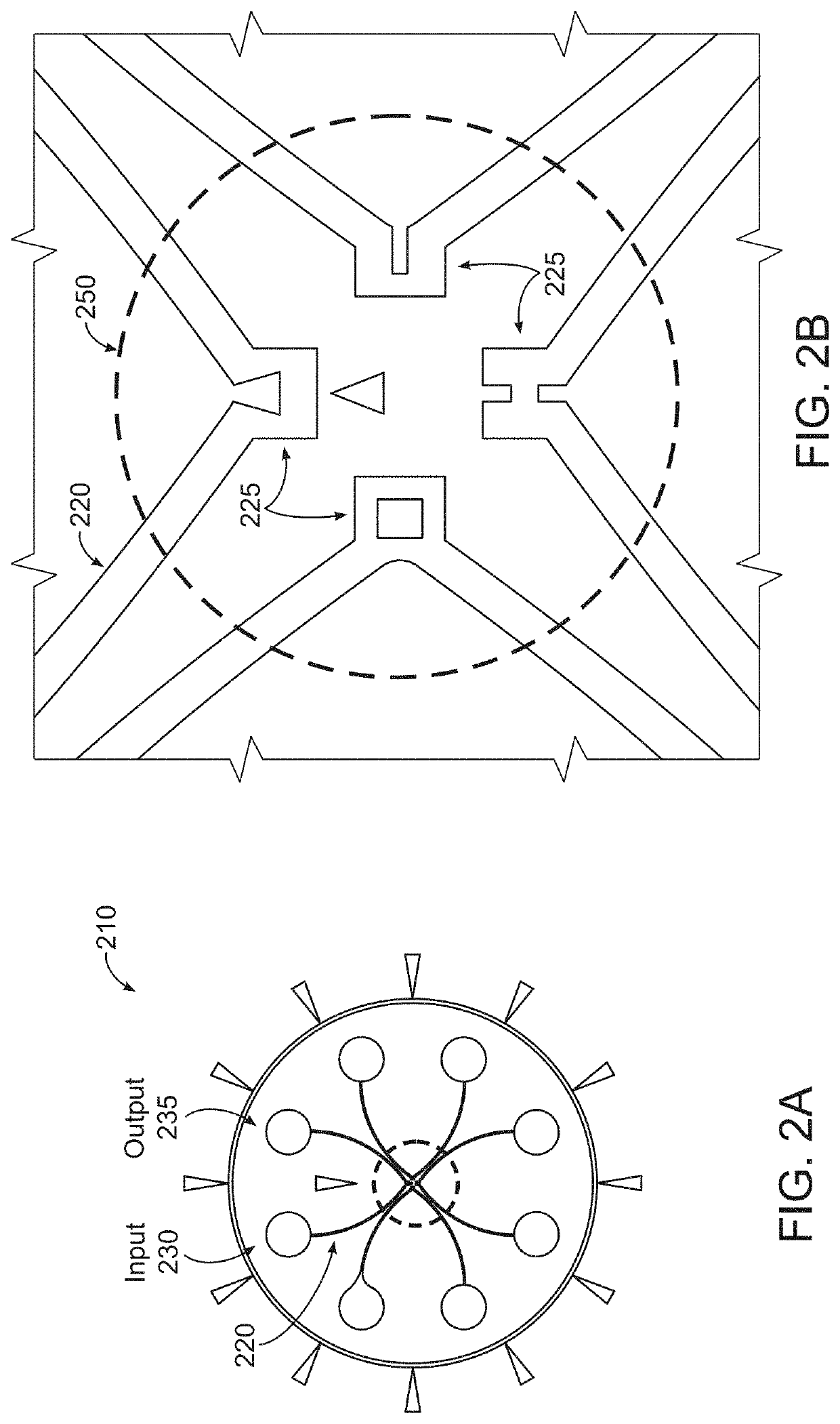
[0185]A general protocol for performing the assay according to an embodiment of the invention is as follows:[0186]a) Add together sample, agonist, + / − calcium, + / − clot detection agent[0187]i. Calcium to a final concentration between 0.005-0.05 M (This concentration is particularly suitable for use with 3.2% buffered sodium citrate. If another anticoagulant is used, the concentration of calcium may not be 0.02 M.)[0188]ii. Clot detection agents can include fluorescent labeled fibrinogen, magnets, beads (may be fluorescent or colored)[0189]b) Load into microfluidic device[0190]i. See, e.g., FIGS. 1A-1D, 2A and 2B for examples of input loading configuration and order[0191]c) Temperature control[0192]i. Room temperature[0193]ii. May increase up to 37° C. (body temperature) (Body temperature is typically 37° C. but the temperature of the assay run can be changed according to the patient's actual temperature. For example, if a patient has a fever, the temperature of the assay run can be ...

example 3

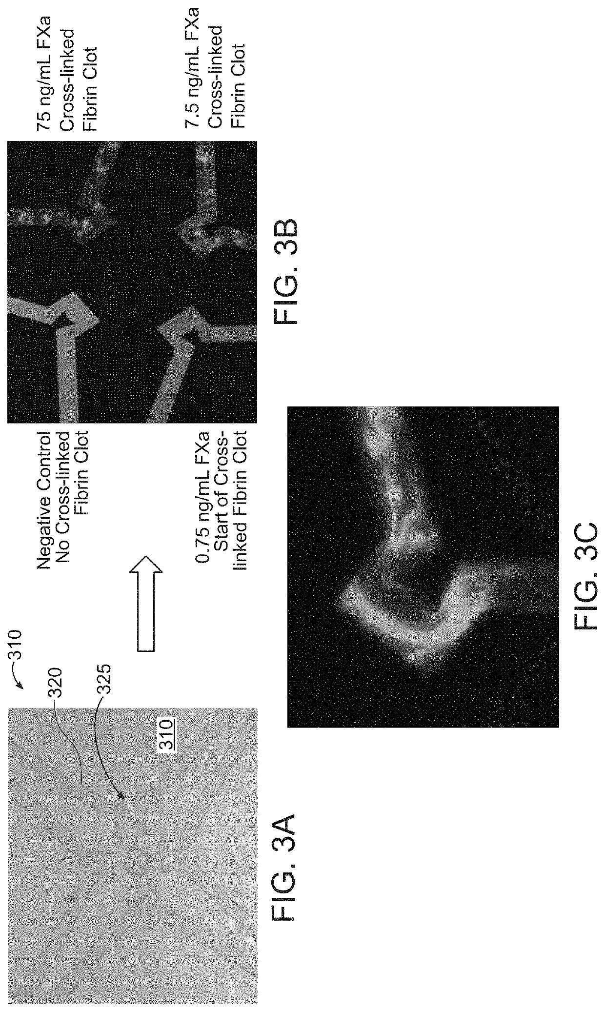
[0196]FIGS. 3A-3C illustrates clot detection using plasma and fluorescent-labeled fibrinogen with a microfluidic device 310 having four channels 320 with clot forming / localizing areas 225 according to an example embodiment. The microfluidic device is similar to the device show in FIGS. 2A and 2B except that all clot forming areas 325 have the same shape. Each clot forming / localizing area 325 includes a protrusion to disrupt sample flow. In this example, as shown in FIG. 3A, the protrusion generally is triangular in shape. Two sides of the protrusion are straight and one side is concave. Each clot forming area 325 causes the flow to change direction four times, including two 90 degree changes in direction.

[0197]In an example, the process of clot detection can include the following procedural steps:[0198]a) A plasma sample is pre-mixed to include: 6 μL plasma+0.6 μL agonist (10% volume to sample)+0.6 μL Calcium (stock 200 mM, 10% volume to sample)+0.6 μL Fibrinogen (this can vary in c...


PUM

PropertyMeasurementUnit
volumeaaaaaaaaaa
volumeaaaaaaaaaa
concentrationaaaaaaaaaa

Abstract

Provided are methods and devices for evaluating coagulation, including the identification of a coagulation impairment such as a factor deficiency or the presence of a factor inhibitor. In various embodiments, the methods and devices measure coagulation of a sample in response to the addition of one or more coagulation factors, added at various concentrations to portions of the sample. Such coagulation measurements can be evaluated to accurately profile coagulation impairments of the sample. In additional various embodiments, point-of-care or bedside testing with a convenient, microfluidic device can be used by minimally trained personnel.

Description

BACKGROUND[0001]The coagulation system is a delicate balance between hemorrhage and thrombosis. There are many disease conditions and clinical situations (such as, for example, cancer, auto-immune disease, infection, trauma, surgery, heart disease, and drug treatments), that can cause a disruption of this balance and result in a patient having severe, and in some cases even life-threatening, bleeding or clotting events. In some cases, a patient may suffer from coagulopathy, which can be congenital and / or hereditary, or acquired. Coagulopathy may result from a deficiency in a coagulation factor; examples of coagulation factor deficiencies that may result in a coagulopathy include a deficiency in Factor VIII, deficiency in Factor IX, and deficiency in Factor XI (e.g., Hemophilia A, B, and C, respectively). While hemophilia is most commonly known as a congenital and / or hereditary disease, there are also acquired causes of hemophilia, such as the development of anti-factor autoantibodie...

Claims


Application Information

Patent Timeline
Patent Type & Authority Applications(United States)
IPC IPC(8): G01N33/86B01L3/00
CPCG01N33/86B01L3/5027B01L2300/088G01N2333/974G01N2333/96463G01N2333/96452G01N2333/9645G01N2333/96458B01L3/502761B01L2300/0803B01L3/502715G01N2333/96444G01N2333/8128G01N2333/4716G01N2333/96461
Inventor FRYDMAN, GALIT H.
Owner FRYDMAN GALIT H
Learn More