Biosensor using exceptional point

Pending Publication Date: 2022-10-13
DAEGU GYEONGBUK INST OF SCI & TECH
View PDF0 Cites 0 Cited by
  • Summary
  • Abstract
  • Description
  • Claims
  • Application Information

AI Technical Summary

Benefits of technology

The patent describes a method to detect a target biomolecule with very high sensitivity by detecting a wavelength difference in light caused by the destruction of an exceptional point.

Problems solved by technology

As for exosomes, their number is very large, amounting to 106 per milliliter, but exosomes are difficult to detect due to their small size and low molecular weight.
However, this assay, which is a multi-stage analysis technique, is labor intensive and is unable to detect a concentration of 1 ng / mL or less due to the very poor sensitivity thereof although fluorescent and luminescent signals are amplified once with an enzyme.
However, the application of this method is limited to the detection of chorionic gonadotrophin (hCG) which is a target produced at a very high concentration in pregnancy, due to its very low sensitivity (LOD (limit of detection) of commercially available kit: 1-5 ng / mL).
An electrochemical immunosensor has been suggested in order to enhance LOD to 10-100 fg / mL in protein biomarkers, but is short in lifespan and works only at a narrow range of temperatures, thus having the problem of ensuring reliability with the resultant limitations imparted into the applications thereof.
Detection methods using nanobeads, surface plasmon resonance (SPR), and surface enhanced Raman spectroscopy (SERS) each have a limit of detection of as high as 1 pg / mL, but is difficult and complicated in terms of usage.
These technologies can detect even one nucleic acid molecule, but requires expensive sequencers, well-trained operators, and a long operation time.
Although drastically reduced in the last five years, the cost for detection by these technologies still amounts to 100 dollars or more per sample (exclusive of price of the sequencer).
In spite of development of biosensors for liquid biopsies, low sensitivity, restricted specificity, labor-intensive and time-consuming work, and high cost still, on the whole, remain as barriers against the clinical application of the biosensors.

Method used


Image

  • Biosensor using exceptional point
  • Biosensor using exceptional point
  • Biosensor using exceptional point

Examples


Embodiment Construction

[0036]To fully understand the configuration and effect of the present disclosure, preferred embodiments of the present disclosure will be described with reference to the accompanying drawings. However, the present disclosure is not limited to the embodiments disclosed below and may be embodied in various forms and various modifications may be applied thereto. The description of the present embodiment is provided so that the disclosure will be complete and will be fully convey the scope thereof to those of ordinary skill in the art to which the present disclosure belongs. In the accompanying drawings, elements are enlarged in size than actual for convenience of description, and ratio of each element may be exaggerated or reduced.

[0037]Terms such as ‘first’ and ‘second’ may be used to describe various elements, but the elements should not be limited by the above terms. The above term may be used only for the purpose of distinguishing one element from another element. For example, with...


PUM


Abstract

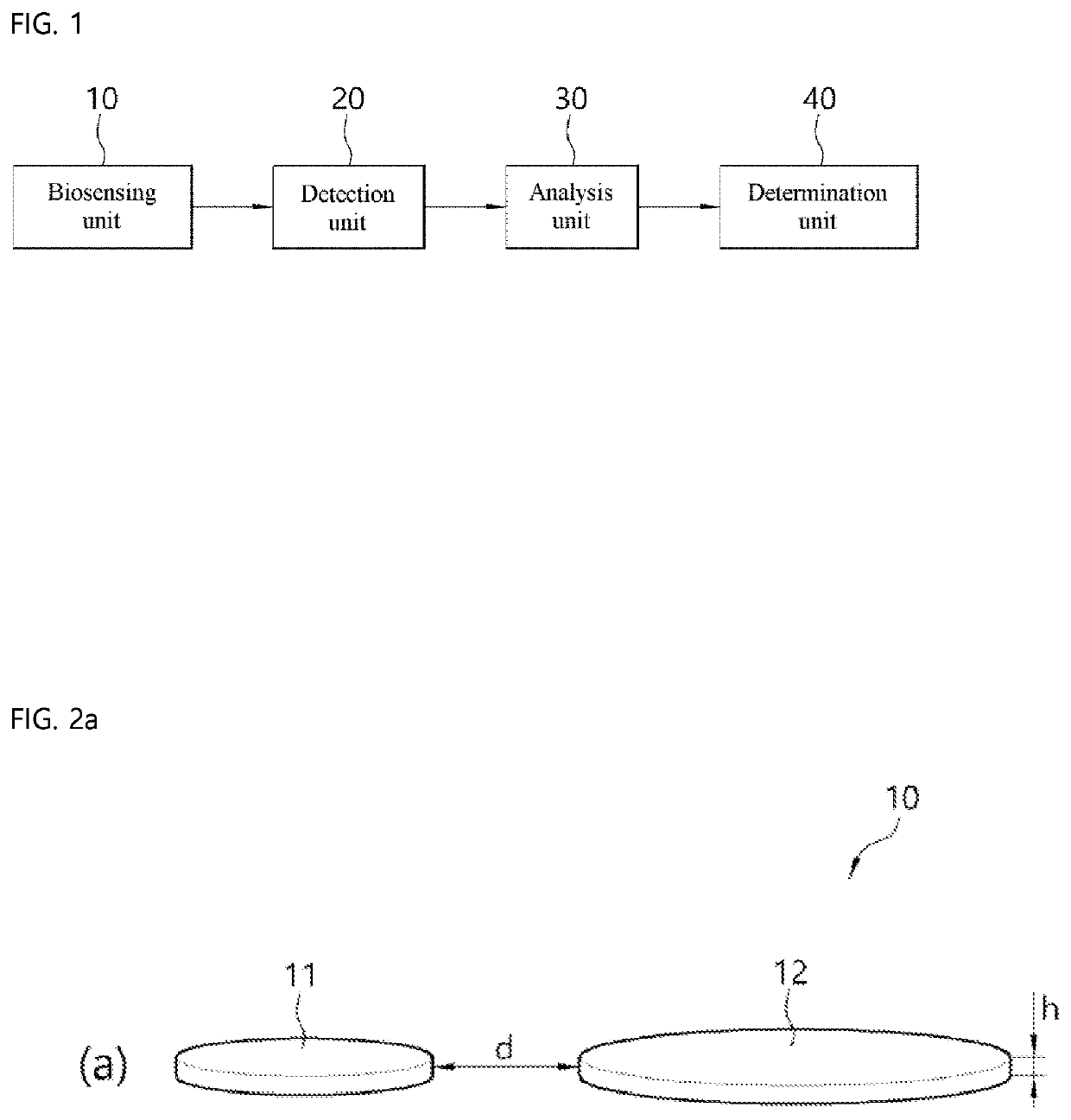
A biosensor using an exceptional point is disclosed. A biosensor according to one embodiment of the present disclosure includes: a biosensing unit configured to output wavelength-separated optical signals from destruction of an exceptional point resulting from attachment of biomolecules; a detection unit configured to convert the wavelength-separated optical signals into wavelength-separated electrical signals; an analysis unit configured to measure a beat frequency resulting from the wavelength-separated electrical signals; and a determination unit configured to determine a wavelength difference resulting from the beat frequency, thereby determining the amount of the biomolecules therefrom.

Description

TECHNICAL FIELD[0001]The present disclosure relates to a biosensor using an exceptional point.BACKGROUND ART[0002]In order to diagnose diseases such as cancer, thorough efforts other than traditional biopsy have recently been focused on the detection of biomolecules in blood. This type of technique, also known as liquid biopsy, is the sampling and analysis of blood to diagnose disease.[0003]Unlike traditional biopsies adapted to sample tumor cells from patients through surgery, such a liquid biopsy has attracted intensive attention because it can sample blood with only a syringe, which may be the minimum surgical tool.[0004]However, blood retains a very small content of target molecules to be detected. Sometimes, for example, even though blood is sampled, circulating cancer cells could not be detected therein because they amount merely to 10 or less in 7.5 mL of blood. As for exosomes, their number is very large, amounting to 106 per milliliter, but exosomes are difficult to detect ...

Claims


Application Information

Patent Timeline
IPC IPC(8): G01N15/06
CPCG01N15/0606G01N2015/0065G01N21/77G01N33/49G01N33/48707G01N33/487G01N2021/7789G01N15/1484G01N15/1429G01N2015/1486G01N2015/1006G01N15/01G01N21/7746G01N33/483G01N21/27H01S3/0627G01N15/075
Inventor KIM, CHIL MINRYU, JIN HYEOKYI, CHANG HWAN
Owner DAEGU GYEONGBUK INST OF SCI & TECH
Why Eureka
Try Eureka