Persons with significant
rotator cuff defects have difficulty raising the affected arm or rotating it out to the side.
Once these lesions begin, it is difficult for them to heal, because of the hostile environment, the compromised vascularity, the large loads, and the large deformations that the healing tissue must endure.
When a
tendon fiber fails, the
muscle fiber to which it attaches produces retraction away from the site of disruption, increasing the gap needing to be closed.
This retraction also places tension on the local vasculature leading to limitation of tendon
blood flow in the area where healing is needed.
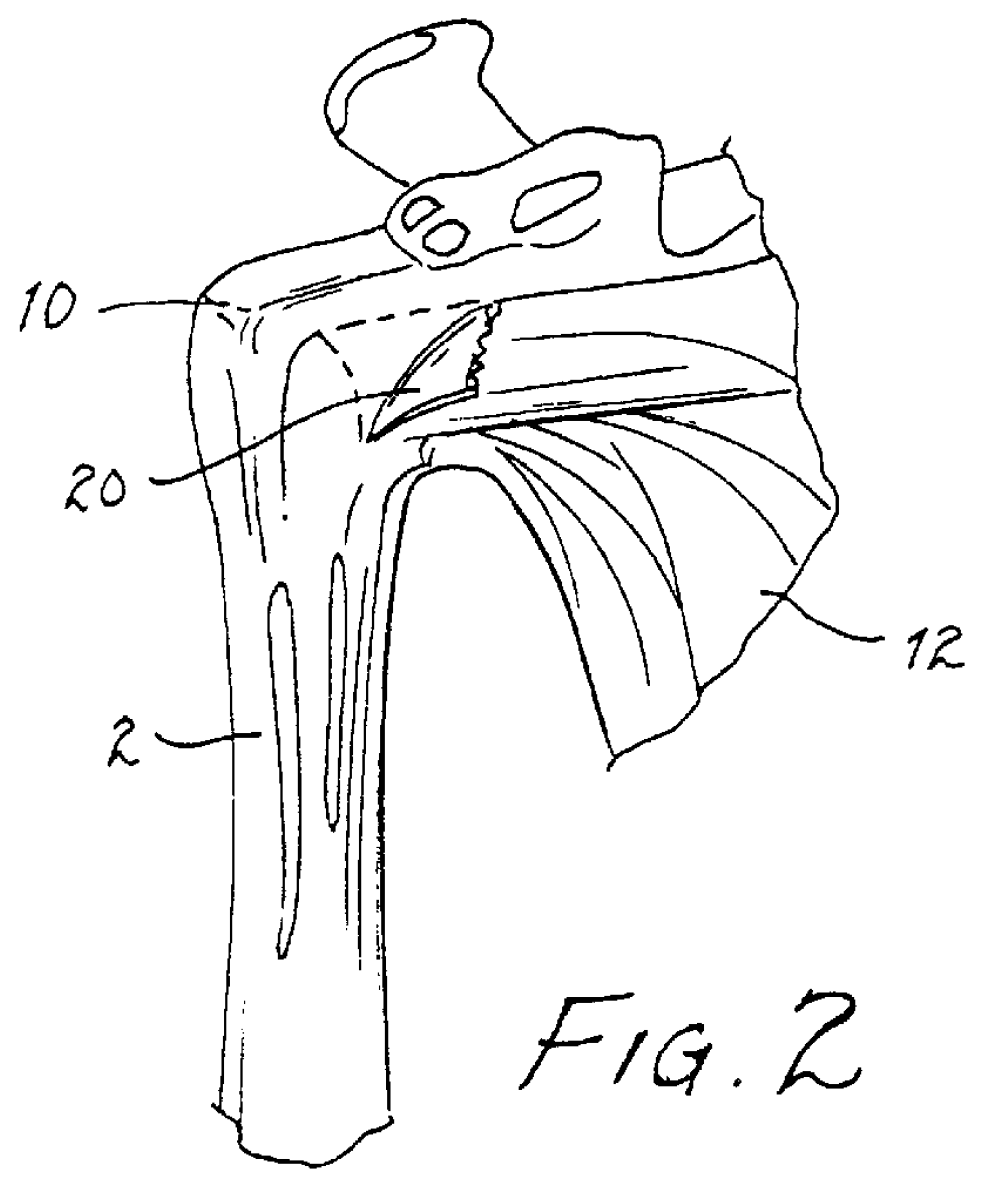
This
full thickness defect tends to concentrate loads at its margin, facilitating additional
fiber failure with smaller loads than those which produced the initial defect.
The concavity compression mechanism is compromised by
cuff disease.
Beginning with the early stages of
cuff fiber failure, the compression of the humeral head becomes less effective in resisting the upward pull of the deltoid.
In turn, this
reflex inhibition along with the absolute loss of strength from fiber detachment makes the
muscle less effective in balance and stability.
Under these circumstances, abrasion occurs with humeroscapular motion, further contributing to
cuff degeneration.
Upward displacement of the head also wears on the upper glenoid lip and labrum, reducing their contributions to the
effective depth of the upper glenoid and to glenohumeral stability from concavity compression.
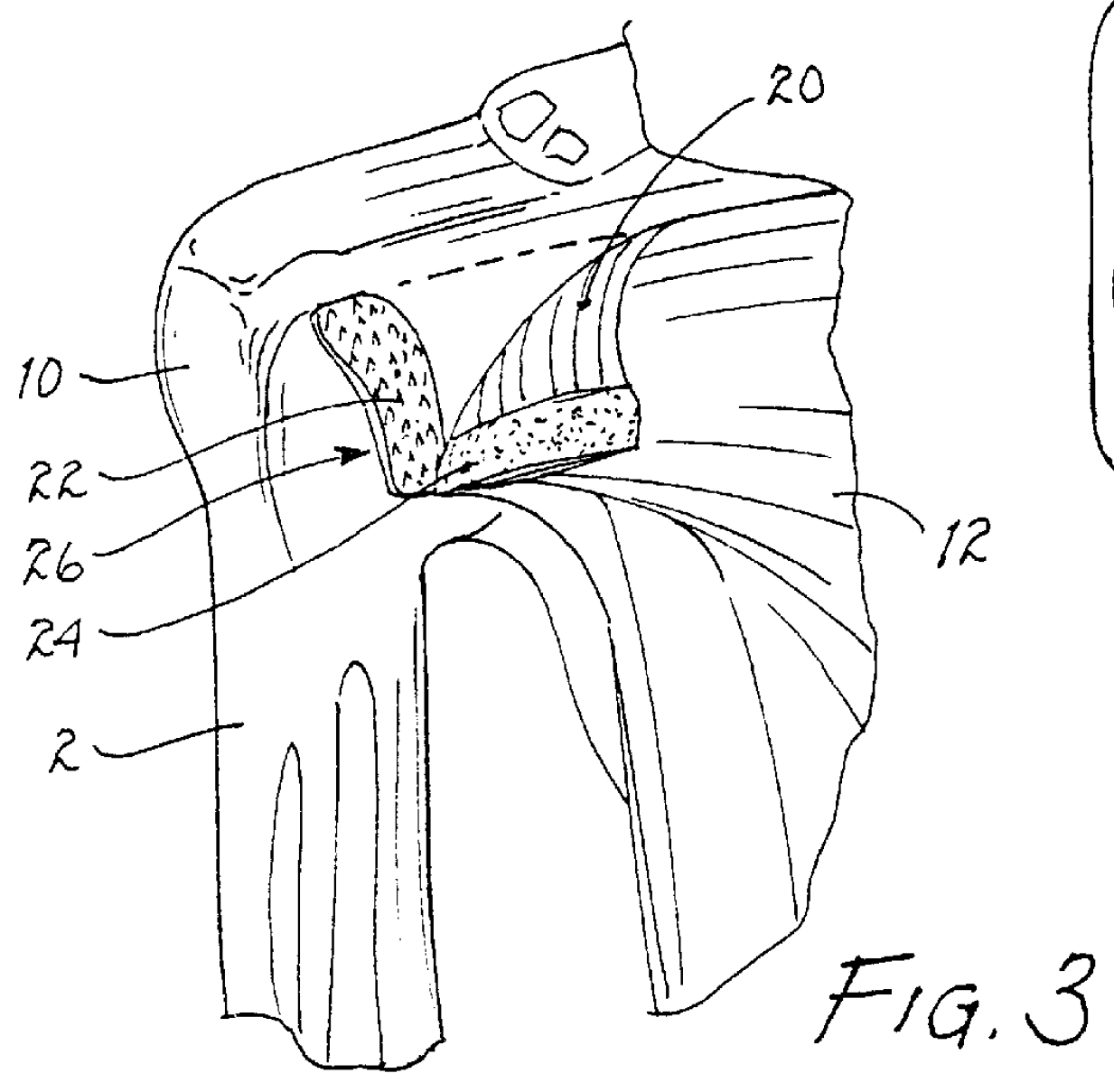
Further deterioration of the cuff allows the tendons to slide down below the center of the humeral head, producing a "boutonniere"
deformity.
It is important to note that cuff defects arising with minimal or no injury suggest that the cuff tissue is of
poor quality and thus is more likely to fail again after
surgical repair.
The disuse of torn tendon leads to scarring and
atrophy of tendon and
muscle.
Loss of cuff material from the degenerative process limits what is available for repair.
Local injections of steroids may further compromise the healing potential of failed cuff fibers.
Once the humeral head has started to subluxate superiorly, increased stretching loads are placed on the residual tendons, tending to exacerbate the cuff defect.
Once the process of superior subluxation is established, stabilization of the humeral head in its
normal position is difficult even if a cuff repair is achieved.
In summary, rotator cuff defects are common causes of shoulder
weakness.
Usually, cuff
tears are associated with degenerative changes, which make the tissue susceptible to failure with low applied loads, especially those applied eccentrically.
Alternatively, cuff
tears can occur in stronger cuff tissue, but these injuries require the application of much greater loads.
Returning to heavy work after a cuff repair risks the integrity of the repair.
By contrast, with chronic massive degenerative
tears the quantity and quality of the cuff are less likely to be optimal for
surgical repair.
The most serious is compromise of the deltoid muscle.
Postoperative function of the deltoid may be compromised by failure to achieve a strong reattachment of this tendon and the anterior muscle fibers after acromioplasty.
This is particularly a problem when a large anterior acromial resection is performed requiring stretch of the deltoid for reattachment.
Unless these releases are carried out, increased tension in the repaired tendon will predispose to tightness of the glenohumeral joint and will additionally challenge the
repair site.
But the rigor of sports added to everyday walking can make the knees extra vulnerable to damage.
Anyone can have knee problems.
Women are especially susceptible because their wider
pelvis tends to make them knock-kneed.
Thus,
cartilage has a limited ability to repair itself.
Unlike the
Achilles tendon rupture, the quadriceps disruption is associated with intense pain.
The patient is often unable to walk, may be unable to extend the knee and may demonstrate a palpable defect.
Sutures are often very difficult to apply and sometimes
surgery beyond making the repair per se is necessary to enable the surgeon to fix and tie the sutures.
The result is increased trauma for the patient and due to the additional
surgery and the time required to perform the operation.
Suture placement is a demanding and exhausting effort for the surgeon.
Staples offer some simplification for certain fastening operations; however, stapled attachments may lack sufficient strength for many procedures and may increase the trauma of the
surgery where staples are placed in bony structures.

