Methods for searching stable docking models of biopolymer-ligand molecule complex

a docking model and docking technology, applied in the field of methods for searching stable docking models of biopolymerligand molecule complexes, can solve the problems of inability to detect stable structures of complexes in experiments, inability to determine the binding modes of all ligand molecules of interest to their target biopolymer by experimental methods such as x-, and large analysis time and analysis effort, so as to reduce the number of combination sets and conformations to be checked, the effect of large conform

Inactive Publication Date: 2001-10-23
INST OF MEDICINAL MOLECULAR DESIGN INC 50
View PDF7 Cites 12 Cited by
  • Summary
  • Abstract
  • Description
  • Claims
  • Application Information

AI Technical Summary

Benefits of technology

In a more preferred embodiment of the present invention, if the generation of a large number of dummy atoms is expected from the biopolymer or if the ligand molecule has a very large conformational flexibility or a large number of heteroatoms, information on the ligand molecule is input in step S9 and, thereafter, the partial structure of the ligand molecule is specified and the process steps S10-S24 is performed on the specified partial structure so as to store the combination sets that provide hydrogen-bond schemes impossible in the partial structure, as well as the information on the hydrogen-bonding heteroatoms in the ligand molecule that do not form any hydrogen-bond with dummy atoms. Any partial structure of the ligand molecule can be specified without structural restrictions. Partial structures containing at least three hydrogen-bonding functional groups are preferred and the conformational flexibility thereof can be handled. Then, the process of steps S10-S31 is per...

Problems solved by technology
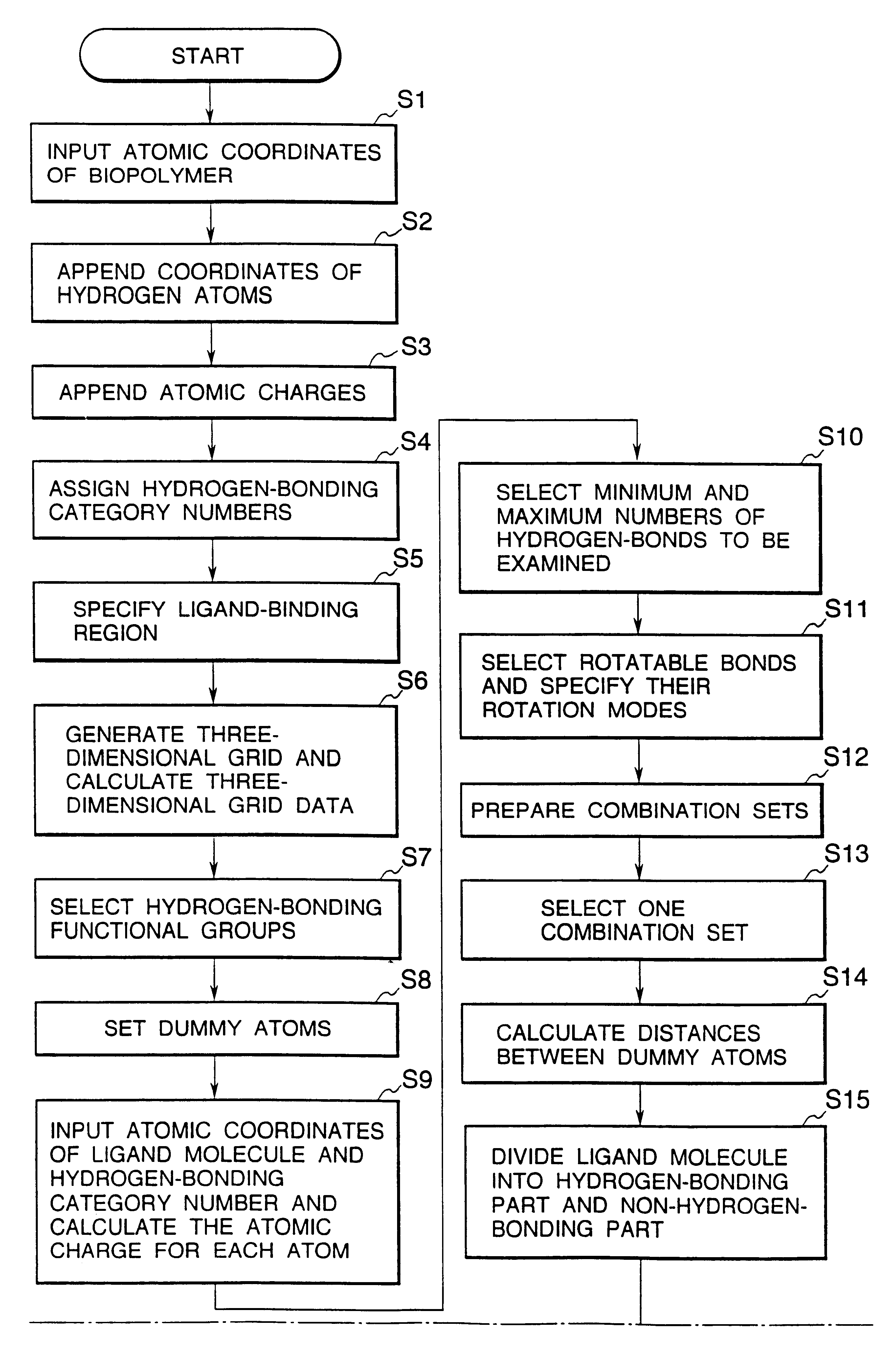
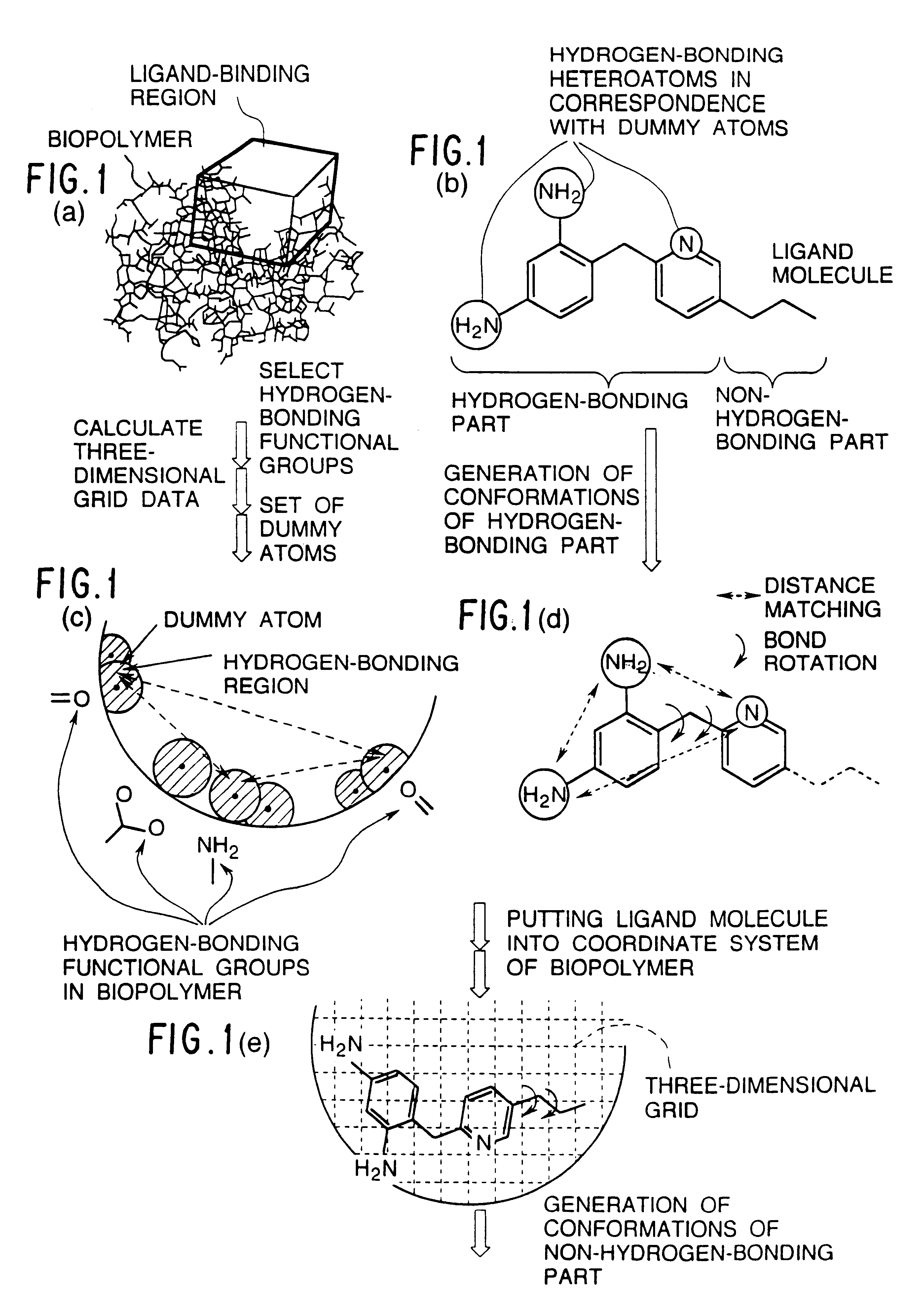
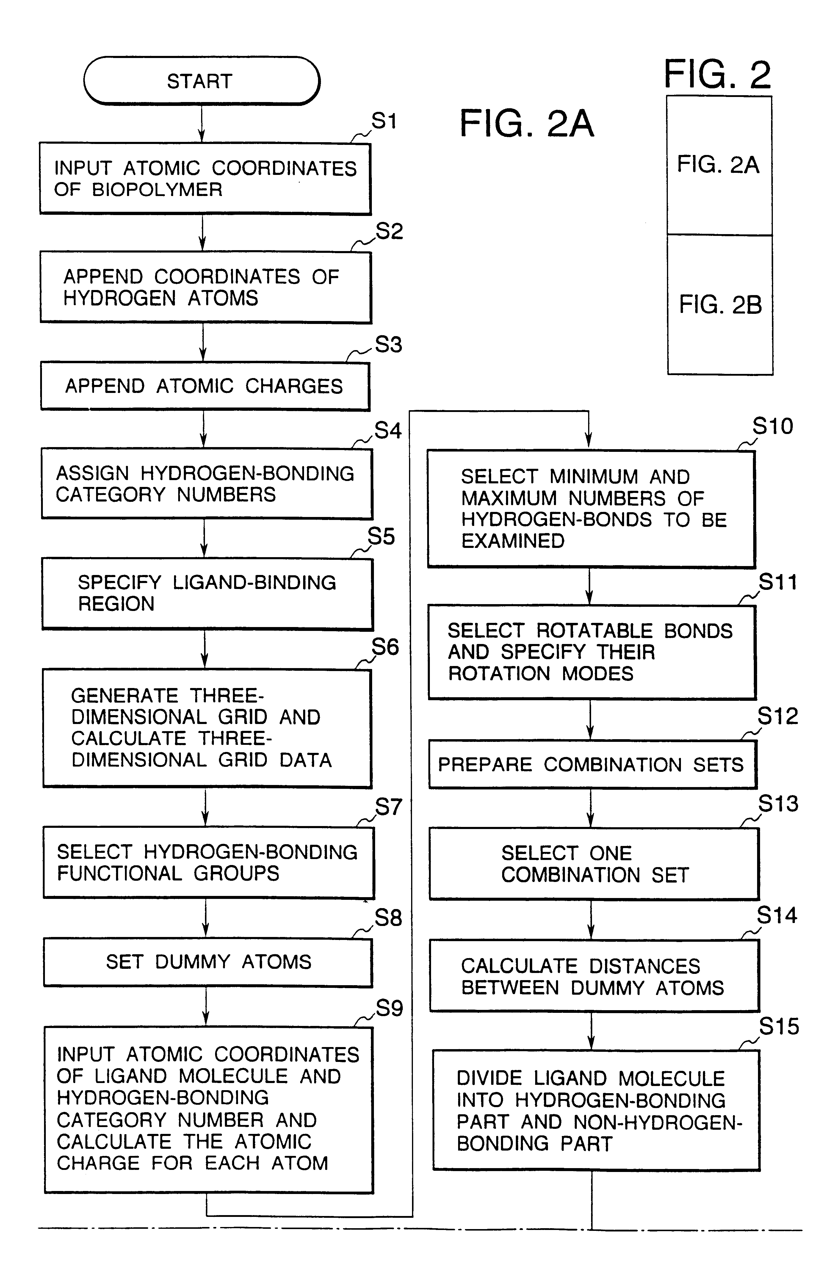
It is impossible to determine the modes of binding of all ligand molecules of interest to their target biopolymer by experimental methods such as X-ray crystallographic analysis.
First, this is because a great effort and time are necessary for analyzing the mode of binding of each ligand molecule to the biopolymer by experimental methods.
In addition, if the ligand molecules are substrates for enzymes, any stable structures of the complex can not be detected in the experiments due to the progress of enzymatic reactions.
Furthermore, the samples of the ligand compounds are often unavailable if they exist.
In the past, the docking study was performed using molecular models, but the use of molecular models had problems in that; it needed much time and effort to construct the molecular models, that the precision of the molecular models and the reproducibility of results were poor and that quantitative results could not be obtained.
However, there are an enormous number of possible docking struct...

Method used


Image

  • Methods for searching stable docking models of biopolymer-ligand molecule complex
  • Methods for searching stable docking models of biopolymer-ligand molecule complex
  • Methods for searching stable docking models of biopolymer-ligand molecule complex

Examples


example 1

Methotrexate Molecule

The terminal carboxyl group of MTX molecule was removed to simplify the example and the following procedure was performed.

As the atomic coordinates of MTX, the atomic coordinates of the unbound crystal structure available from Cambridge Crystallographic Database were input. The atomic charge on each atom of MTX was calculated by the MNDO method in the MOPAC program. Since the nitrogen at position 1 in the pteridine ring of MTX was susceptible to protonation, the atomic charge thereon was calculated on the assumption that it was protonated. In the structures shown in FIG. 3, the encircled heteroatoms were selected as the hydrogen-bonding heteroatoms and the hydrogen-bonding category numbers were assigned thereto. In the MTX molecular structure shown in FIG. 3, bond a was rotated at intervals of 60.degree. in the range of 0.degree.-360.degree., bond b was rotated at intervals of 60.degree. in the range of 0.degree.-180.degree., bond c was assigned as either 0.degr...

example 2

Dihydrofolic Acid Molecule

Although the binding mode of DHFR to its substrate DHF has not yet been identified by X-ray analysis, it has been predicted from the stereospecifity of tetrahydrofolic acid that is the product of enzymatic reaction for the following reasons that the DHF molecule binds to the enzyme in a different mode from that of binding with MTX: It is known that the hydrogen at position C6 in the tetrahydrofolic acid that is produced by the reducing action of DHFR is derived as a hydride ion from coenzyme NADPH. If it is assumed that the binding mode of the DHFR-DHF complex is the same as that of the MTX and DHFR molecules in the crystal structure of the ternary DHFR-MTX-NADPH complex obtained by X-ray analysis, tetrahydrofolic acid with opposite chirality should be produced. This strongly indicated that pteridine of DHF molecule is reversed from that of MTX (see FIG. 6).

In Example 2, the structures of stable DHFR-DHF complexes were searched without any preconception by ...


PUM


Abstract

Methods for searching stable docking models of biopolymer-ligand molecule complex, which comprise the steps of: (1) searching possible hydrogen-bond schemes between a biopolymer and a ligand molecule by preparing possible combination sets of hydrogen-bonding heteroatoms in the ligand molecule with dummy atoms located at the positions of heteroatoms that can be hydrogen-bond partners to hydrogen-bonding functional groups in the biopolymer; (2) estimating the possible the hydrogen-bond schemes between the biopolymer and ligand molecule and the possible conformations of a hydrogen-bonding part of the ligand molecule at the same time by comparing the distances between the dummy atoms with the distances between the hydrogen-bonding heteroatoms; and (3) obtaining the possible docking models of the biopolymer-ligand molecule complex by changing the coordinates of all atoms of the ligand molecule into the coordinate system of the biopolymer, according to the correspondences between the hydrogen-bonding heteroatoms in the ligand molecule and the dummy atoms in combination sets for each of the hydrogen-bond schemes and conformations obtained in the second step are provided. According to the methods of the present invention, the structures of stable biopolymer-ligand molecule complex can be searched efficiently and precisely in a short time.

Description

The present invention relates to methods for searching stable docking models of biopolymer-ligand molecule complex that can be utilized in designing the structures of drugs, pesticides, and other biologically active compounds.Strong binding of drugs and biologically active substances to their target biopolymers is required for the appearances of their bioactivities. The biopolymers include pharmacological receptors which take part in signal transmission between cells, as well as enzymes, cytokines and other proteins, complexes comprising these proteins as main components, and nucleic acids. Since 1960, the steric structures of a large number of biopolymers have been revealed at the atomic level by X-ray crystallographic analyses and the structural data thereof have been stored in the Protein Data Bank and published.Small molecules which can be bound to these biopolymers are called ligand molecules. The ligand molecules include candidate molecules for drugs such as drug molecules to ...

Claims


Application Information

Patent Timeline
IPC IPC(8): G06F17/30G16B15/30
CPCG06F19/16G16B15/00G16B15/30
Inventor ITAI, AKIKOMIZUTANI, MIHO
Owner INST OF MEDICINAL MOLECULAR DESIGN INC 50
Why Eureka
Try Eureka