Friction transmission with axial loading and a radiolucent surgical needle driver

Inactive Publication Date: 2002-06-04
THE JOHN HOPKINS UNIV SCHOOL OF MEDICINE
View PDF2 Cites 148 Cited by
  • Summary
  • Abstract
  • Description
  • Claims
  • Application Information

AI Technical Summary

Benefits of technology

An object of the present invention is to provide a simple and effective method and system for radi

Problems solved by technology

However, percutaneous needle access of the surgical target may be difficult, and usually requires an extensive amount of experience and skill on the part of the surgeon.
The above problem is exacerbated by the fact that prior art radiological image guidance techniques and associated imaging devices do not provide effective three dimensional information to the surgeon regarding needle insertion.
Although the above systems successfully address issues of image-to-robot registration and provide convenient means for defining target anatomy, they can nevertheless be expensive and cumbersome in an operating room environment.
Moreover, for the implementation of the active robot mentioned above, the radiological profile of the end-effector, or needle, may interfere with a clear view of the target.
A disadva

Method used


Image

  • Friction transmission with axial loading and a radiolucent surgical needle driver
  • Friction transmission with axial loading and a radiolucent surgical needle driver
  • Friction transmission with axial loading and a radiolucent surgical needle driver

Examples


Embodiment Construction

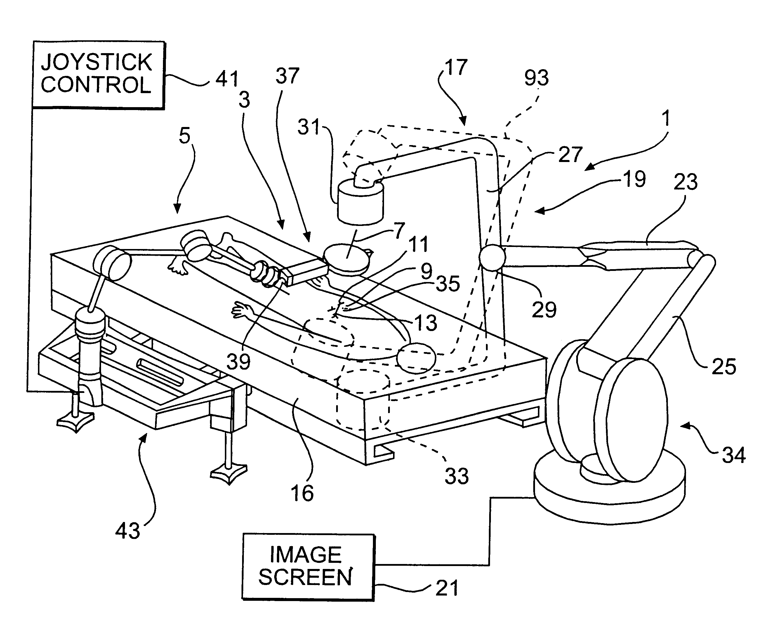
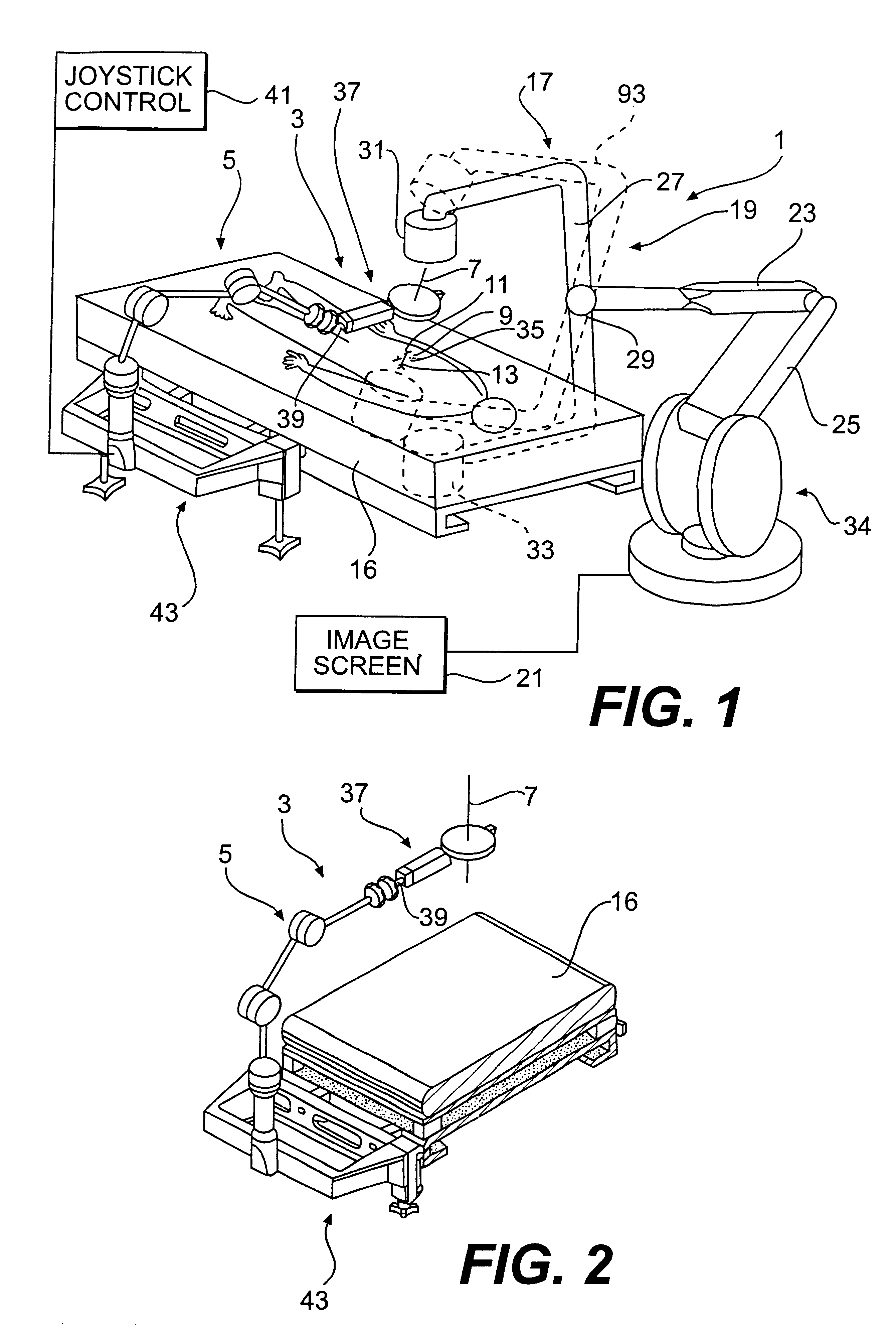
As seen in FIG. 1, a system 1 for radiological image guidance in percutaneous surgery is shown. The system is disposed in an area suitable for surgery, such as an operating room. A novel needle insertion mechanism 3 comprises a passive needle manipulator 5 which maintains the needle 7 in position above a patient 9, and is effective in minimizing the surgeon's radiation exposure and disturbances in the needle trajectory during the insertion of the needle through insertion site 11 toward target 13. System 1 requires neither a fully actuated robot nor position feedback sensors by virtue of using a superimposed registration technique as described previously, thus minimizing costs.

As further shown in FIG. 1, the system further includes an operating room table 16 for the patient, and a conventional C-arm imaging device 17 including a C-arm 19 and an image screen 21. The C-arm imaging device may, for example, comprise the X-ray system disclosed in U.S. Pat. No. 5,549,439. Thus, by way of e...


PUM


Abstract

A method for performing radiological-image-guided percutaneous surgery with a system which includes a radiological image generating device for generating an image of a target anatomy of a patient, and a needle insertion mechanism disposed adjacent the image generating device and having a needle adapted to be inserted into the patient. The method includes the steps of: determining a needle trajectory of the needle by positioning the image generating device for aligning, in the image generated by the image generating device, a desired skin insertion site of the patient with a target region of the target anatomy; locking the needle in a direction of the needle trajectory; and repositioning the image generating device to obtain a lateral view of the needle trajectory for viewing an insertion depth and path of the needle during its insertion into the patient. Moreover, a motion transmission mechanism includes an output shaft and an output shaft driver which has two rotational components having respective contact faces between which the output shaft is pressed for frictional engagement therewith. The frictional engagement creates a force between the output shaft and the rotational components which is parallel to the rotational axis of the rotational components for allowing the rotational components to impart a translational motion to the output shaft by virtue of their rotational motion.

Description

BACKGROUND OF THE INVENTION1. Field of the InventionThe present invention pertains to a method and system for radiological image guidance in percutaneous surgery. The invention further pertains to a friction transmission mechanism with axial loading.2. Description of the Related ArtAs an alternative to traditional open surgery, percutaneous surgery has been found to significantly reduce morbidity and post-operative recovery time.However, percutaneous needle access of the surgical target may be difficult, and usually requires an extensive amount of experience and skill on the part of the surgeon. The above problem is exacerbated by the fact that prior art radiological image guidance techniques and associated imaging devices do not provide effective three dimensional information to the surgeon regarding needle insertion.In order to overcome the above problem, several robotic systems have been proposed to date to assist in needle placement.According to one solution, a stereopair of two...

Claims


Application Information

Patent Timeline
IPC IPC(8): A61B19/00
CPCA61B19/52A61B2017/00902A61B90/36
Inventor STOIANOVICI, DANKAVOUSSI, LOUIS R.WHITCOMB, LOUIS L.TAYLOR, RUSSELL H.CADEDDU, JEFFREY A.DEMAREE, ROGER D.BASILE, STEPHEN A.
Owner THE JOHN HOPKINS UNIV SCHOOL OF MEDICINE
Why Eureka
Try Eureka