Method and system for knowledge guided hyperintensity detection and volumetric measurement

a hyperintensity detection and volumetric measurement technology, applied in the field of magnetic resonance imaging, can solve the problems of low sensitivity, low specificity, and reduced hippocampal volume, and achieve the effect of high sensitivity

Inactive Publication Date: 2002-08-06
UNIV OF SOUTH FLORIDA
View PDF4 Cites 305 Cited by
  • Summary
  • Abstract
  • Description
  • Claims
  • Application Information

AI Technical Summary

Benefits of technology

It is, therefore, a feature and advantage of the present invention to provide a method and/or system for detecting hyperintense regions in MRI images which addresses the problems associated with human subjectivity and existing thresholding techniques. In particular, the present invention introduces the concept of the use of knowledge guided rules and methods for automatically locating certain anatomical regions and detecting hyperintensities associated with same, which in turn are candidates for possible lesions.
It is another feature and advantage of the present invention to provide a method and/or system for detecting white matter hyperintensities and subcortical hyperintensity regions in MRI images which is designed for high sensitivity, detection and monitoring of subtle (small) brain lesions in patients with neurodegenerative diseases.
It is also a feature and advantage of the present invention to provide an automated method and/or automated system for detecting hyperintensities in MRI images which requires no a priori knowledge of the intensity distributions of image pixels and which does not require any additional direction from an expert operator to perform its task

Problems solved by technology

Unfortunately, hippocampal volume is also reported to decline in other clinical conditions.
Thus, while hippocampal volumes may have good sensitivity in detecting dementias, by itself, specificity is optionally restricted given the other clinical conditions where reduced hippocampal volume is reported.
However, simple measurements of volumes alone may not have much specificity, owing to multiple conditions in which the same volumetric changes are found.
Studies attempting to link the occurrence of HYPs to neuropsychological changes indicate that these HYPs may be associated with a decline of cognitive performance especially with respect to performance on timed attentional and visuopractical tasks (Rao et al, 1996).
Visual interpretation and categorical assignment can provide insight to lesion load within distinct anatomical regions of the brain, but lacks objectivity or the ratio scale measurement required by parametric statistics.
In spite of efforts to standardize MRI imaging of CERAD subjects and the utilization of a uniform rating scale, the frequency, locations and severity of white matter lesions resulted in an unsatisfactory intraclass correlation.
While the discrepancies may be due in part to the use of different MRI

Method used


Image

  • Method and system for knowledge guided hyperintensity detection and volumetric measurement
  • Method and system for knowledge guided hyperintensity detection and volumetric measurement
  • Method and system for knowledge guided hyperintensity detection and volumetric measurement

Examples


Embodiment Construction

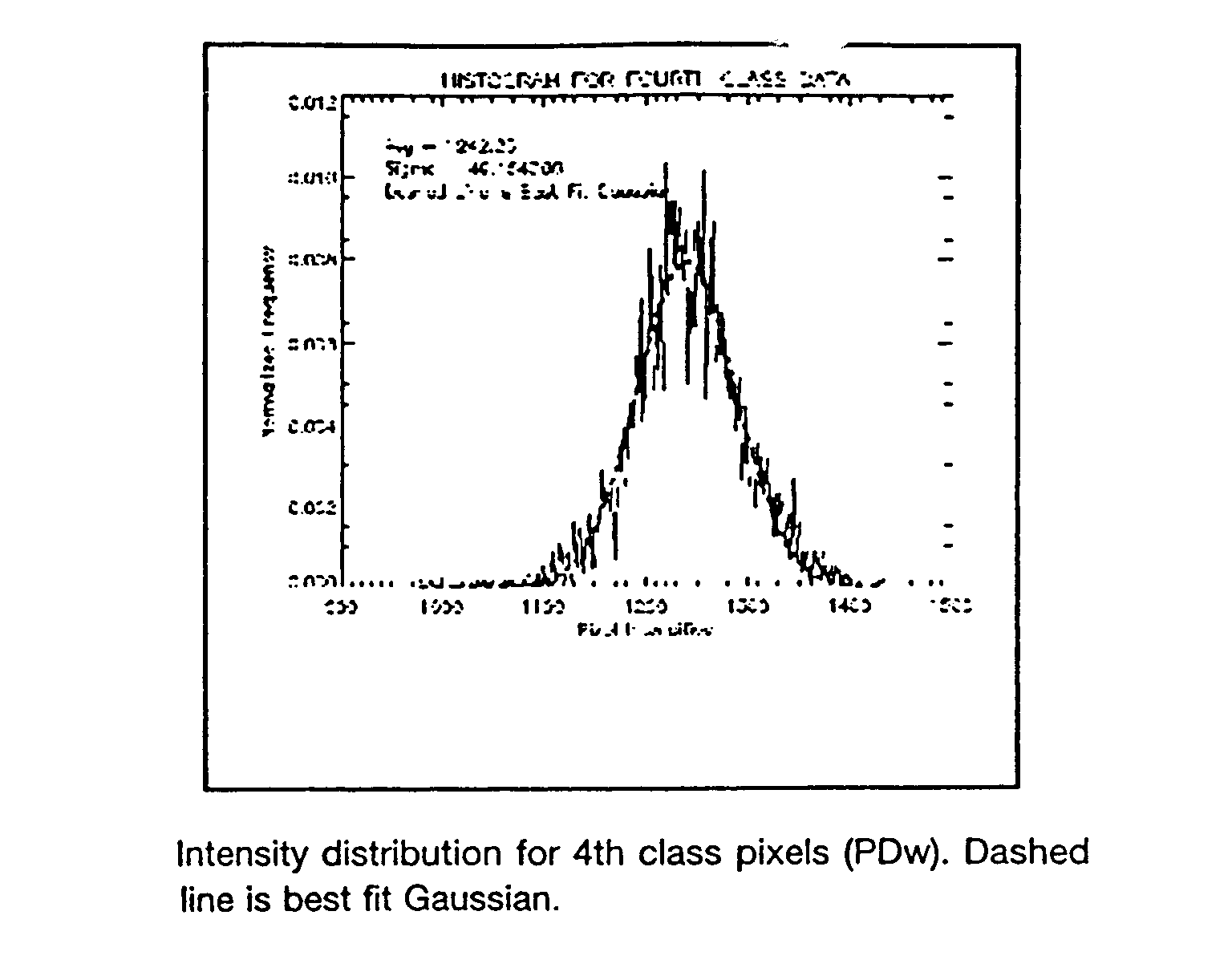
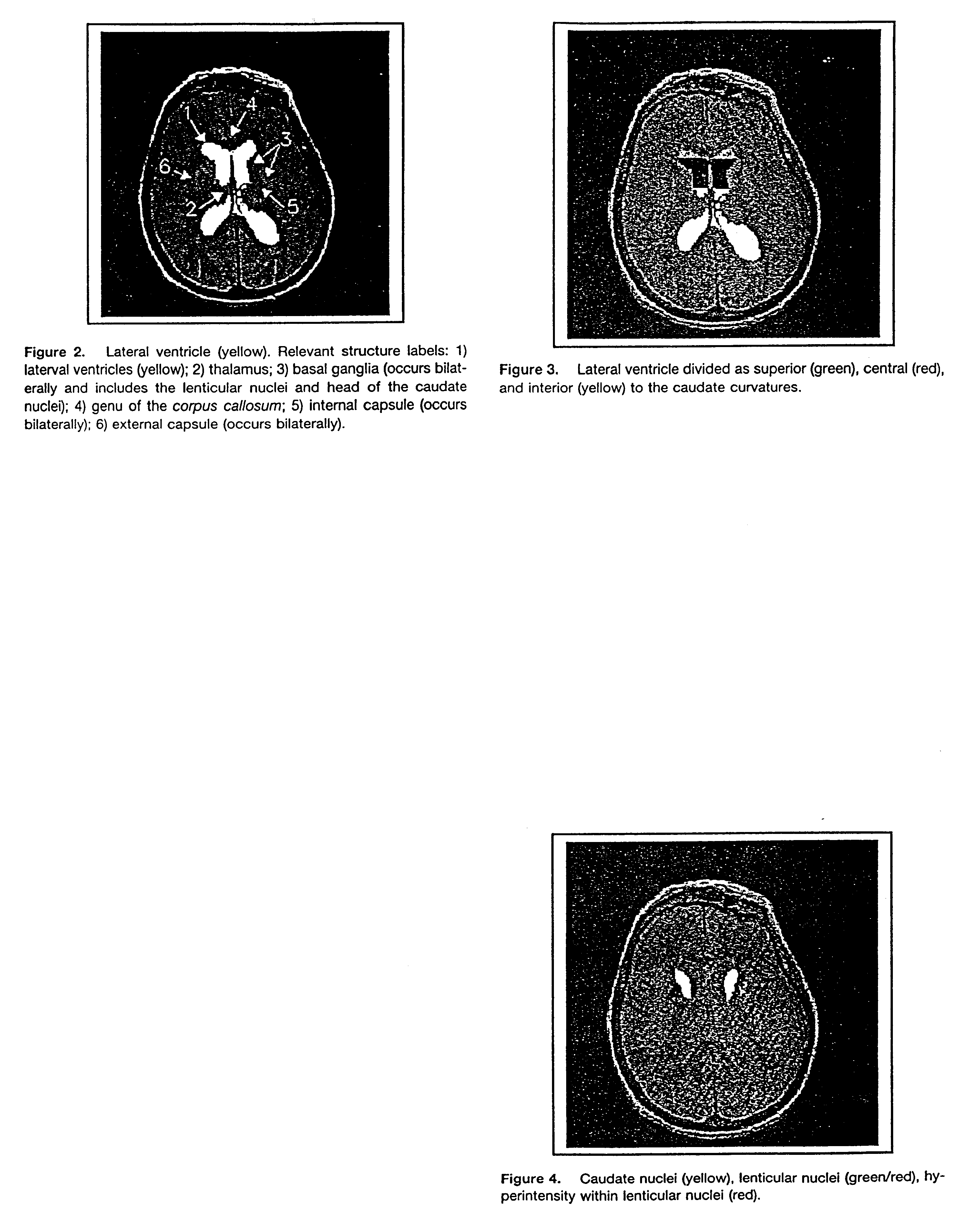
In accordance with an embodiment of the present invention, an automated method of identifying suspected lesions in a brain is provided, which comprises: (a) providing a magnetic resonance image ("MRI") of a patient's head, including a plurality of slices of the patient's head, which MRI comprises a multispectral data set that can be displayed as an image of varying pixel intensities; (b) identifying a brain area within each slice to provide a plurality of masked images of intracranial tissue; (c) applying a segmentation technique to at least one of the masked images to classify the varying pixel intensities into separate groupings, which potentially correspond to different tissue types; (d) refining the initial segmentation into the separate groupings of at least the first masked image obtained from step (c) using one or more knowledge rules that combine pixel intensities with spatial relationships of anatomical structures to locate one or more anatomical regions of the brain; (e) i...


PUM


Abstract

An automated method and/or system for identifying suspected lesions in a brain is provided. A processor (a) provides a magnetic resonance image (MRI) of a patient's head, including a plurality of slices of the patient's head, which MRI comprises a multispectral data set that can be displayed as an image of varying pixel intensities. The processor (b) identifies a brain area within each slice to provide a plurality of masked images of intracranial tissue. The processor (c) applies a segmentation technique to at least one of the masked images to classify the varying pixel intensities into separate groupings, which potentially correspond to different tissue types. The processor (d) refines the initial segmentation into the separate groupings of at least the first masked image obtained from step (c) using one or more knowledge rules that combine pixel intensities with spatial relationships of anatomical structures to locate one or more anatomical regions of the brain. The processor (e) identifies, if present, the one or more anatomical regions of the brain located in step (d) in other masked images obtained from step (c). The processor (f) further refines the resulting knowledge rule-refined images from steps (d) and (e) to locate suspected lesions in the brain.

Description

2. FIELD OF THE INVENTIONThis invention relates generally to the field of magnetic resonance imaging ("MRI") and, in particular, to the automation of the interpretation of information present in MRI images to detect brain lesions. More specifically, the present invention relates to a method and / or system of detecting hyperintense regions in MRI images that are suspected of being related to various brain pathologies, such as Alzheimer's Disease, Multiple Sclerosis and like neurodegenerative conditions and / or determining volumetric measurements of cerebral anatomical regions.3. BACKGROUND OF THE INVENTIONPresent day computerized methods of hyperintensity identification in brain magnetic resonance images either rely heavily on human intervention, or on simple thresholding techniques. Consequently, these methods lead to considerable variation in the quantification of brain hyperintensities depending on image parameters such as contrast.A review of a number of the published computerized ...

Claims


Application Information

Patent Timeline
IPC IPC(8): G06T7/00G06T5/00
CPCG06T7/0012G06T7/0081G06T7/0091G06T2207/10088G06T2207/30016Y10S128/92Y10S128/923Y10S128/925G06T7/11G06T7/155
Inventor GOSCHE, KAREN M.
Owner UNIV OF SOUTH FLORIDA
Why Eureka
Try Eureka