
Core structure of gp41 from the HIV envelope glycoprotein

a glycoprotein and core structure technology, applied in the field of core structure of gp41 from the hiv envelope glycoprotein, can solve the problems of not being able to obtain a detailed structure of gp41, and achieve the effect of difficult resistance and potent antiviral activity

Inactive Publication Date: 2003-01-14
WHITEHEAD INST FOR BIOMEDICAL RES
View PDF12 Cites 47 Cited by
  • Summary
  • Abstract
  • Description
  • Claims
  • Application Information

AI Technical Summary

Benefits of technology

CD4 binding also induces conformational changes in gp41, as inferred from changes in antibody binding and sensitivity to limited proteolysis (Sattentau, Q. J. et al., Phil. Trans. Royal Soc. B 342:59-66 (1993); Sattentau, Q. J. et al., J. Virol. 67:7383-7393 (1993)). Moreover, addition of low levels of soluble CD4 enhances the infectivity of some viral isolates, suggesting that the gp120 / gp41 conformational changes induced by CD4 play a role in membrane fusion (Allan, J. S. et al, Science 247:1084-1088 (1990); Sullivan, N. et al., J. Virol. 69:4413-4422 (1995)). These conformational changes are thought to expose the hydrophobic, glycine-rich fusion-peptide region of gp41 that is essential for membrane-fusion activity.
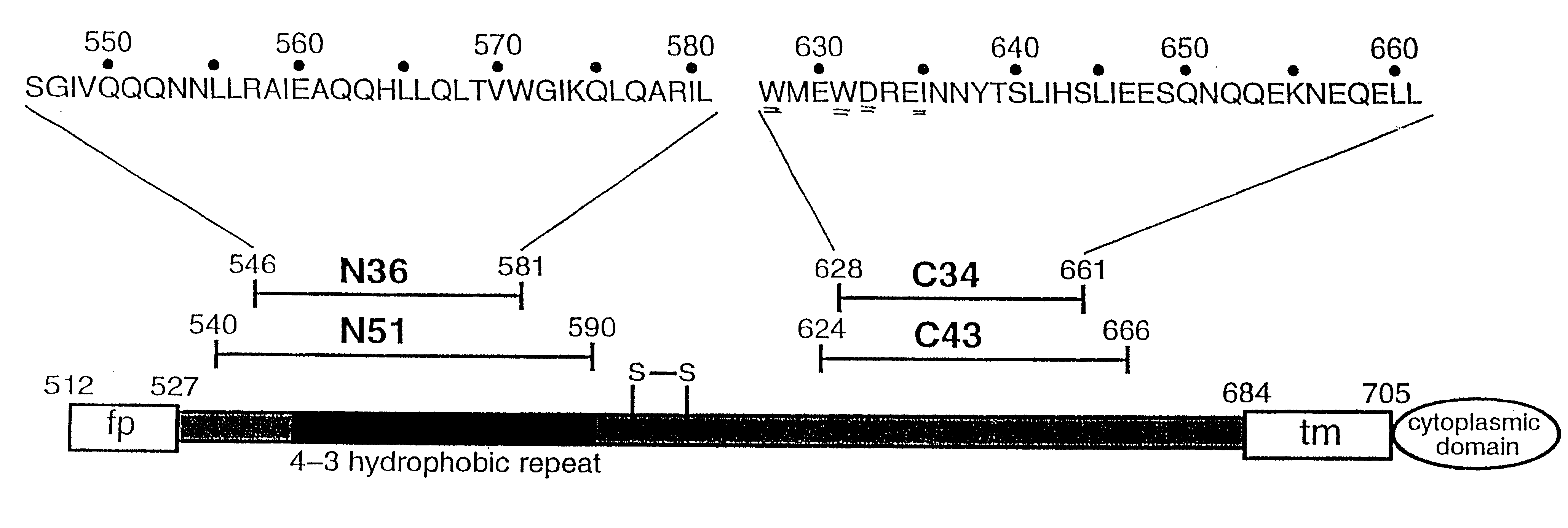
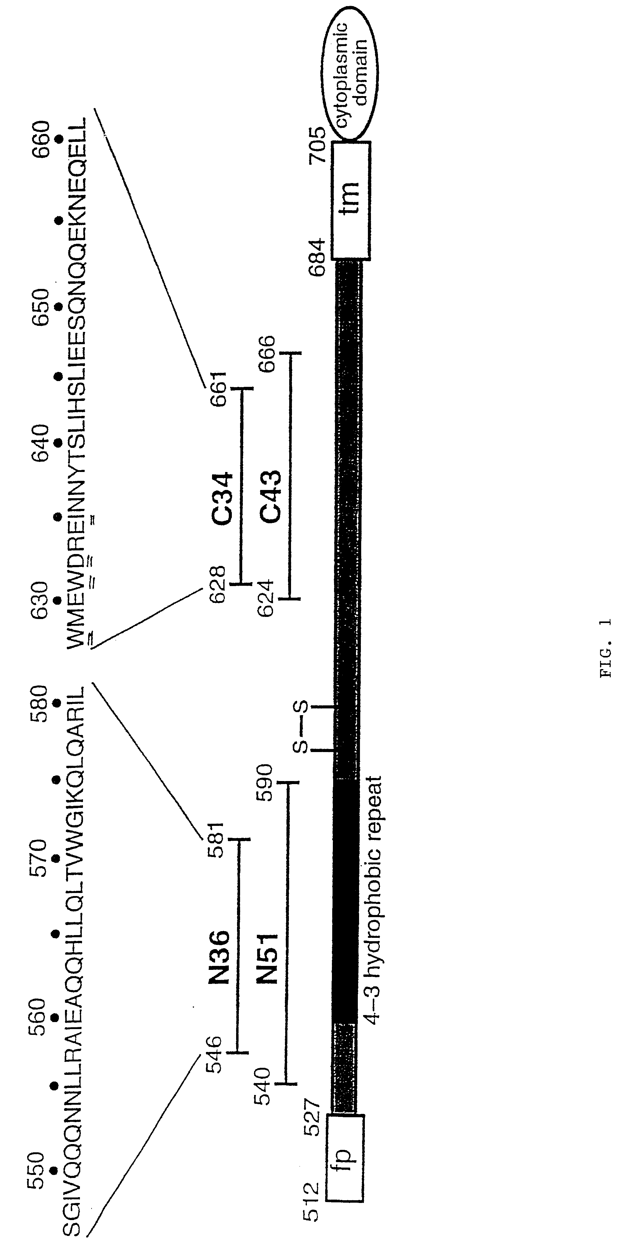
Interestingly, isolated peptides that overlap, or are derived from, the N51 and C43 regions of gp41 can have potent anti-viral activity (Wild, C. T. et al., Proc. Natl. Acad. Sci., USA 89:10537-10541 (1992); Wild, C. T. et al, Proc. Natl. Acad. Sci., USA 91:9770-9774 (1994); Jiang, S. et al., Nature 365:113 (1993)). Peptides from the C-terminal region of the ectodomain have the highest activity. Consistent with these studies, both N51 and C43 are capable of inhibiting HIV envelope-mediated cell fusion; the C43 peptide exhibits 10-fold greater activity than N5 1 (Lu, M. et al., Nature Struct. Biol. 2:1075-1082 (1995)). The inhibitory activity of the C43 peptide, however, is markedly reduced when stoichiometric amounts of N51 are present, suggesting that the C43 peptide inhibits membrane fusion in a dominant-negative manner, by associating with an N51 region within intact gp41 (Lu, M. et al., Nature Struct. Biol. 2:1075-1082 (1995)). Thus, in addition to providing insights into the mechanism of membrane fusion, determining the structural basis for interaction between the N51 and C43 regions will assist anti-viral drug-development efforts.
Both the N- and C-peptide classes of inhibitors are effective against a wide range of HIV strains, including laboratory-adapted strains and primary isolates. Wild, C. T. et al., Proc. Natl. Acad. Sci., USA 89:10537-10541 (1992); Jiang, S. et al., Nature 365:113 (1993); Wild, C. T. et al., Proc. Natl. Acad. Sci., USA 91:9770-9774 (1994). In contrast, soluble CD4 and many neutralizing antibodies are typically effective only on a limited subset of HIV strains (e.g., Daar, E. S. et al., Proc. Natl. Acad. Sci., USA 87:6574-6578 (1990); Palker, T. J. et al., Proc. Natl. Acad. Sci., USA 85:1932-1936 (1988); Nara, P. L. et al., J. Virol. 62:2622-2628 (1988); Moore, J. P. et al., J. Virology 69:101 -109 (1995). There is a striking conservation of residues involved in interactions between the N-peptide and C-peptide, comparing gp41 from HIV-1 and SIV. The broad neutralizing effects of the N- and C-peptides derive from the strong sequence conservation of these residues.

Problems solved by technology

It has not yet been possible to obtain a detailed structure for gp41, either alone or in complex with gp120.

Method used


Image

  • Core structure of gp41 from the HIV envelope glycoprotein
  • Core structure of gp41 from the HIV envelope glycoprotein
  • Core structure of gp41 from the HIV envelope glycoprotein

Examples


example 1

Production of Crystals of N36 / C34

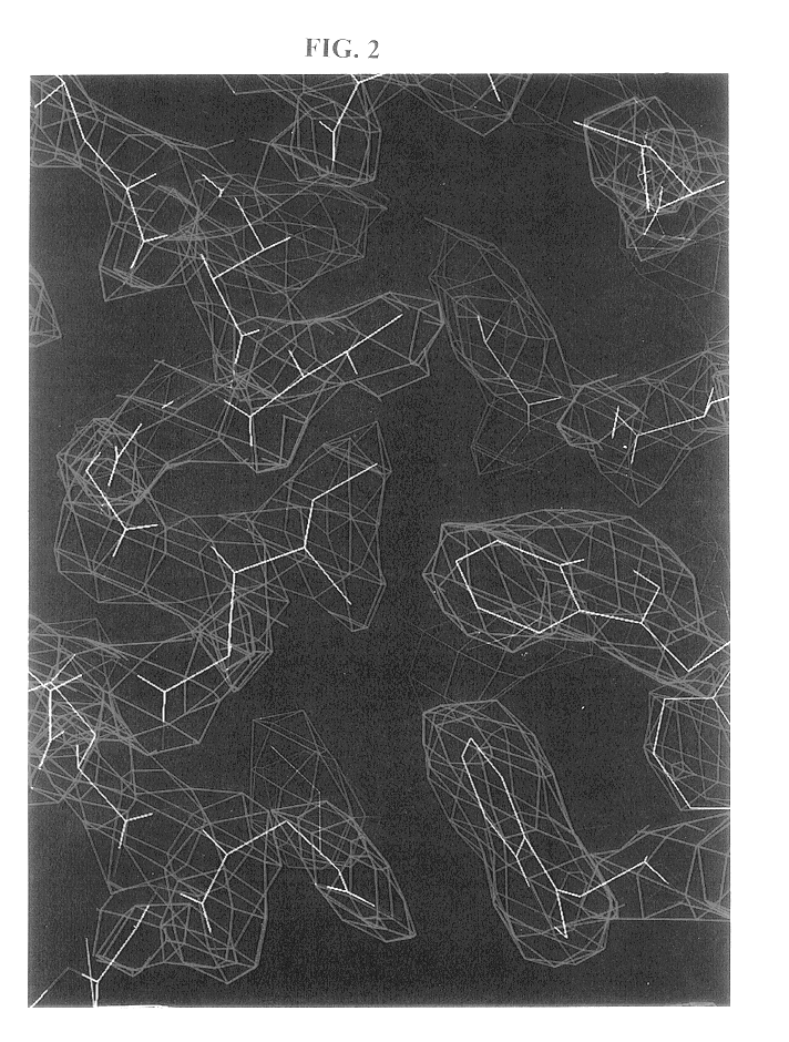
Crystals of N36 / C34 were grown by sitting-drop vapor diffusion (see Methods). An initial model of the complex was built into an electron density map generated by multi-wavelength anomalous dispersion (MAD) analysis (Hendrickson, W. A., Science 254:51-58 (1991)) of an osmium-derivatized crystal. Details of data collection and MAD phasing statistics are listed in the Table. A representative portion of the solvent-flattened electron density map used for building the initial model is shown in FIG. 2. The structure was refined against data to 2.0 .ANG. from a native crystal to yield an R.sub.free of 0.266 and an R.sub.cryst of 0.238 (Table).

example 2

Assessmetn of the Structure of the N36 / C34 Complex

The N36 / C34 complex is a six-stranded helical bundle (FIG. 3). The center of this bundle consists of a parallel, trimeric coiled coil of three N36 helices wrapped in a gradual left-handed superhelix. Three C34 helices wrap antiparallel to the N36 helices in a left-handed direction around the outside of the central coiled-coil trimer. The complex is a cylinder measuring .about.35 .ANG. in diameter and .about.55 .ANG. in height.

As in other naturally-occurring coiled coils (Cohen, C. et al., Proteins 7:1-15 (1990)), the interior residues at the a and d positions of the N36 heptad repeat are predominantly hydrophobic, although occasional buried polar interactions are also present in the central three-stranded coiled coil (FIG. 4). A sequence comparison of HIV-1 (HXB2 strain) and SIV (Mac239 strain) gp41 shows that the residues at these two heptad-repeat positions are highly conserved (FIG. 4). The characteristic "knobs-into-holes" packin...

example 3

Determination of Interactions Between the N- and C-Peptide Helices

Three C34 helices pack obliquely against the outside of the N36 coiled-coil trimer in an antiparallel orientation. These C34 helices interact with N36 mainly through hydrophobic residues in three grooves on the surface of the central coiled-coil trimer. Sequence comparisons between HIV and SIV gp41 shows that the residues lining these grooves are highly conserved. In contrast, the N36 residues flanking the C34 helices are divergent between HIV and SIV.

This pattern of sequence conservation is also apparent on a helical wheel representation of three N36 helices and one C34 helix (FIG. 4). In this diagram, the residue positions in C34 are depicted as ellipses to indicate the oblique tilt of the C34 helix relative to the N36 superhelix and to emphasize that C34 is not part of a coiled coil. Residues at the e and g positions of the N36 helices lie on the outside of the central coiled coil and point into the triangular inte...


PUM

PropertyMeasurementUnit
heightaaaaaaaaaa
diameteraaaaaaaaaa
melting temperatureaaaaaaaaaa

Abstract

Described are the crystal structure of the alpha-helical domain of the gp41 component of HIV-1 envelope glycoprotein which represents the core of fusion-active gp41, methods of identifying and designing drugs which inhibit gp41 function and drugs which do so.

Description

GOVERNMENT SUPPORTWork described herein was funded by the Howard Hughes Medical Institute.The surface glycoproteins of enveloped viruses play critical roles in the initial events of viral infection, mediating virion attachment to cells and fusion of the viral and immune response in infected hosts. Envelope glycoproteins are also major targets for the anti-viral immune response in infected hosts. The human immunodeficiency virus type 1 (HIV-1) envelope glycoprotein consists of two noncovalently associated subunits, gp120 and gp41, that are generated by proteolytic cleavage of a precursor polypeptide, gp160. Luciw, P. A., In Fields Virology, Third Edition, B. N. Fields et al., eds., Lippincott-Raven Publishers, Philadelphia, pp. 1881-1952 (1996); Freed, E. O. et al., J. Biol. Chem. 270: 23883-23886 (1995). gp120 directs target-cell recognition and viral tropism through interaction with the cell-surface receptor CD4 and one of several co-receptors that are members of the chemokine rece...

Claims


Application Information

Patent Timeline
Patent Type & Authority Patents(United States)
IPC IPC(8): C07K14/16C07K14/005A61K38/00
CPCC07K14/005A61K38/00C12N2740/16122
Inventor CHAN, DAVID C.FASS, DEBORAHLU, MINBERGER, JAMES M.KIM, PETER S.
Owner WHITEHEAD INST FOR BIOMEDICAL RES
Learn More