Method and system for selective or isolated integrate cerebral perfusion and cooling

a technology of applied in the field of selective or isolated integration cerebral perfusion and cooling, can solve the problems of releasing significant amounts of atheromatous debris, avoiding such intravascular techniques, and the brain and cerebral vasculature are at the greatest risk of embolization and ischemia

Inactive Publication Date: 2004-05-18
BARBUT DENISE & PATTERSON RUSSEL +1
View PDF47 Cites 81 Cited by
  • Summary
  • Abstract
  • Description
  • Claims
  • Application Information

AI Technical Summary

Benefits of technology

Optionally, in addition to improving cerebral perfuision, the methods of the present invention may combine or otherwise rely on cooling of the patient's head and cerebral vasculature in treatment of both global and focal cerebral ischemia to inhibit tissue damage resulting from lack or limitation of cerebral blood circulation. Usually, the oxygenated medium which is circulated as part of the methods of the present invention will be cooled in order to cool the brain tissue and reduce the risk of ischemic damage. Further optionally, the patient's head may be cooled even prior to initiating perfusion of externally oxygenated, optionally cooled blood. In some instances, the cooled blood can be used to externally cool the patient's head during the treatment protocol, e.g., by passing the blood through a helmet or other structure which permits the blood to selectively cool the head. This selective isolated cooling of the head and / or cerebral vasculature is desirable and preferred over systemic cooling, since coagulopathy, poor healing, cardiac arrhythmia and cardiac arrest can ensue as a result of systemic cooling.
In still other embodiments, methods, systems, and kits are provided for isolating and perfusing the cerebral vasculature, usually to facilitate access to a patient's aorta, during performance of a diagnostic or interventional procedure on the aorta, more usually during performance of an open surgical interventional procedure on the aorta, such as repair of an aortic aneurysm, dissections, reconstruction of the aorta, endarterectomy, or the like. The heart will usually be arrested during open surgical procedures where the aorta is opened and procedure is performed within the lumen of the aorta. In some instances, however, the heart may remain beating while the procedure is performed intravascularly, i.e. through using catheters and other instruments introduced from the peripheral vasculature and into the aorta. The methods of the present invention will serve primarily to isolate the cerebral vasculature and prevent gaseous and atheromatous emboli from entering the cerebral vasculature while the vasculature is perfused with an oxygenated medium.
In a presently preferred procedure, occluding balloons will be positioned within the brachiocephalic artery, the left common carotid artery, and the left subdlavian artery. Both the right subdlavian artery and the left subdlavian artery will be blocked, preferably with external tourniquets on the arms. Blood or other oxygenated medium will then be perfused into the arterial cerebral vasculature to points immediately upstream of each of the occluding balloons, preferably using lumens or other infusion components incorporated within the occluding devices themselves. Inhibition of blood flow down into the arms is beneficial since it redirects the blood or other oxygenated medium back into the cerebral arterial vasculature. While this approach may be optimal in many ways, the present invention can be carried out in other ways as well. Most simply, internal occlusion of the right brachiocephalic artery and perfusion of oxygenated medium distal to the point of occlusion may be sufficient in some cases by itself.
In many cases, it will be desirable to occlude the arteries at a point as close to the aortic arch as possible. In particular, this is true of the brachiocephalic artery, the left carotid artery, and the left subdlavian artery which branch directly from the aortic arch. Occlusion close to the aortic arch (i.e., immediately above the branch or within 3 cm thereof) is of benefit primarily because it enables the surgeon to access the artery and initiate the occlusion with minimal aortic dissection toward the neck. In other cases, of course, it will be possible to access any one of the brachiocephalic artery at a point close to the aortic arch and to intravascularly advance an occluding balloon or other devise to a desired point of occlusion. In some instances, it may even be desirable to deliver and position devices carrying multiple occluding balloons and / or lumens for delivering oxygenated medium to the cerebral arterial vasculature.
The methods of the present invention will-find their greatest use in open and thoracoscopic surgical procedures where the aorta is exposed and surgically opened to permit performance of the desired procedure. In such cases, the heart will be arrested and the perfusion of the oxygenated medium will be relied on to achieve adequate oxygenation of the brain tissue and to avoid deleterious ischemia. Generally, the flow rates and temperatures set forth above will be sufficient to both achieve adequate perfusion and avoid ischemia. After the open procedure is completed, and the aorta is surgically closed, heart function may be reestablished. In order to avoid the release of emboli from the aorta into the cerebral vasculature, occlusion of carotid artery(ies) will be maintained for a minimum amount of time after heart function has been reestablished, typically for at least about 2 minutes, preferably for at least about 5 minutes, in order to permit atheromatous debris and air to be cleared from the aorta and away from the brain.

Problems solved by technology

The use of clamps, however, is generally not preferred since they can cause the release of significant amounts of atheromatous debris when released.
Thus, in many instances, it will be undesirable to use such intravascular techniques since they will lie within the regions where the procedure is being performed.
The brain and cerebral vasculature are at greatest risk from embolization and ischemia during the performance of aortic procedures that require arresting of the heart.

Method used


Image

  • Method and system for selective or isolated integrate cerebral perfusion and cooling
  • Method and system for selective or isolated integrate cerebral perfusion and cooling
  • Method and system for selective or isolated integrate cerebral perfusion and cooling

Examples


Embodiment Construction

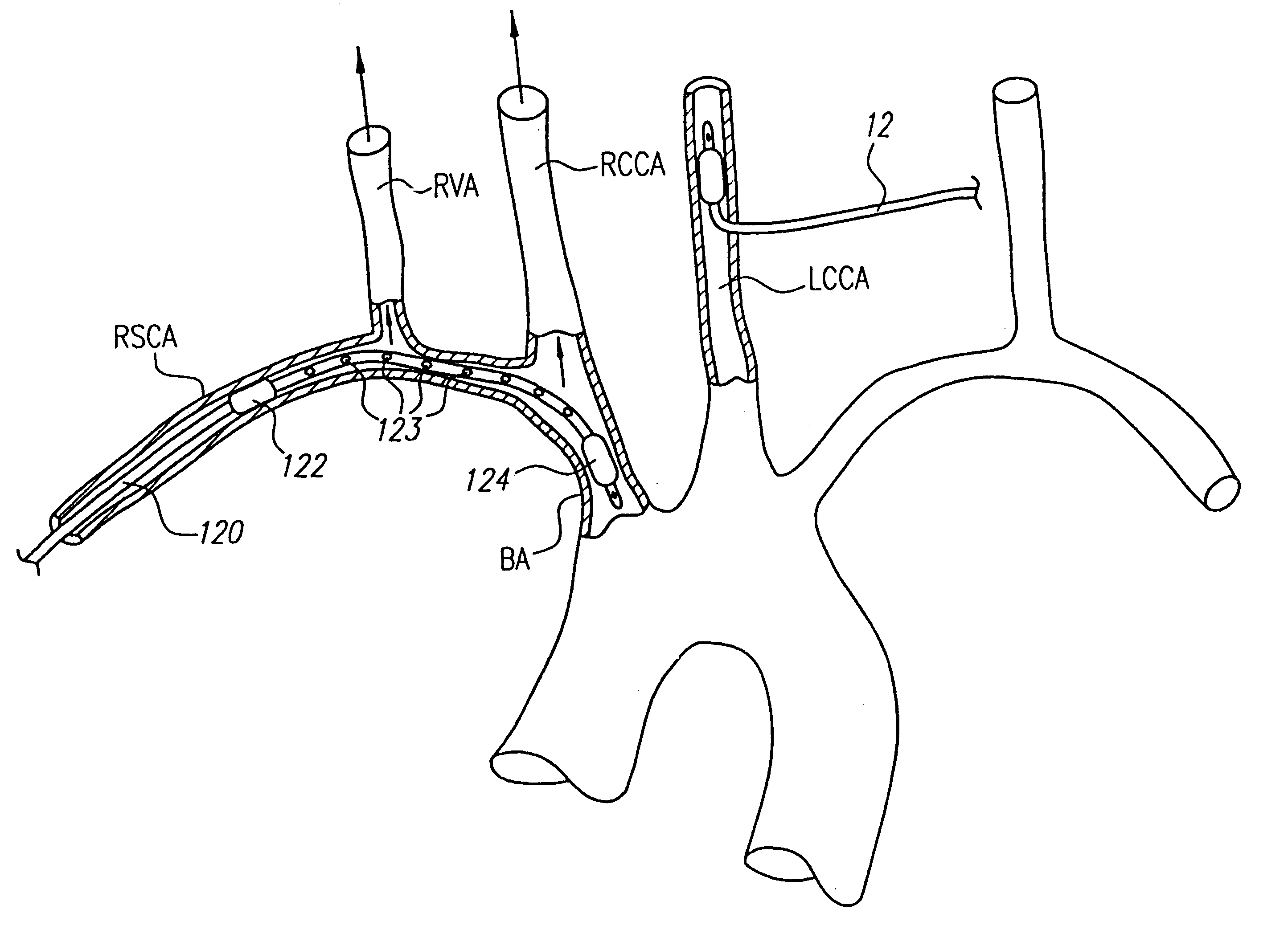
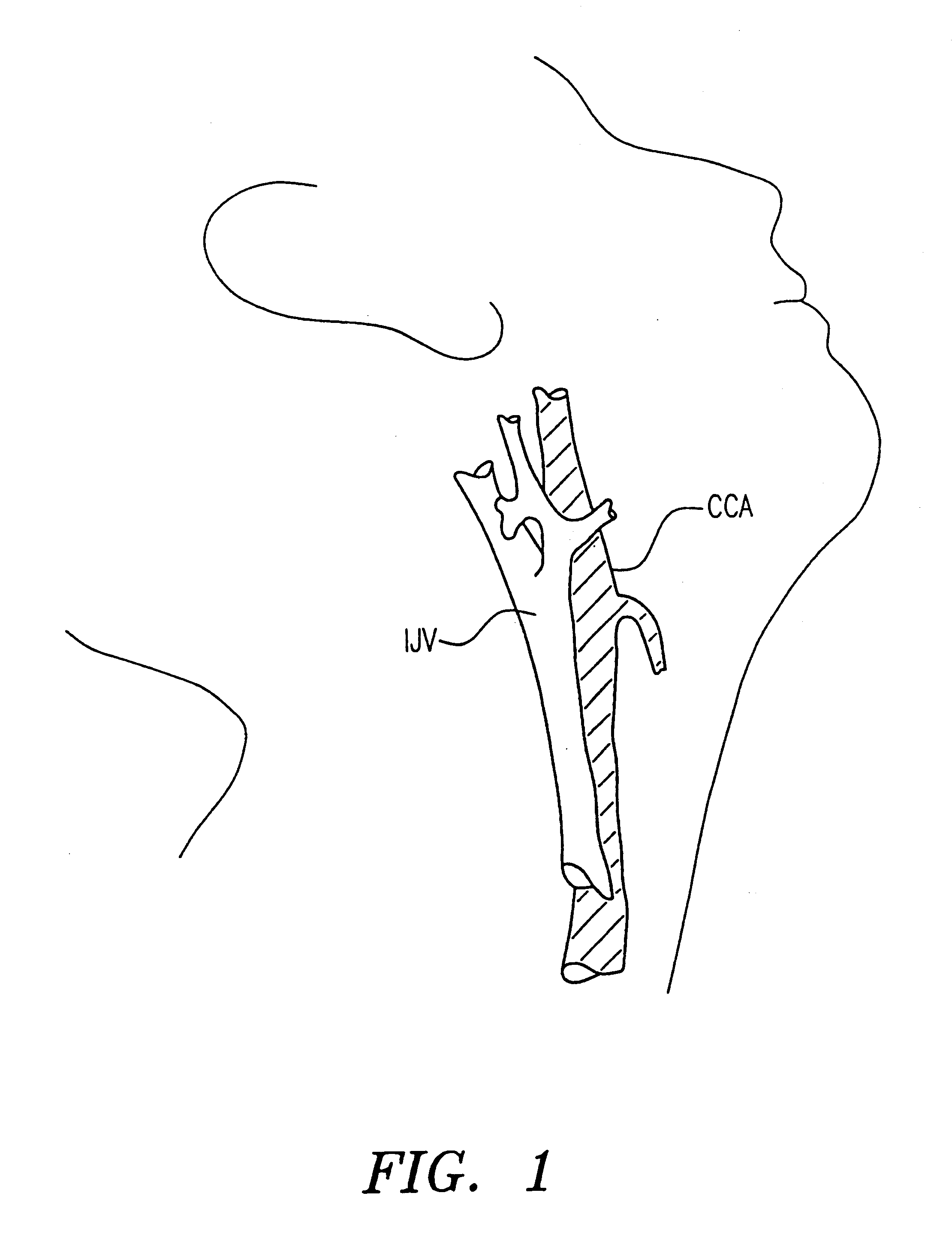
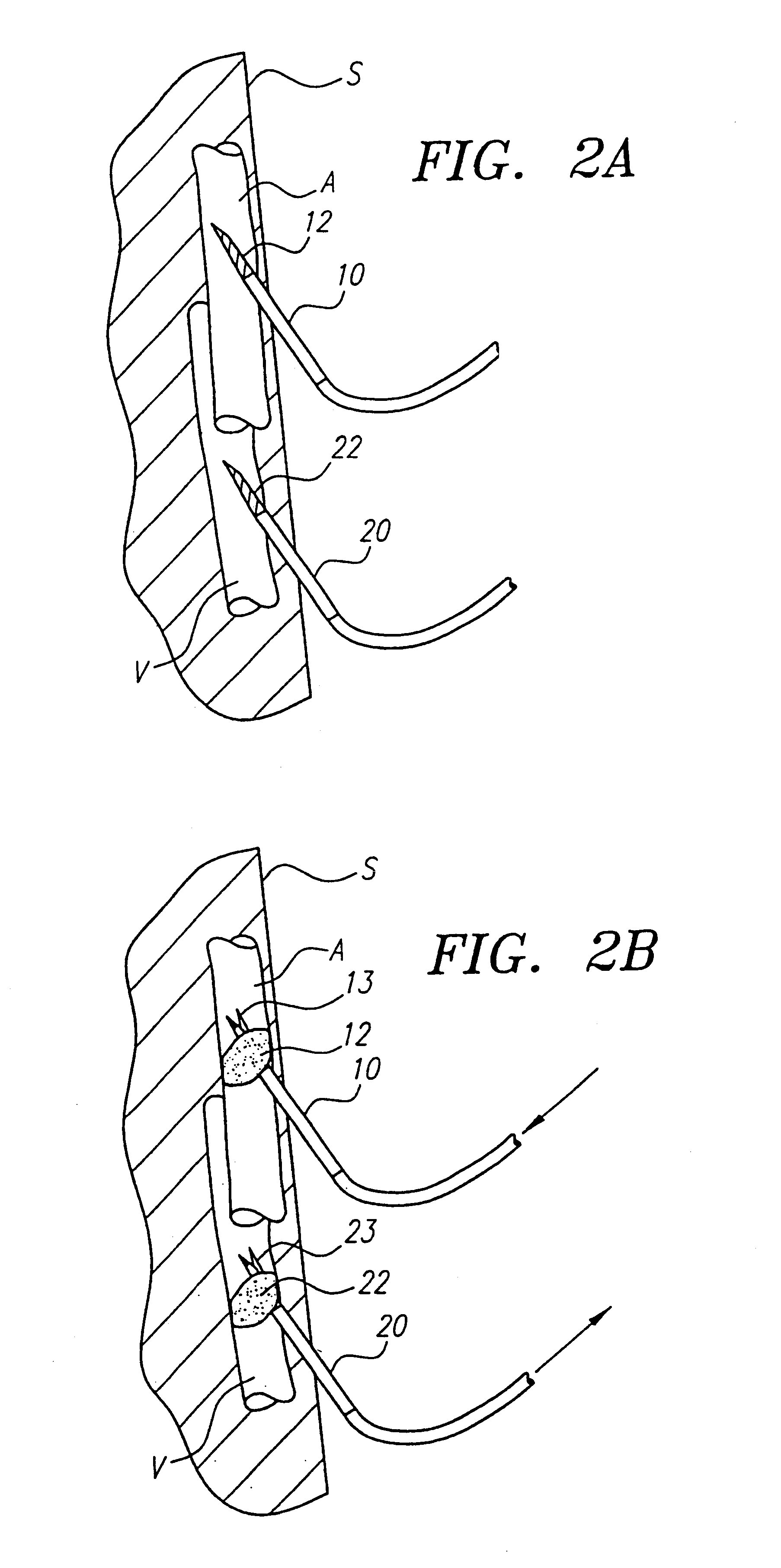
The present invention provides methods, systems, and kits for perfusing the cerebral vasculature of a patient with an oxygenated medium. For the purposes of the present invention, the cerebral vasculature includes all arteries and veins leading into or from the patient's head, particularly including the common carotid arteries, the external and internal carotid arteries, and all smaller arteries which branch from the main arteries leading into the head. In some cases, particularly in open surgical procedures, access may be established in the aortic arch and innominate (brachycephalic trunk) artery as well. Cerebral veins include the external and internal jugular veins, the superior vena cava, and the smaller veins which feed into the primary veins draining the cerebral vasculature. Preferred access points should be at locations in the vasculature which permit relatively direct percutaneous introduction of a needle, cannula, or other access conduit through which the flow of oxygenate...


PUM


Abstract

Patients having diminished circulation in the cerebral vasculature as a result of cardiac arrest or from other causes are treated by flowing an oxygenated medium through an arterial access site into the cerebral vasculature and collecting the medium through an access site in the venous site of the cerebral vasculature. In addition to oxygenation, the recirculating blood may also be cooled to hypothermically treat and preserve brain tissue. Isolation and cooling of cerebral vasculature in patients undergoing aortic and other procedures is achieved by internally occluding at least the right common carotid artery above the aortic arch. Blood or other oxygenated medium is perfused through the occluded common carotid artery(ies) and into the arterial cerebral vasculature. Usually, oxygen depleted blood or other medium leaving the cerebral vasculature is collected, oxygenated, and cooled in an extracorporeal circuit so that it may be returned to the patient.

Description

The present invention relates generally to medical devices and methods. More particularly, the present invention relates generally to methods, systems, and kits for perfusing and optionally cooling the cerebral vasculature of a patient with oxygenated blood or other media.Cerebral ischemia, i.e., reduction or cessation of blood flow to the cerebral tissue, can be characterized as either focal or global. Focal cerebral ischemia refers to reduced perfusion to the cerebral tissue resulting from a partial or complete occlusion in the intracranial or extracranial cerebral arteries, e.g., stroke, subarachnoid hemorrhage spasms, iatrogenic vasospasm. Global cerebral ischemia refers to reduced perfusion to the cerebral tissue resulting from systemic circulatory failure caused by, e.g., cardiac arrest, shock, circulatory arrest, and septicemia.Cardiac arrest is a major contributor to global cerebral ischemia. Cardiac arrest refers to cessation or significant reduction of a patient's cardiac ...

Claims


Application Information

Patent Timeline
Patent Type & Authority Patents(United States)
IPC IPC(8): A61M1/36
CPCA61M1/3621A61M1/3603A61M1/3613
Inventor BARBUT, DENISE R.PATTERSON, RUSSEL H.
Owner BARBUT DENISE & PATTERSON RUSSEL
Why Eureka
Try Eureka