
Surgical probe for supporting inflatable therapeutic devices in contact with tissue in or around body orifices and within tumors

Inactive Publication Date: 2005-01-04
BOSTON SCI SCIMED INC
View PDF61 Cites 121 Cited by
  • Summary
  • Abstract
  • Description
  • Claims
  • Application Information

AI Technical Summary

Benefits of technology

Accordingly, the general object of the present inventions is to provide a device that avoids, for practical purposes, the aforementioned problems. In particular, one object of the present inventions is to provide a device that can be used to create circumferential lesions in or around the pulmonary vein and other bodily orifices in a more efficient manner than conventional apparatus.
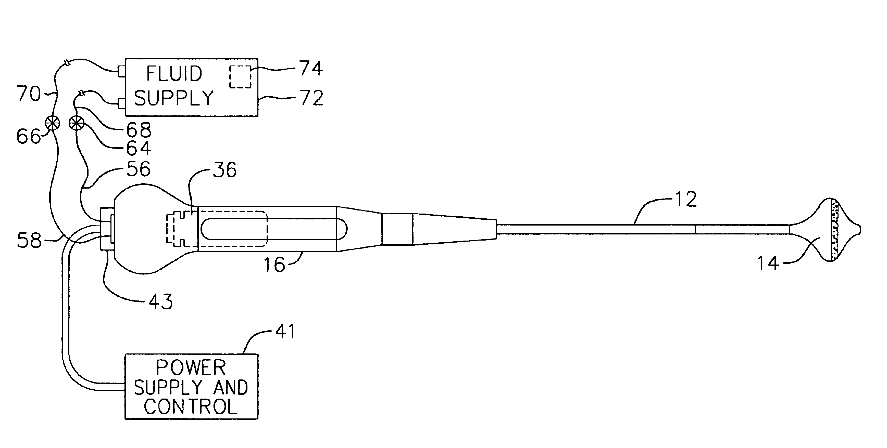
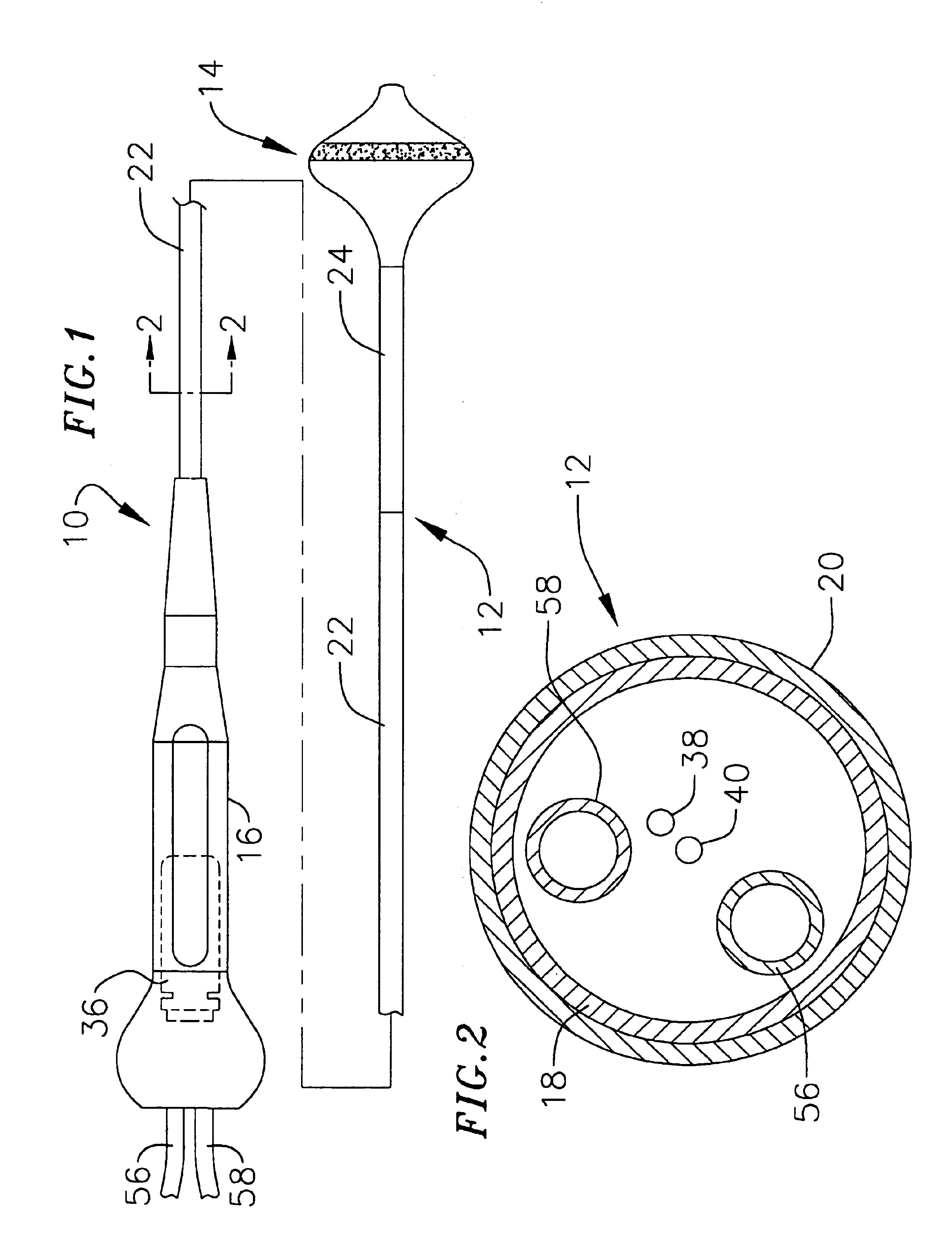
Such a probe provides a number of advantages over conventional apparatus. For example, the present surgical probe may be used during open heart surgery or in less invasive procedures where access to the heart is obtained via a thoracostomy, thoracotomy or median sternotomy. The relatively short shaft and manner in which access is obtained allows the therapeutic element to be easily inserted into the heart and placed against the target tissue with the desired level of contact, thereby eliminating many of the problems associated with catheter-based procedures. Moreover, the present therapeutic element may be used to form lesions in an annular region of tissue within or around the pulmonary vein (or other orifice in other procedures) in one step, thereby eliminating the need to either drag a tip electrode around an annular region or form a number of interconnected curvilinear lesions that is associated with catheter-based procedures.
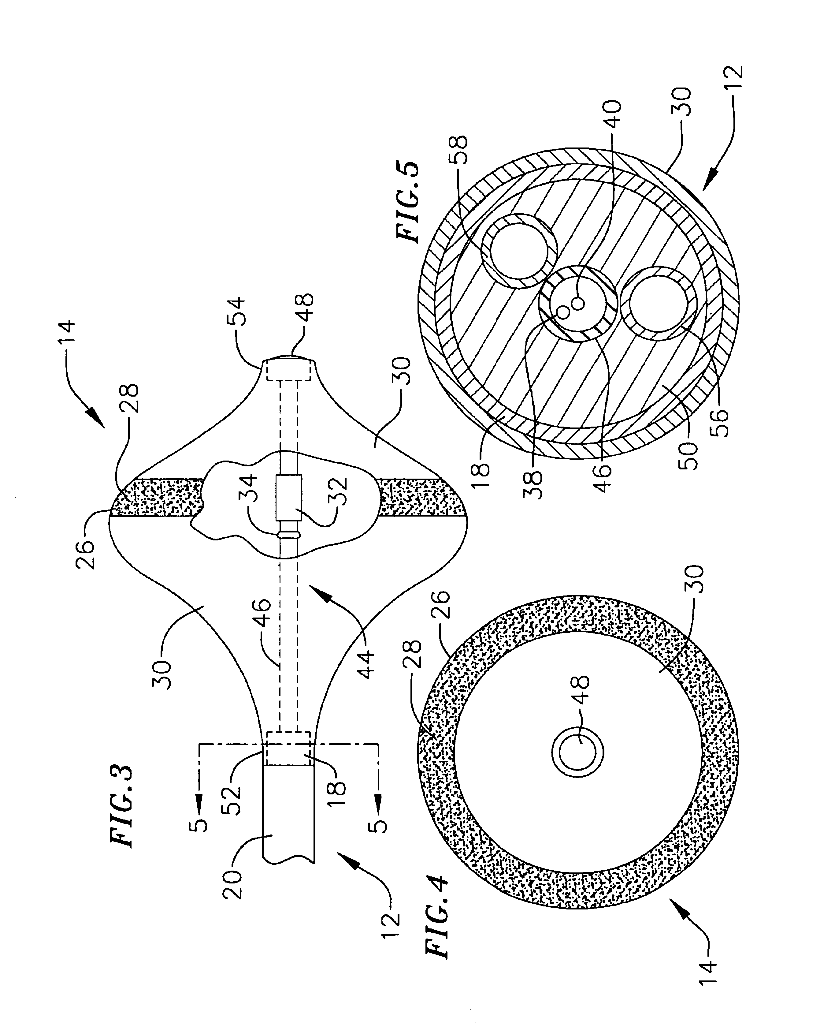
Additionally, in accordance with a preferred embodiment, the flexibility of the inflatable therapeutic element may be varied as appropriate. This allows the physician to achieve the appropriate level of tissue contact, even when the shaft is not perfectly perpendicular to the target tissue area, the target tissue area is somewhat uneven, or the target tissue has become rigid due to calcification.
In accordance with another preferred embodiment, the inflatable therapeutic element will be configured such that it can be inserted into a tumor (or other target location), inflated and then used to uniformly coagulate the entire tumor (or a large volume of tissue associate with the other location) without charring. Once the coagulation procedure is complete, the inflatable therapeutic element can be deflated and removed from patient without the difficulty and trauma associated with needle-like electrodes.

Problems solved by technology

These wavelets, called “reentry circuits,” disrupt the normally uniform activation of the left and right atria.
Because of a loss of atrioventricular synchrony, the people who suffer from atrial fibrillation and flutter also suffer the consequences of impaired hemodynamics and loss of cardiac efficiency.
They are also at greater risk of stroke and other thromboembolic complications because of loss of effective contraction and atrial stasis.
However, the maze procedure is technically difficult to do.
Nevertheless, the inventors herein have determined that catheter-based procedures are not appropriate in every situation and that conventional catheters are not capable of reliably forming all types of lesions.
For example, one lesion that has proven to be difficult to form with conventional catheter devices is the circumferential lesion that is used to isolate the pulmonary vein and cure ectopic atrial fibrillation.
Such techniques have proven to be less than effective because they are slow and gaps of conductive tissue can remain after the procedure.
It can also be difficult to achieve the adequate tissue contact with conventional catheters.
Heretofore, tumors have been treated with highly toxic drugs that have proven to have severe side effects.
Most notably, the needle-like electrodes produce non-uniform, shallow lesions and / or spot lesions and also fail to coagulate the entire volume of tumor tissue.
This failure can ultimately result in the tumor growing to be even larger than its original size.
The needle-like electrodes can also cause tissue charring.
Moreover, tissue tends to shrink around the needle-like electrodes during the coagulation process.
This makes it very difficult to withdraw the electrodes from the patient and often results in tissue trauma.

Method used


Image

  • Surgical probe for supporting inflatable therapeutic devices in contact with tissue in or around body orifices and within tumors
  • Surgical probe for supporting inflatable therapeutic devices in contact with tissue in or around body orifices and within tumors
  • Surgical probe for supporting inflatable therapeutic devices in contact with tissue in or around body orifices and within tumors

Examples


Embodiment Construction

The following is a detailed description of the best presently known modes of carrying out the inventions. This description is not to be taken in a limiting sense, but is made merely for the purpose of illustrating the general principles of the inventions.

This specification discloses a number of probe structures, mainly in the context of cardiac ablation, because the structures are well suited for use with myocardial tissue. For example, the present inventions are designed to produce intimate tissue contact with target substrates associated with arrhythmias such as atrial fibrillation. One application is the creation of lesions within or around the pulmonary vein to treat ectopic atrial fibrillation. Nevertheless, it should be appreciated that the structures are applicable for use in therapies involving other types of soft tissue. For example, various aspects of the present inventions have applications in procedures concerning other regions of the body such as the prostate, liver, br...


PUM


Abstract

A probe that facilitates the creation of lesions in bodily tissue. The probe includes a relatively short shaft and an inflatable therapeutic element.

Description

BACKGROUND OF THE INVENTIONS1. Field of InventionsThe present inventions relate generally to surgical probes that support therapeutic devices in contact with body tissue.2. Description of the Related ArtThere are many instances where diagnostic and therapeutic elements must be inserted into the body. One instance involves the treatment of cardiac conditions such as atrial fibrillation and atrial flutter which lead to an unpleasant, irregular heart beat, called arrhythmia.Normal sinus rhythm of the heart begins with the sinoatrial node (or “SA node”) generating an electrical impulse. The impulse usually propagates uniformly across the right and left atria and the atrial septum to the atrioventricular node (or “AV node”). This propagation causes the atria to contract in an organized way to transport blood from the atria to the ventricles, and to provide timed stimulation of the ventricles. The AV node regulates the propagation delay to the atrioventricular bundle (or “HIS” bundle). Th...

Claims


Application Information

Patent Timeline
IPC IPC(8): A61B18/14A61B5/0408A61B5/042A61B17/34A61B18/04A61F2/958
CPCA61B18/1492A61B5/0422A61B2018/00065A61B2018/00083A61B2018/00095A61B2218/002A61B2018/0022A61B2018/00238A61B2018/00577A61B2018/00839A61B2019/465A61B2018/00214A61B2090/065A61B5/287
Inventor KOBLISH, JOSEF V.HEGDE, ANAT V.SWANSON, DAVID K.
Owner BOSTON SCI SCIMED INC
Learn More