Methods for inhibiting rejection of transplanted tissue

Inactive Publication Date: 2005-01-18
THE GENERAL HOSPITAL CORP
View PDF6 Cites 25 Cited by
  • Summary
  • Abstract
  • Description
  • Claims
  • Application Information

AI Technical Summary

Benefits of technology

The invention features a method for inhibiting rejection by a recipient animal of a transplanted tissue. The method involves modifying, eliminating, or masking an antigen which, when present on the surface of a cell of the tissue, is capa

Problems solved by technology

Conjugate formation is followed by T-cell-mediated lysis, resulting in donor cell de

Method used


Image

  • Methods for inhibiting rejection of transplanted tissue
  • Methods for inhibiting rejection of transplanted tissue
  • Methods for inhibiting rejection of transplanted tissue

Examples


example 1

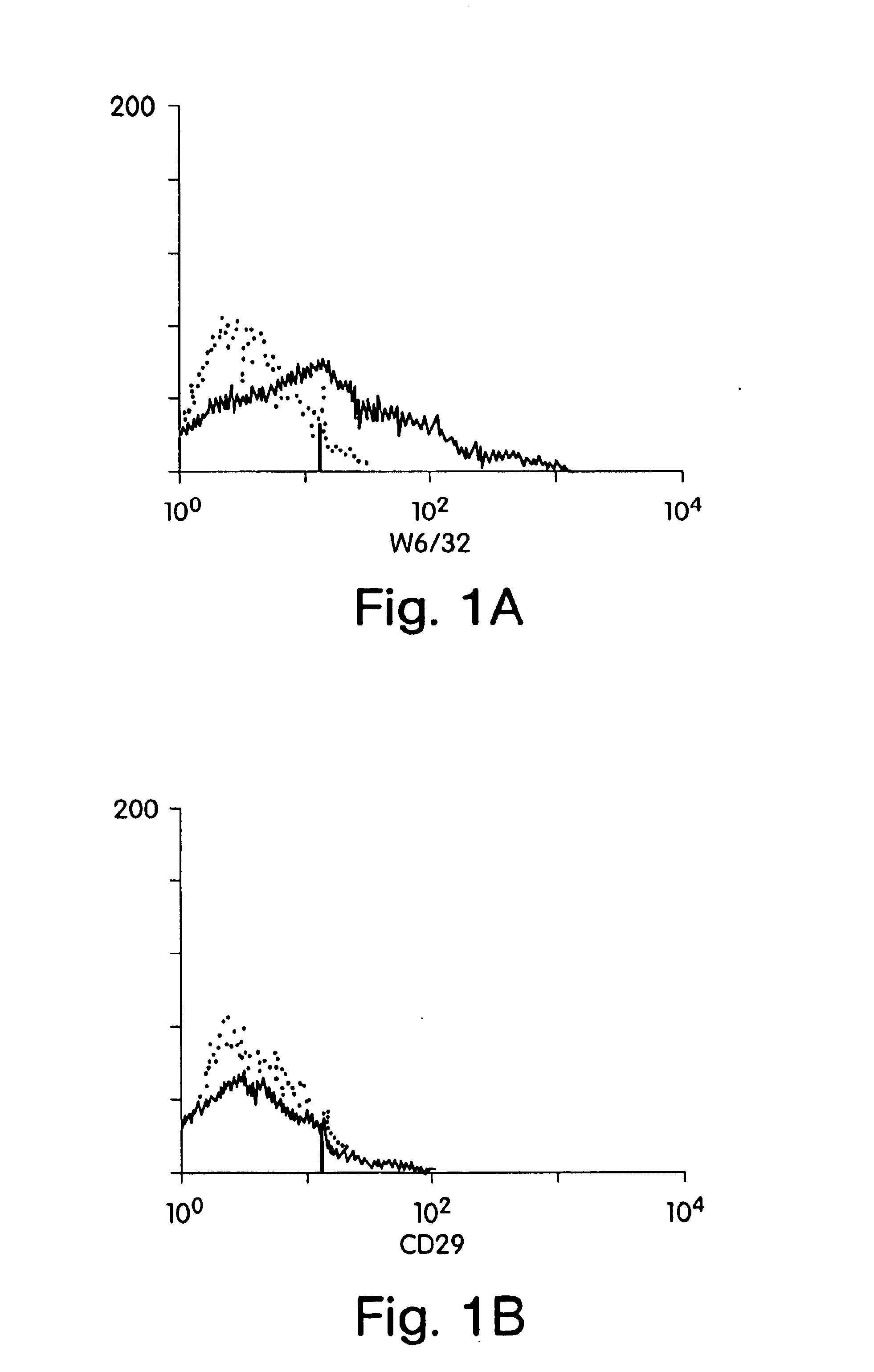
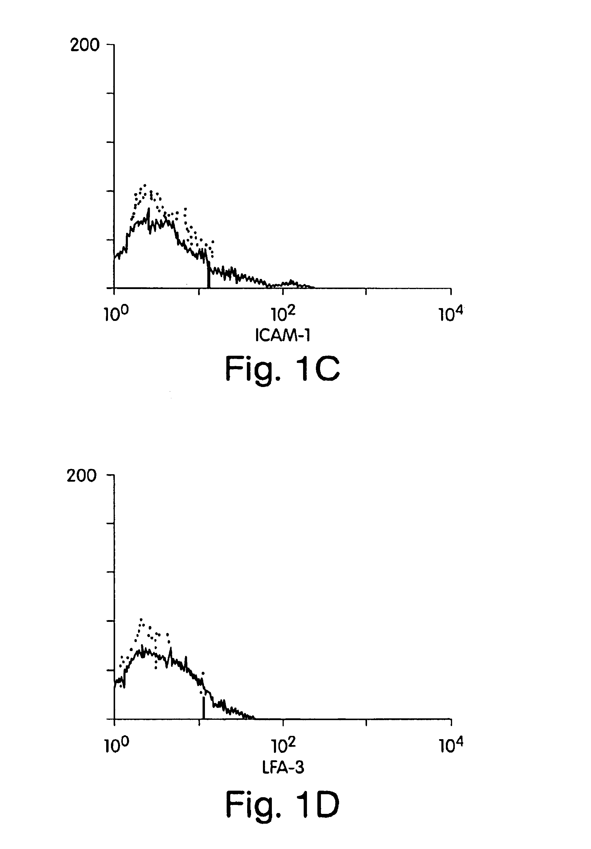
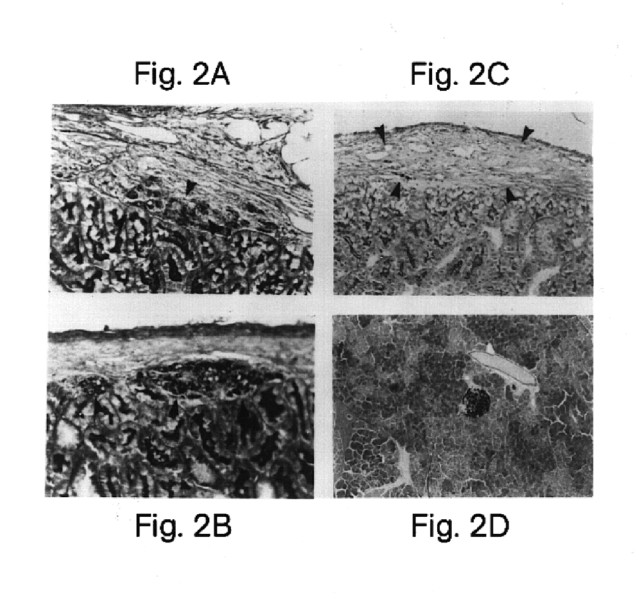
This example involves xenogeneic transplantation of HLA class I positive human islet cells into nonimmunosuppressed Balb / c mouse recipients. Freshly isolated human islets were pretreated prior to transplantation with whole monoclonal antibody or F(ab′)2 monoclonal antibody fragments to conceal (“mask”) donor antigens. F(ab′)2 fragments lack the Fc antibody region, thus circumventing complement-mediated killing after antigen binding. Intact immunoglobulin was used as a control. Human islets were treated with relevant HLA class I monoclonal antibody (W6 / 32) (American Tissue Culture Society) (Barnstable et al. (1978) Cell, 14:9-20) or irrelevant CD29 monoclonal antibody (Coulter Corporation, Hialeah, Fla.). Clean human islet preparations, free of contaminating endothelial and fibroblast overgrowth, are negative for ICAM-1 expression, negative for CD29 expression, have low LFA-3 expression, and are positive for HLA class I antigens (FIG. 1). Therefore, islets, unlike other cytotoxic T-l...

example 2

This example involves the xenogenenic transplantation of rat insulinoma tumor cells (RIN) into nonimmunosupressed Balb / c mouse recipients to investigate the possibility of graft specific tolerance with growth of a transplanted tissue pre-treated with polyclonal F(ab′)2.

RIN tumor cells are an established rat insuloma tumor cell line (Meflasson et al., 1983 J. Biol. Chem. 258:2094-2097). Polyclonal mouse anti-RIN serum was produced and F(ab′)2 antibody fragments were generated as described above in Example 1. As expected, xenogeneic RIN cells (approx. 5,000 cells per recipient) transplanted under the kidney capsule of nonimmunosuppressed BALB / c mice were uniformly rejected when evaluated by histology with aldehyde fuscin staining (n=4) (Table 3). In addition, pretreatment of RIN cells with intact polyclonal mouse anti-RIN antibody, without removal of the complement-fixing Fc region, prior to transplantation also failed to protect grafts from recipient mediated rejection (n=4). In cont...

example 3

The effectiveness of F(ab′)2 HLA class I antibody coating in preventing rejection of non-tumorgeneic human liver cells in xenogeneic transplants was also investigated. Approximately 5,000 fresh human liver cells from the parenchymal tissue of the liver were injected into the subscapular space of the kidney capsules of nonimmunosuppressed mouse recipients. Histological examination using PAS staining of the subscapular sites indicated that all 5 transplant recipients of F(ab′)2-treated liver cells demonstrated easily located viable liver cells at the subscapular renal site 30 days after transplantation. As expected, untreated human liver cells were uniformly rejected in all five mice by day 30 after transplantation.

It is clear from the results of Examples 1 and 3 that the simple interruption of recipient T cell recognition by masking of foreign HLA class I determinants allows prolonged xenograft survival up to 200 days. This new strategy eliminates recipient treatment, thus preserving...


PUM


Abstract

A method for inhibiting rejection by a recipient animal of a transplanted tissue, said method comprising modifying, eliminating, or masking an antigen which, when present on the surface of a cell of said tissue, is capable of causing a T-lymphocyte-mediated response in said animal, to inhibit antigen-mediated interaction between said cell and a T-lymphocyte of said animal without causing lysis of said cell.

Description

BACKGROUND OF THE INVENTIONThis application is a continuation-in-part of Faustman U.S. Ser. No. 07 / 575,150, filed Aug. 30, 1990. This invention relates to transplantation of tissues, e.g., islet cells, muscle cells, and whole organs, into hosts in need of such tissues, e.g., patients who have or are at risk of developing diabetes mellitus, have muscular dystrophy, or are in need of an organ transplant.Diabetes mellitus is a prevalent degenerative disease, characterized by insulin deficiency, which prevents normal regulation of blood glucose levels, and which leads to hypergylycemia and ketoacidosis.Insulin, a peptide hormone, promotes glucose utilization, protein synthesis, formation and storage of neutral lipids, and the growth of some cell types. Insulin is produced by the β cells within the islets of Langerhans of the pancreas. Early-onset diabetes (10-20% of cases) is caused by an auto-immune reaction that causes complete destruction of β cells. Adult-onset diabetes has a number...

Claims


Application Information

Patent Timeline
IPC IPC(8): A61K35/12A61K35/39A61K35/407A61K35/34A61K35/37C07K16/18C07K16/28A61K38/00A61K35/14A61K35/22A61K35/42A61K39/00A61K39/395A61K48/00A61P3/10A61P37/06C12N5/071C12N5/10C12N15/09
CPCA61K35/34A61K35/407C07K16/2833C12N5/0676A61K35/22A61K35/42A61K38/00A61K2039/505Y10S424/81C07K2317/54Y10S424/809C07K2316/96C07K2317/70A61P37/06A61P3/10
Inventor FAUSTMAN, DENISE
Owner THE GENERAL HOSPITAL CORP
Why Eureka
Try Eureka