
Method for treating diseased or damaged organs

a tissue graft and organ technology, applied in the field of tissue engineering, can solve the problems of unsatisfactory long-term and unpractical use of the patient's own vessels, and achieve the effect of facilitating bioremodeling and adding strength to the gra

Inactive Publication Date: 2005-05-10
ORGANOGENESIS
View PDF141 Cites 19 Cited by
  • Summary
  • Abstract
  • Description
  • Claims
  • Application Information

AI Technical Summary

Benefits of technology

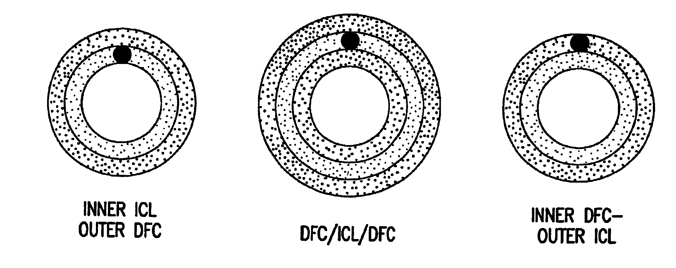
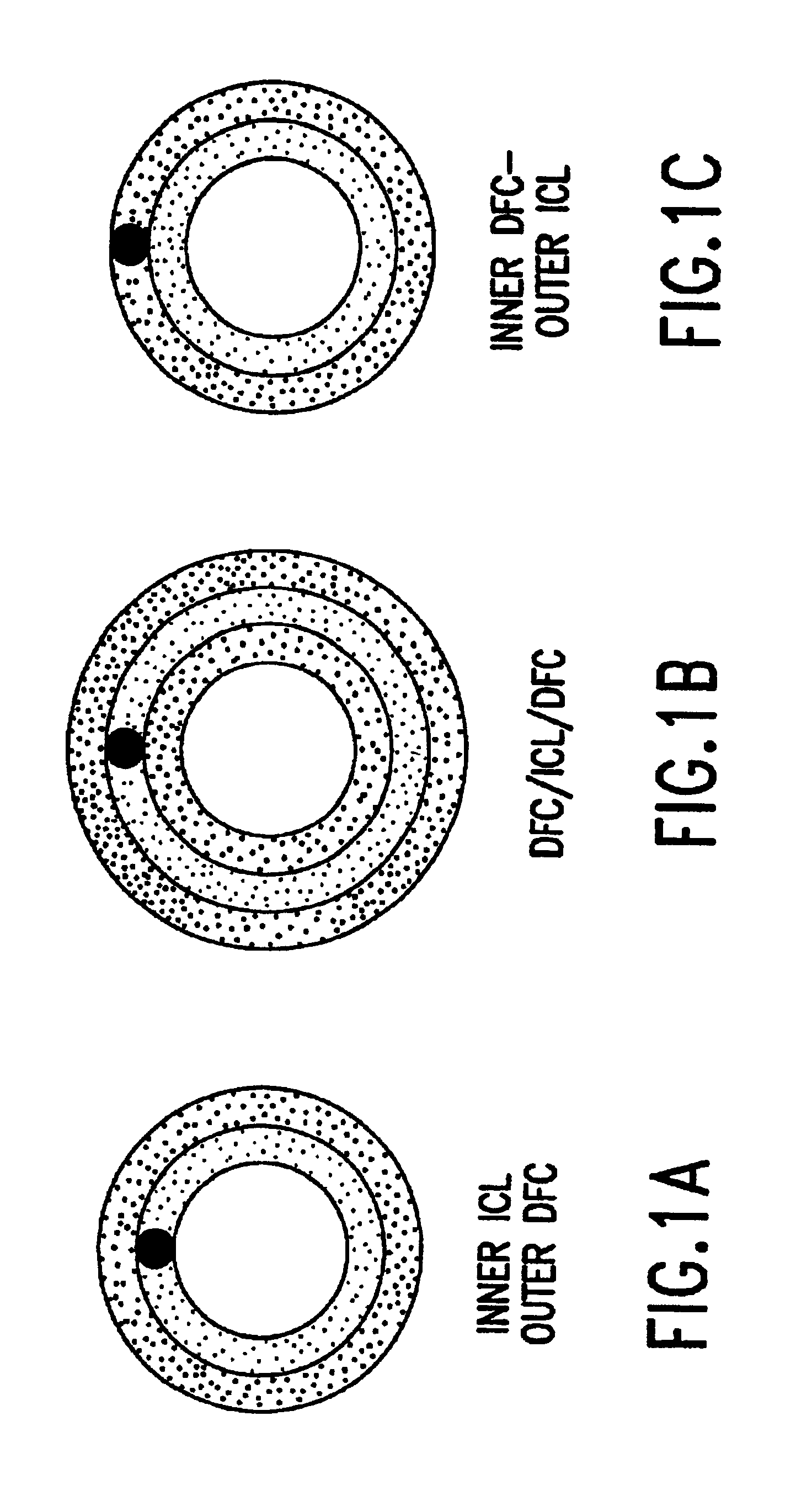
[0004]This invention is directed to a method for treating damaged or injured organs, particularly blood vessels, by replacing, or repairing, a section of the organ in a patient with a bioremodelable collagen graft prostheses. This prosthesis, when implanted into a mammalian host, undergoes controlled biodegradation accompanied by adequate living cell replacement, or neo-tissue formation, such that the original implanted prosthesis is bioremodeled by the host's cells before it is digested by host enzymes. The prosthesis of this invention comprises at least two layers: (a) at least one layer is composed of collagen or a collagenous material; and (b) at least one layer is composed of material which provides structural stability, and is pliable, semi-permeable, and suturable. In the preferred embodiment of this invention, the two-layered prosthesis has an inner (luminal) layer which provides a smooth, thrombosis-resistant flow surface and an outer structural layer which provides structural stability, and is pliable, semi-permeable, and suturable. In another preferred embodiment of this invention the prosthesis has three layers: an inner (luminal) layer which acts as a smooth, thrombosis-resistant flow surface; a middle structural layer which provides structural stability, and is pliable, semi-permeable, and suturable; and, an outer (abluminal) layer. The outer layers of both the two-layer or the three-layer prosthesis add strength to the graft and allow the patient's host cells to attach and grow into the graft thereby facilitating the bioremodeling.

Problems solved by technology

However, in many cases, the use of the patient's own vessels is not practical because the veins are either damaged, diseased or are not available.
In these cases, synthetic materials are used, but with unsatisfactory long-term results.

Method used


Image

  • Method for treating diseased or damaged organs
  • Method for treating diseased or damaged organs
  • Method for treating diseased or damaged organs

Examples


example 1

Two and Three Layer Tubular Prosthesis

[0058]The small intestine of a pig was harvested and mechanically stripped so that the tunica submucosa is delaminated from the tunica muscularis and the luminal portion of the tunica mucosa of the section of small intestine. (The machine was a striper, crusher machine for the mechanical removal of the mucosa and muscularis layers from the submucosal layers using a combination of mechanical action (crushing) and washing using hot water.) This was accomplished by running the intact intestine through a series of rollers that strip away successive layers. The intestinal layer was machine cleaned so that the submucosa layer solely remains. The submucosa was decontaminated or sterilized with 0.1% peracetic acid for 18 hours at 4° C. and then washed after the peracetic acid treatment.

[0059]This machine cleaned intestinal collagen layer (ICL) was mounted and stretched on a frame so that it was under slight tension both radially and longitudinally. Coar...

example 2

Remodeling of the Collagen Graft

Long Term Implant Histology

[0067]Three-layer prosthesis were implanted in the infra-renal aorta of rabbits using standard surgical techniques. Proline, 7-0, was used to construct end to end anastomoses to the adjacent arteries. The grafts were 1.5 cm in length and 3 mm in diameter. No anti-platelet medications were administered post-operatively.

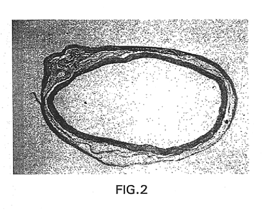
[0068]Following pressure perfusion with McDowells-Trump fixative, the grafts were explanted, and submitted for light and electron microscopy. Specimens from 30, 60, 90, 120, and 180 day implants were available. Materials were examined with H / E, VonGieson elastica, Masson's Trichrome, g-Actin, Factor VIII, and Ram-11 (macrophage) stains, and polarized microscopy. Qualitative morphometric comparisons were made to stained non-implanted retention samples.

[0069]Histological evaluations demonstrated that the graft was readily invaded by host cells. The luminal collagen was resorbed and remodeled with the production o...

example 3

Comparison of Three-Layer Prosthesis and e-PTFE

[0076]Both two-layer and three-layer small diameter prosthesis were implanted and evaluated over time for patency and remodeling.

[0077]FIGS. 4-9 show the results of a comparison of three-layer prothesis with a similarly configured contra-lateral reference material, e-PTFE, in a canine femoral artery study. The grafts were implanted in canines as femoral interposition prosthesis. Grafts were explanted from 30 to 256 days.

[0078]Histological evaluation of the three-layer collagen graft demonstrated cellular ingrowth into the graft at 30 days, with more than 90 percent of the graft collagen remodeled by 90 days; and a mature ‘neo-artery’ at 180 days. Host tissue bridged the anastomosis by 60 days with the anastomosis only demarcated by the non-resorbable sutures. The predominant cell type in the neo-artery was a positive g-Actin staining smooth muscle like cell. The surface of the remodeled graft was lined by endothelial cells as demonstrat...


PUM

PropertyMeasurementUnit
Thicknessaaaaaaaaaa
Thicknessaaaaaaaaaa
Diameteraaaaaaaaaa

Abstract

A bioremodelable prosthesis for treating a patient with a diseased or damaged organs comprising a first layer that contains acid-extracted fibrillar or non-fibrillar collagen, and a second layer derived from the tunica submucosa of the small intestine that provides structural stability, is pliable and is semi-permeable,pe1 59564443.npc wherein the prosthesis undergoes controlled biodegradation occurring with adequate living cell replacement such that the original prosthesis is replaced by the patient's living cells.

Description

CROSS REFERENCE TO RELATED APPLICATIONS[0001]This application is a continuation of U.S. patent application Ser. No. 08 / 889,079, filed Jul. 7, 1997 now U.S. Pat. No. 6,334,872, which is a continuation-in-part of U.S. patent application Ser. No. 08 / 461,756, filed Jun. 5, 1995, now abandoned, which is a continuation of U.S. patent application Ser. No. 08 / 198,062, filed Feb. 18, 1994, now abandoned; each of which is in its entirety hereby incorporated by reference.FIELD OF THE INVENTION[0002]This invention is in the field of tissue engineering. The present invention is a resilient, biocompatible two- or three-layered tissue graft which can be engineered in flat sheets or in tubes with various luminal diameters and thicknesses. At least one layer is composed of collagen or a collagenous material. The present invention is gradually degraded and bioremodeled by the host's cells which replace the implanted prosthesis and assume its shape.BACKGROUND OF THE INVENTION[0003]Each year approximat...

Claims


Application Information

Patent Timeline
IPC IPC(8): A61F2/06A61L27/58A61L27/00A61L27/24A61L27/34A61F2/00A61F2/02
CPCA61L27/34A61L27/58A61L27/24A61F2/06A61F2/02C08L89/06A61F2002/30062A61F2002/3024A61F2002/30971A61F2210/0004A61F2230/0069A61F2240/001Y10S623/917A61F2002/30235
Inventor TERMIN, PAUL L.CARR, JR., ROBERT M.CONDON, KIMBERLIE D.
Owner ORGANOGENESIS
Learn More