Method of detecting DNA single strand breaks
- Summary
- Abstract
- Description
- Claims
- Application Information
AI Technical Summary
Benefits of technology
Problems solved by technology
Method used
Image
Examples
example 1
Cell Culture
[0050]XRCC1-proficient and -deficient CHO cells (Taylor, et al. (2002) Mol. Cell. Biol. 22:2556-2563) were cultured as monolayers in alpha-minimal essential medium (INVITROGEN™, Carlsbad, Calif.) supplemented with 10% fetal bovine serum (Sigma-Aldrich, St. Louis, Mo.), 100 μg / mL penicillin, and 100 μg / mL streptomycin. Immortalized mouse embryonic fibroblasts derived from PART-1+ / + and PARP-1− / − were maintained in Dulbecco's modified Eagle's medium, 4.5 g / Liter glucose medium (INVITROGEN™, Carlsbad, Calif.) supplemented with 10% fetal bovine serum and 0.5% gentamicin (Schreiber, et al. (2002) J. Biol. Chem. 277:23028-23036). Human lymphoblastoid cells (GM15030, GM15061, GM15237, GM15268, GM15339, GM15349, GM15365, and GM15380; Coriell Cell Repositories, Camden, N.J.) were grown in RPMI 1640 medium (INVITROGEN™, Carlsbad, Calif.) supplemented with 15% fetal bovine serum (Sigma-Aldrich, St. Louis, Mo.), 100 μg / mL penicillin, and 100 μg / mL streptomycin. The cells were mainta...
example 2
Detection of Intracellular NAD(P)H
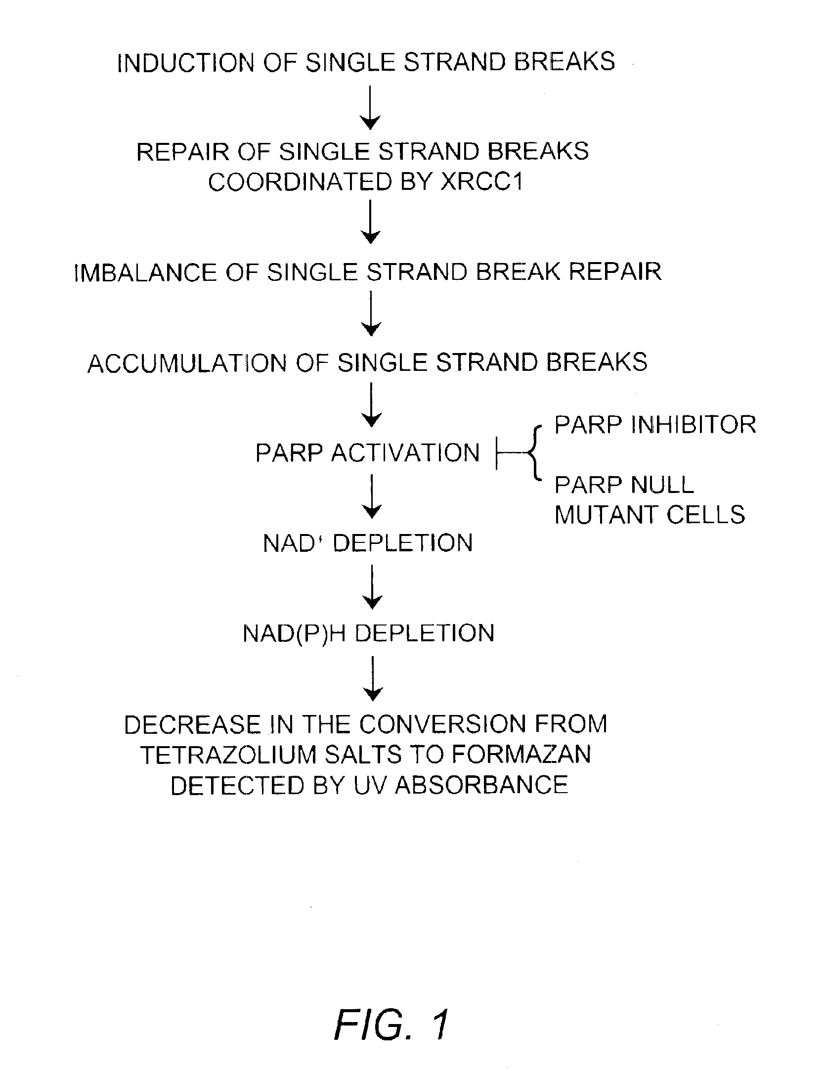
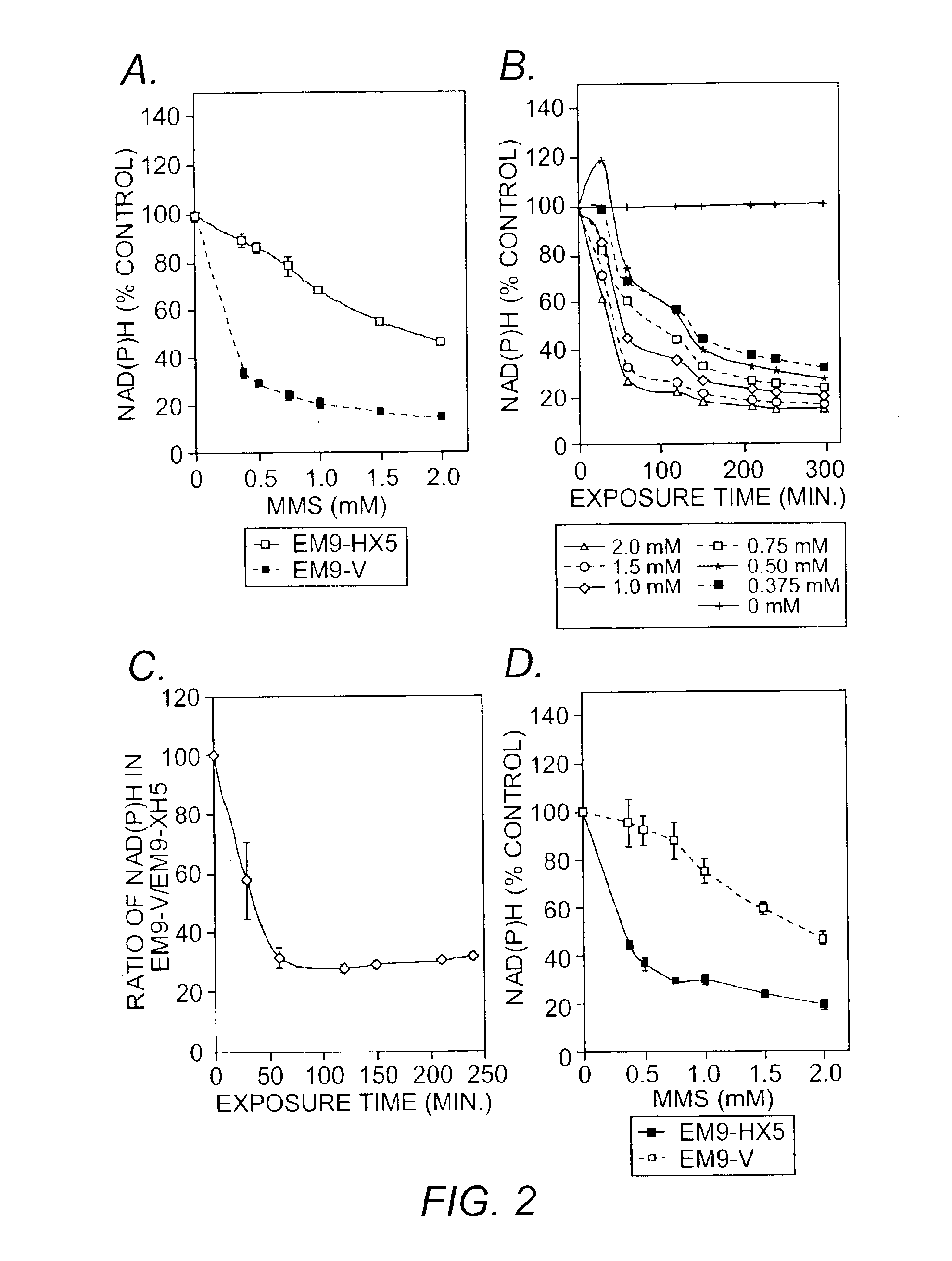
[0051]A water-soluble tetrazolium salt was used to measure the amount of intracellular NAD(P)H through the reduction to a formazan dye. The total amount of NAD(P)H within viable cells in the medium was determined periodically by a spectrophotometer. Cells were seeded into 96-well plates (CHO cells and mouse fibroblasts: 5×10³ cells / well; lymphoblastoid cells: 15×103 cells / well) and were cultured in 100 μL of medium with fetal bovine serum and antibiotics as described. After a 30-minute incubation, cells were treated with MMS at indicated concentrations and {fraction (1 / 10)} volume of CCK-8 solution (Dojindo Molecular Technology, Gaithersburg, Md.) which consisted of a water-soluble 2-(2-methoxy-4-nitrophenyl)-3-(4-nitrophenyl)-5-(2,4-disulfophenyl)-2H-tetrazolium monosodium salt (WST-8; 5 mM) and 1-Methoxy-5-methylphenazinium methylsulfate (1-Methoyx PMS; 0.2 mM) as an electron mediator. Cells in each well were further cultured for up to 4 hours. WS...
example 3
NAD(P)H Reduction in the Presence of PARP Inhibitors
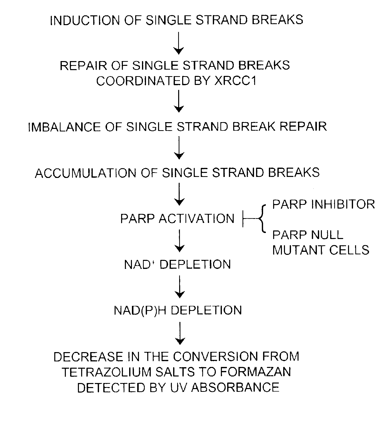
[0055]PARP transfers hundreds of branched chains of ADP-ribose to a variety of nuclear proteins through its activation by DNA single strand breaks (Kubota, et al. (1996) supra; Caldecott, et al. (1994) supra). Under massive DNA damage, activation of PARP depletes its substrate, NAD+ (Taylor, et al. (2002) supra). Since NAD(P)H are generated from NAD(P) by the reaction of dehydrogenase and its substrate, the decrease in the amount of NAD(P)H depletes NAD+. To determine whether the reduction in NADH was due to a reduction of mitochondrial function or due to the depletion of NAD+ by PARP activation, CHO cells were co-exposed to MMS and specific PARP inhibitors. Specific PARP inhibitors, 3-aminobenzamide (3-AB; Sigma-Aldrich, St. Louis, Mo.)(10 mM) and 3,4-dihydro-5-[4-(1-piperidinyl)butoxy]-1(2H)-isoquinolinone (DPQ, Sigma-Aldrich, St. Louis, Mo.)(90 μM) were applied 1 to 2 hours prior to the MMS treatment and kept in the medium durin...
PUM
| Property | Measurement | Unit |
|---|---|---|
| Time | aaaaa | aaaaa |
| Solubility (mass) | aaaaa | aaaaa |
| Absorbance | aaaaa | aaaaa |
Abstract
Description
Claims
Application Information
 - R&D
- Intellectual Property
- Life Sciences
- Materials
- Tech Scout
- Unparalleled Data Quality
- Higher Quality Content
- 60% Fewer Hallucinations
Browse by: Latest US Patents, China's latest patents, Technical Efficacy Thesaurus, Application Domain, Technology Topic, Popular Technical Reports.
© 2025 PatSnap. All rights reserved.Legal|Privacy policy|Modern Slavery Act Transparency Statement|Sitemap|About US| Contact US: help@patsnap.com


