Magnetic resonance angiography data

Inactive Publication Date: 2005-08-02
LANTHEUS MEDICAL IMAGING INC
View PDF35 Cites 12 Cited by
  • Summary
  • Abstract
  • Description
  • Claims
  • Application Information

AI Technical Summary

Benefits of technology

[0009]The invention provides a method of visualizing MRI data taken from two data sets, one set that can comprising high spatial resolution anatomical image data, and the other that can comprise physiological data which is not necessarily of as high a resolution as the other data set. The method then combines the two data sets to produce a clinically useful MR image. The methods disclo

Problems solved by technology
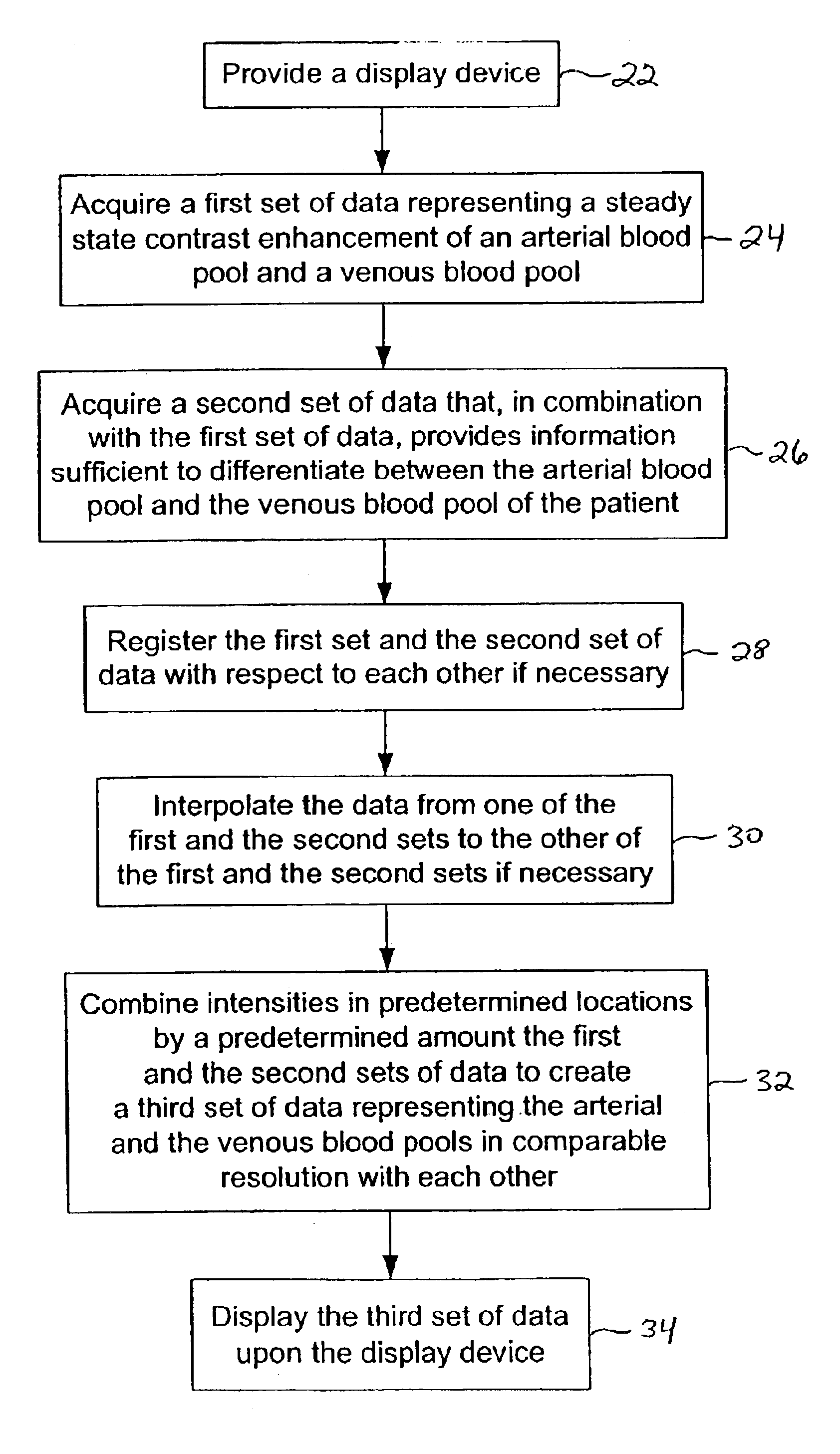
Cardiovascular disease is a primary health threat.
Until intravascular agents were introduced, MRA image acquisition was typically limited to the first pass of the contrast bolus, much like XRA, due to the extensive leakage of the contrast agent into the extracellular fluid.
This longer persistance in the blood, however, introduces a problem in visualization.
Because of simultaneous enhancement of both the arterial and venous blood pools, the vessels obscure each other when using projection methods (such as maximum intensity projection (MIP)), and can potentially confuse the reader even when viewing the source data.
Each of these methods, however, disadvantageously remove a significant amount of potentially useful information and/or have reliability issues.
Additionally, reliance on capturing a contrast bol

Method used


Image

  • Magnetic resonance angiography data
  • Magnetic resonance angiography data
  • Magnetic resonance angiography data

Examples


Example

EXAMPLE

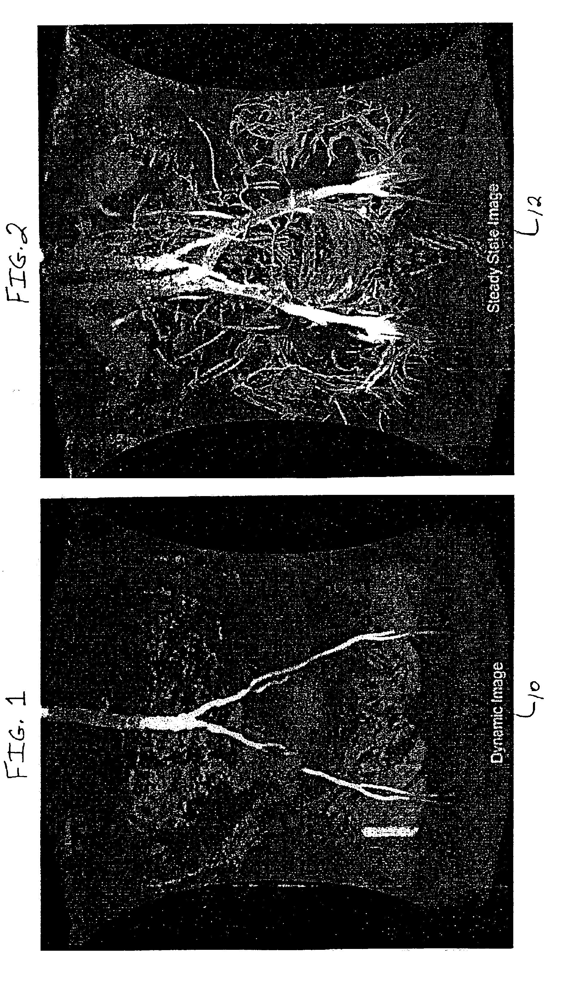
[0053]An adult human subject was administered MS-325 by intravenous injection and a series of dynamic MRA scans were collected of the thorax during the first pass of contrast agent with the following imaging parameters:[0054]Instrument: 1.5 T GE Medical Systems[0055]Pulse sequence: T1-weighted SPGR, TE=1.8, TR=8.7, flip angle=40[0056]Resolution: 1.8×1.8×3.6 mm[0057]Dimensions: 512×192×52[0058]Field of View: 440×330 mm

An MIP of this data set is presented in FIG. 1. During the steady state the patient was imaged again to generate a second data set with the following modifications to the above parameters:[0059]Pulse sequence: T1-weighted SPGR, TE=2.1, TR=18.7, flip angle=30[0060]Resolution: 0.9×0.9×1.8 mm[0061]Dimensions: 512×512×128

An MIP of this data set is presented in FIG. 2. The data set from the dynamic scan was interpolated up to the resolution of the steady state scan and the two were combined according to equation I (vide supra), with α=0.75 and β=0.25, by the computer ...


PUM


Abstract

A method is provided for improved visualization of vascular structures contained within magnetic resonance angiograms, particularly steady-state contrast-enhanced magnetic resonance angiograms. The disclosed methods include manipulating the relative enhancement levels of the arterial and venous blood pools images with respect to each other and to the surrounding anatomy. This manipulation is meant to optimize visualization of one or both of the individual blood pools without the necessity of completely obscuring or removing the supporting information given by presentation of the other blood pool and/or the surrounding anatomy.

Description

[0001]This application is a continuation of U.S. Ser. No. 09 / 778,585, filed on Feb. 7, 2001, now U.S. Pat. No. 6,549,798, which issued on Apr. 15, 2003, the entirety of which is incorporated herein by reference.FIELD OF THE INVENTION[0002]The application relates generally to the use of magnetic resonance imaging (MRI) data. More specifically, the application relates to the improved visualization of steady-state contrast-enhanced MRI anatomical data with superimposed physiologic data. Most specifically, the application relates to improving the visualization of the arterial and venous blood pools in magnetic resonance angiography (MRA).BACKGROUND OF THE INVENTION[0003]Cardiovascular disease is a primary health threat. Therefore, it is essential to detect cardiovascular disease in its early stages. One manner to detect cardiovascular disease is through the use of magnetic resonance angiography (MRA). The primary goal of MRA is to non-invasively acquire necessary clinical information vi...

Claims


Application Information

Patent Timeline
IPC IPC(8): G01R33/563G01R33/54G01R33/48A61B5/055G06T5/50G06T7/00
CPCG01R33/563G06T5/50G06T7/38G06T5/008A61B5/055G06T2207/10088G06T2207/30061G06T2207/30101G06T2207/30048
Inventor STEFANCIK, ROBERT M.EARNEST, RANDY J.WEISSKOFF, ROBERT M.
Owner LANTHEUS MEDICAL IMAGING INC
Why Eureka
Try Eureka