Method for binding basophils and mast cells

a basophil and mast cell technology, applied in the field of basophil and mast cell binding methods, can solve the problems of not being able to recognize basophils and mast cells simultaneously, and no antibody is known to date, and achieve the effects of facilitating analysis of basophil differentiation, reducing the number of mast cells, and increasing sample throughpu

Inactive Publication Date: 2005-09-27
UNIV TUBINGEN
View PDF9 Cites 8 Cited by
  • Summary
  • Abstract
  • Description
  • Claims
  • Application Information

AI Technical Summary

Benefits of technology

[0028]Using antibody 97A6, it is readily possible to isolate the surface structure to which the antibody binds, and to produce both monoclonal and polyclonal antibodies against it.
[0069]The advantage of such a reagent is that cells bound to it can be detected and selected. A further advantage of such a reagent is that it can also contain, in addition to an antibody according to the present invention, further constituents that allow the detection of bound antibodies or the selection of antibody-bound cells. These can be, for example, fluorescent-labeled antibodies or antibodies coupled to magnetic beads, which bind to the antibody according to the present invention.

Problems solved by technology

No antibody is hitherto known, however, to simultaneously recognize basophils, mast cells, as well as their precursor cells.

Method used


Image

  • Method for binding basophils and mast cells
  • Method for binding basophils and mast cells
  • Method for binding basophils and mast cells

Examples


example 1

[0076]Isolation and detection of basophils from peripheral blood of normal subjects and leukemia patients

[0077]It is known that the number of basophils in the peripheral blood of patients with CML rises with the transition from the chronic to the accelerated phase.

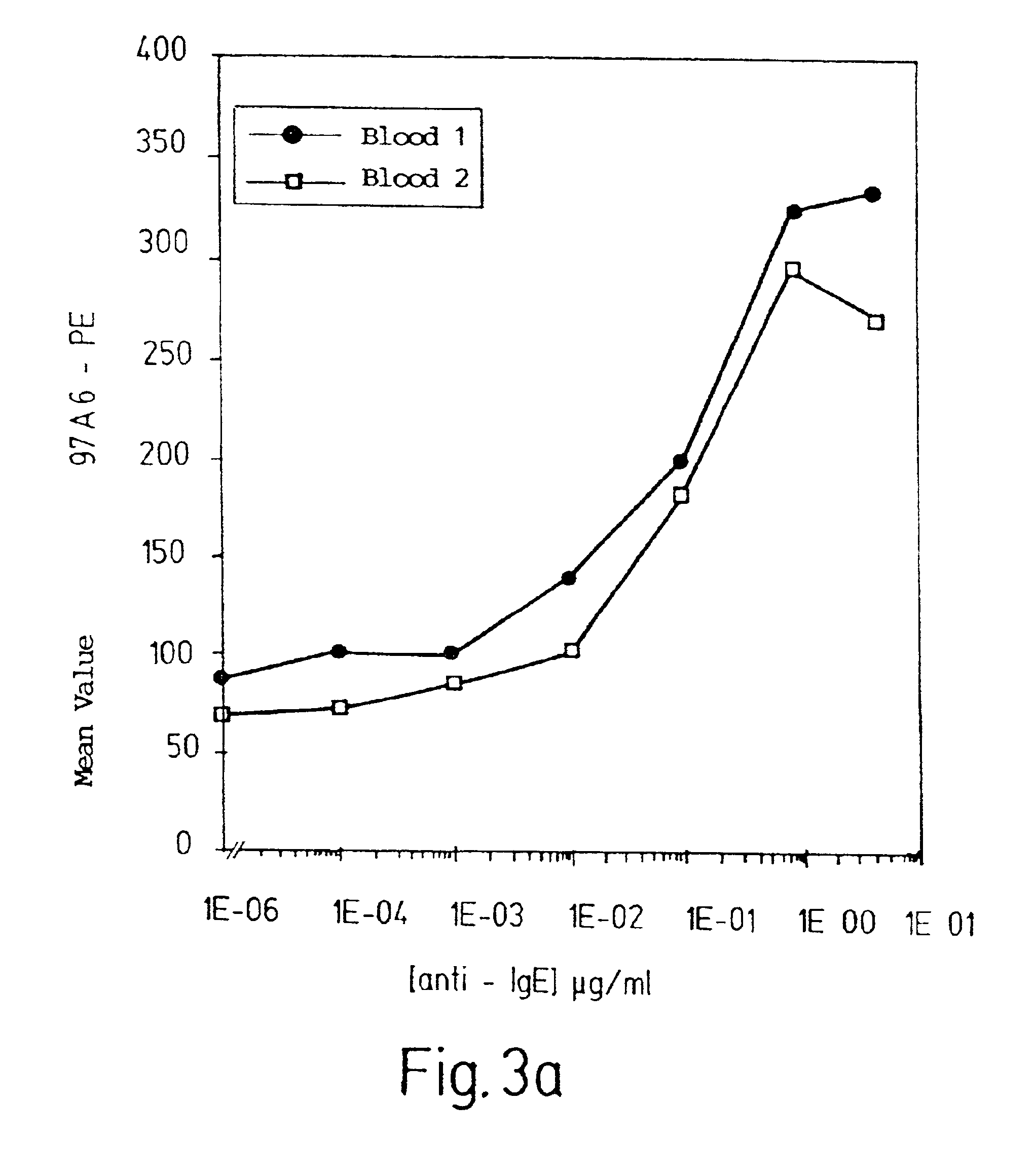
[0078]In order to test the specificity of antibody 97A6 for basophils, mononuclear leukocytes were separated, using Ficoll Hypaque, from other constituents of Buffy Coats from the peripheral blood of normal subjects or patients with CML in the acceleration phase. The interphase cells were then incubated with biotin-labeled antibody 97A6, washed three times, and incubated with anti-IgG MACSbeads (Miltenyi, Bergisch Gladbach, Germany). 97A6-positive cells were then isolated by MACS and then, for preparation monitoring purposes, labeled with phycoerythrin-labeled (PE-labeled) streptavidin and a PE-labeled 97A6 antibody, and analyzed in a flow cytometer.

[0079]The purity of the 97A6-positive cells was 99%.

[0080]For morphologica...

example 2

[0082]Detection of Tissue Mast Cells Using Antibody 97A6

[0083]Lung, foreskin, or uterine tissue was minced, washed in a buffer with 200 mg / l KCl, 50 mg / l NaH2PO4×H2O, 8 g / l NaCl, and 1 g / l glucose, and then incubated for 90 to 180 minutes at 37 ° C. with 2 mg / ml collagenase type II (Sebak, Suben, Austria). The dispersed cells were then centrifuged, washed, and incubated for 30 minutes with human AB serum. After being washed again, the cells were incubated for 30 minutes at 4° C. with antibody 97A6, washed, and then incubated for 30 minutes at 4° C. with a fluorescein-labeled goat F(ab′)2 IgG anti-mouse antibody. The cells were fixed for 1 minute in 0.025% glutaraldehyde, washed, and incubated for 8-12 minutes with 0.0125% toluidine blue. After washing, toluidine blue-stained mast cells were identified by their morphology using a fluorescence microscope (Olympus, Vienna, Austria) in brightfield mode, and then examined by fluorescent light.

[0084]It was found that only mast cells from ...

example 3

Isolation and Cultivation of Precursor Cells

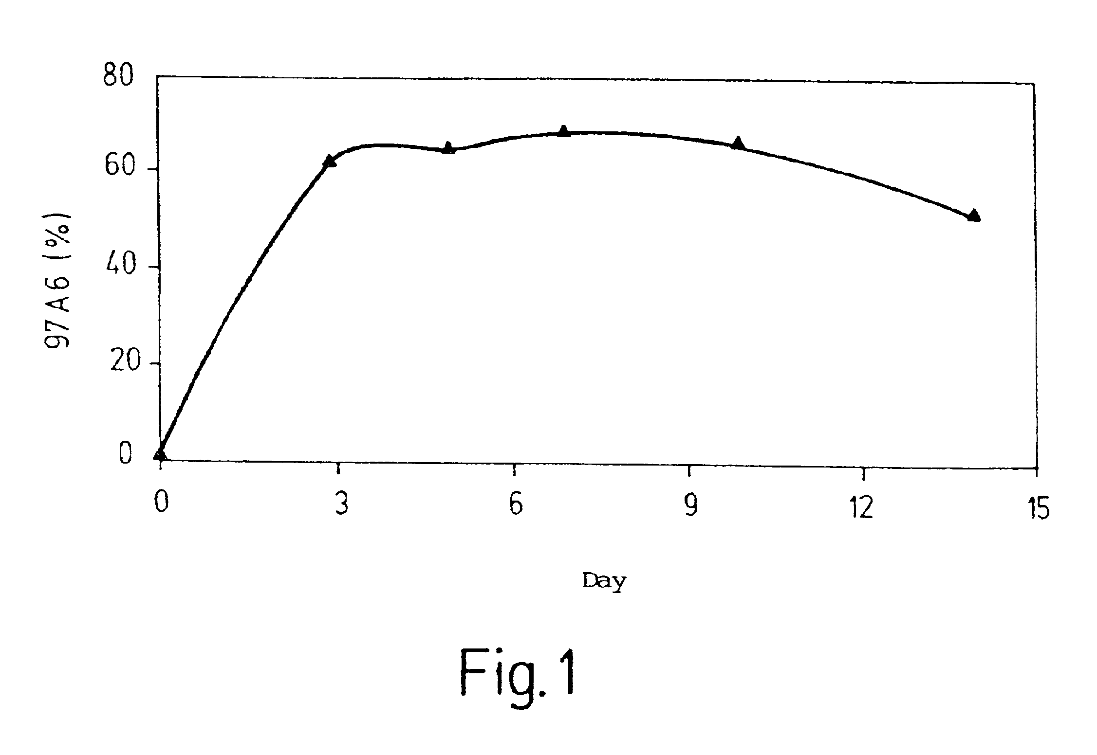
[0085]Cells from bone marrow samples were centrifuged on Ficoll Hypaque that had a density of 1.077 g / cm3. The interphase cells were isolated, and cells that were positive for CD34 (a marker for hematopoietic precursor cells) were isolated using MACS. These cells were incubated with a FITC-labeled antibody against CD34 and with PE-labeled antibody 97A6. Doubly positive cells were selected by FACS and seeded in a semi-solid ready-made medium containing 0.9% methyl cellulose and a mixture of growth factors including IL-3 (a differentiation factor for basophils and eosinophils) (CellSystems, Remagen, Germany).

[0086]After incubation for 16 days at 37° C. in a humidified atmosphere (5% CO2), the resulting colonies were counted, transferred with a Pasteur pipette onto microscope slides, and stained with May-Grünwald-Giesma for morphological examination. In addition to the predominating pure basophil colonies, this examination also revealed mixed...


PUM

PropertyMeasurementUnit
densityaaaaaaaaaa
concentrationaaaaaaaaaa
fluorescentaaaaaaaaaa

Abstract

The present invention relates to an antibody for the detection quantification, or isolation of basophils, mast cells, the precursor cells of basophils or mast cells, or a surface structure of basophils or mast cells. This antibody corresponds to an antibody with the designation 97A6, produced and released by hybridoma cells that were deposited in accordance with the Budapest Treaty on Feb. 12, 1997 under accession number DSM ACC 2297 at Deutsche Sammlung von Mikroorganismen und Zellkulturen GmbH (DSMZ).

Description

RELATED APPLICATION[0001]This is a continuation of International Patent Application PCT / EP00 / 04468, filed May 17, 2000 and published in German, which claims priority to German application number 19922863.9, filed May 19, 1999, and German application number 19926879.7, filed Jun. 12, 1999. The German applications are incorporated herein by reference.BACKGROUND OF THE INVENTION[0002]1. Field of the Invention[0003]The present invention relates to the use of an antibody for the detection and / or quantification and / or isolation of basophils and / or mast cells and / or of the precursor cells of basophils and / or mast cells and / or of a surface structure of said cells.[0004]The invention further concerns[0005]a method for studying allergies;[0006]a method for providing hematopoietic precursor cells that can differentiate into mast cells or basophils;[0007]a substantially pure population of basophils and / or mast cells and / or of the precursor cells of basophils and / or mast cells; as well as[0008]a...

Claims


Application Information

Patent Timeline
Patent Type & Authority Patents(United States)
IPC IPC(8): C07K14/435C07K16/18C07K16/28C07K14/705G01N33/569G01N33/53C12N5/07C12N5/0789G01N33/577
CPCC07K14/705C07K16/28G01N33/56972
Inventor BUHRING, HANS-JORGVAN AGTHOVEN, JOHANNES ANDREASJAROSSAY, DAVID
Owner UNIV TUBINGEN
Why Eureka
Try Eureka