Method and apparatus for detecting an abnormality within a host medium utilizing frequency-swept modulation diffusion tomography

a technology of diffusion tomography and frequency-swept modulation, which is applied in the field of detection methods and apparatus, can solve the problems of insufficient quality of images obtained by x-ray mammography techniques to detect masses in the patient's breast, high cost of magnetic resonance imaging machines, and inability to meet the needs of patients, so as to facilitate breast imaging, eliminate deleterious edge effects, and improve image quality

Inactive Publication Date: 2006-02-28
MEDICAL OPTICAL IMAGING
View PDF99 Cites 50 Cited by
  • Summary
  • Abstract
  • Description
  • Claims
  • Application Information

AI Technical Summary

Benefits of technology

[0020]In order to move the pair of plates in order to appropriately compress the patient's breast, the apparatus of the present invention also includes means for moving the plates relative to one another. In addition, the apparatus can include a separation detector for measuring the distance by which the pair of plates are separated. The apparatus of this embodiment also includes an adjustable belt extending between the plates proximate the breast. The adjustable belt is capable of being tightened about the breast such that the breast fills a region defined by the pair of plates and the adjustable belt, thereby facilitating imaging of the breast.
[0021]Preferably, the apparatus of this embodiment also includes an opaque material that fills a region defined by the plates that is unfilled by the breast. In this regard, the adjustable belt can be operably connected to the opaque material, such as by extending through the opaque material, such that the opaque material is drawn about the breast as the adjustable belt is tightened. In order to identify any regions of separation between the opaque material and the breast, the apparatus of this embodiment can also include a background light to specifically illuminate the regions of separation. By tightening the belt about the breast such that the breast substantially fills the region defined by the pair of plates and the adjustable belt and also covering those portions of the plate that are unfilled by the breast with an opaque material, the apparatus and method of this embodiment of the present invention can obtain an improved image of the breast by substantially eliminating deleterious edge effects.
[0022]Accordingly, the method and apparatus of the present invention permits abnormalities to be detected within a host medium in a reliable and cost effective manner. In addition, the method and apparatus of the present invention permits physiological parameters that at least partially define the abnormality to be determined in order to characterize the abnormality. As such, the method and apparatus of the present invention can be advantageously utilized to provide early detection of suspicious lesions within a patient's breast and to appropriately characterize the suspicious lesions, such as being either malignant or benign, based upon physiological parameters that are determined from the detected signals without requiring a biopsy or other invasive procedure. By facilitating early detection and characterization of suspicious lesions within the patient's breast, the patient's chances of survival are substantially increased.

Problems solved by technology

Unfortunately, x-ray mammography exposes the patient to ionizing radiation which is a known cause of cancer.
X-ray mammography also generally requires that the patient's breast be greatly compressed, such as to 4 centimeters, which can be quite painful.
In addition, the images obtained by x-ray mammography techniques are not always of a sufficiently high quality to detect masses in the patient's breast, particularly for patients having radiodense breast tissue.
Unfortunately, each of these imaging techniques suffers from a number of shortcomings.
For example, while magnetic resonance imaging generally provides acceptable images, a magnetic resonance imaging machine is extremely expensive and is therefore not commonly utilized for breast cancer diagnosis.
Unfortunately, the strongly diffusive nature of light propagation in breast tissue significantly reduces the contrast and resolution of the optical images obtained by most optical imaging techniques.
As such, optical imaging techniques have had difficulty consistently detecting and characterizing suspicious lesions, such as tumors, that are relatively small and / or deep within the breast.
Unfortunately, continuous wave transillumination techniques generally suffered from relatively low sensitivity and / or a relatively high number of false positive results.

Method used


Image

  • Method and apparatus for detecting an abnormality within a host medium utilizing frequency-swept modulation diffusion tomography
  • Method and apparatus for detecting an abnormality within a host medium utilizing frequency-swept modulation diffusion tomography
  • Method and apparatus for detecting an abnormality within a host medium utilizing frequency-swept modulation diffusion tomography

Examples


Embodiment Construction

[0029]The present invention now will be described more fully hereinafter with reference to the accompanying drawings, in which preferred embodiments of the invention are shown. This invention may, however, be embodied in many different forms and should not be construed as limited to the embodiments set forth herein; rather, these embodiments are provided so that this disclosure will be thorough and complete, and will fully convey the scope of the invention to those skilled in the art. Like numbers refer to like elements throughout.

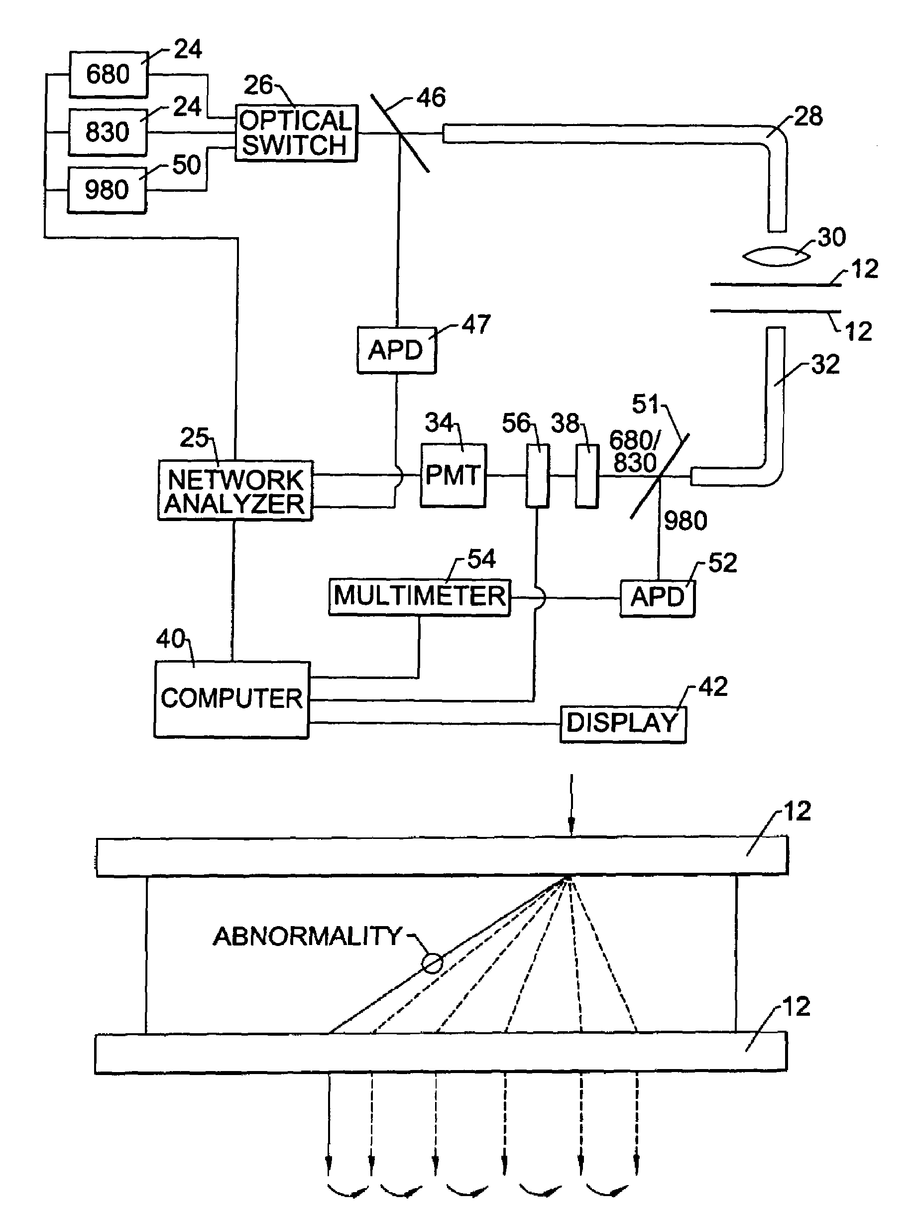
[0030]A method and apparatus for detecting an abnormality in a host medium will now be described in conjunction with the present invention. The method and apparatus of the present invention will be primarily described in conjunction with the detection of a tumor and, more particularly, the detection of a tumor or other mass in a patient's breast. However, the method and apparatus of the present invention can also be employed to detect abnormalities in a nu...


PUM

PropertyMeasurementUnit
wavelengthaaaaaaaaaa
poweraaaaaaaaaa

Abstract

Methods and apparatus are provided for detecting an abnormality in a host medium, such as a tumor within a patient's breast. The host medium is initially illuminated at a plurality of different positions with light of at least two different wavelengths. Following the propagation of the signals through the host medium, the amplitude and phase of the signals are detected and, based upon the detected signals, a shadow image can be created in which the abnormality is depicted as a suspicious region. Once a suspicious region has been identified, at least that portion of the host medium that contains the suspicious region is again illuminated with frequency-swept modulated signals of at least two different wavelengths generated by the light source and modulated by a modulator, such as a network analyzer. Based upon the detected signals that have propagated through at least that portion of the host medium that includes the suspicious region, a P-criteria can be determined that is dependent upon the coefficient of absorptivity of the host medium and embedded abnormalities. Likewise, an Svar-criteria can be determined based upon concentrations of oxygenated hemoglobin and deoxygenated hemoglobin. Based upon at least these criteria which reflect physiological parameters of the host medium and embedded abnormalities as well as the possible comparison of the shadow images to an x-ray image of the host medium, the abnormality can be characterized. In addition, an apparatus is provided that facilitates the compression of a patient's breast in order to improve the resulting images.

Description

RELATED APPLICATIONS[0001]The present application claims priority from U.S. Provisional Application No. 60 / 177,335 filed Jan. 21, 2000, the contents of which are incorporated herein.GOVERNMENT LICENSE RIGHTS[0002]The U.S. Government may have rights in this invention as provided for by the terms of SBIR Grant No. DMS-9704923 awarded by the National Science Foundation.FIELD OF THE INVENTION[0003]The present invention relates generally to detection methods and apparatus and, more particularly, to methods and apparatus for detecting an abnormality within a host medium.BACKGROUND OF THE INVENTION[0004]Imaging systems are widely utilized to construct an image or model of a structure which is otherwise unobservable to the eye. Typically, imaging systems are designed to detect abnormalities, foreign objects or other structures which are embedded within a host medium and which alter or perturb the signal propagation properties of the host medium. For example, x-ray tomography and other medic...

Claims


Application Information

Patent Timeline
Patent Type & Authority Patents(United States)
IPC IPC(8): G06K9/00A61B1/06H01S3/10G01N21/17A61B5/00A61B5/145A61B5/1455A61B6/00A61B10/00G01N21/35G01N21/47G06T1/00
CPCA61B5/0091G01N21/4795A61B5/4312
Inventor ZEYLIKOVICH, IOSIF SEMENKLIBANOV, MICHAEL VICTORKHARISOV, ANDREY ANATOLYEVICH
Owner MEDICAL OPTICAL IMAGING
Why Eureka
Try Eureka