Near-infrared spectroscopic tissue imaging for medical applications

a tissue imaging and near-infrared technology, applied in the field of medical diagnostics, can solve the problems of short photon penetration depth in the tissue, time-consuming and bulky diagnostic equipment, and extraction of information, and achieve the effect of cost-effectiveness

Inactive Publication Date: 2006-03-21
LAWRENCE LIVERMORE NAT SECURITY LLC
View PDF15 Cites 108 Cited by
  • Summary
  • Abstract
  • Description
  • Claims
  • Application Information

AI Technical Summary

Benefits of technology

[0010]Accordingly, the present tissue imaging system and method provides a portable, cost effective, non-invasive arrangement, capable of directing substantially monochromatic electromagnetic radiation and capable of providing spectrally processed images in the much desired need for differentiating components in human and / or animal tissues.

Problems solved by technology

Diagnostic medical equipment typically includes time-consuming, bulky, expensive apparatus that often exposes human tissue components to potentially harmful radiation and or chemicals.
The disadvantage of these wavelengths is their short photon penetration depth in tissues, which leads to extraction of information only from superficial tissue layers.

Method used


Image

  • Near-infrared spectroscopic tissue imaging for medical applications
  • Near-infrared spectroscopic tissue imaging for medical applications
  • Near-infrared spectroscopic tissue imaging for medical applications

Examples


Embodiment Construction

[0017]Referring now to the drawings, specific embodiments of the invention are shown. The detailed description of the specific embodiments, together with the general description of the invention, serves to explain the principles of the invention.

General Description

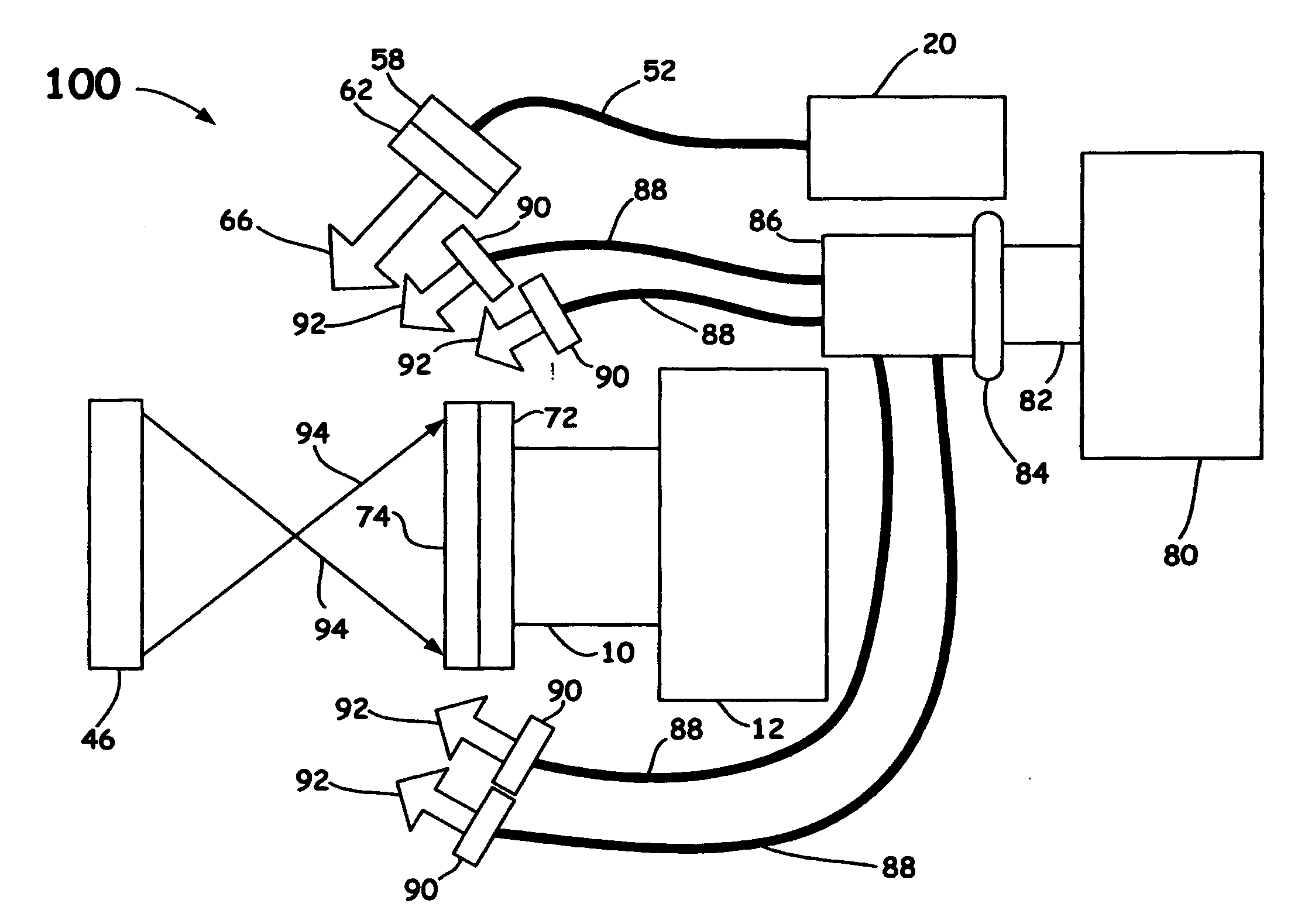
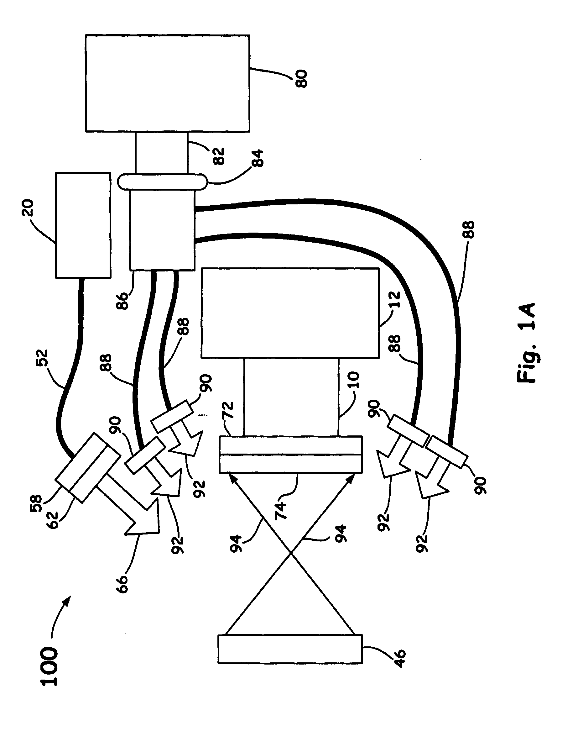
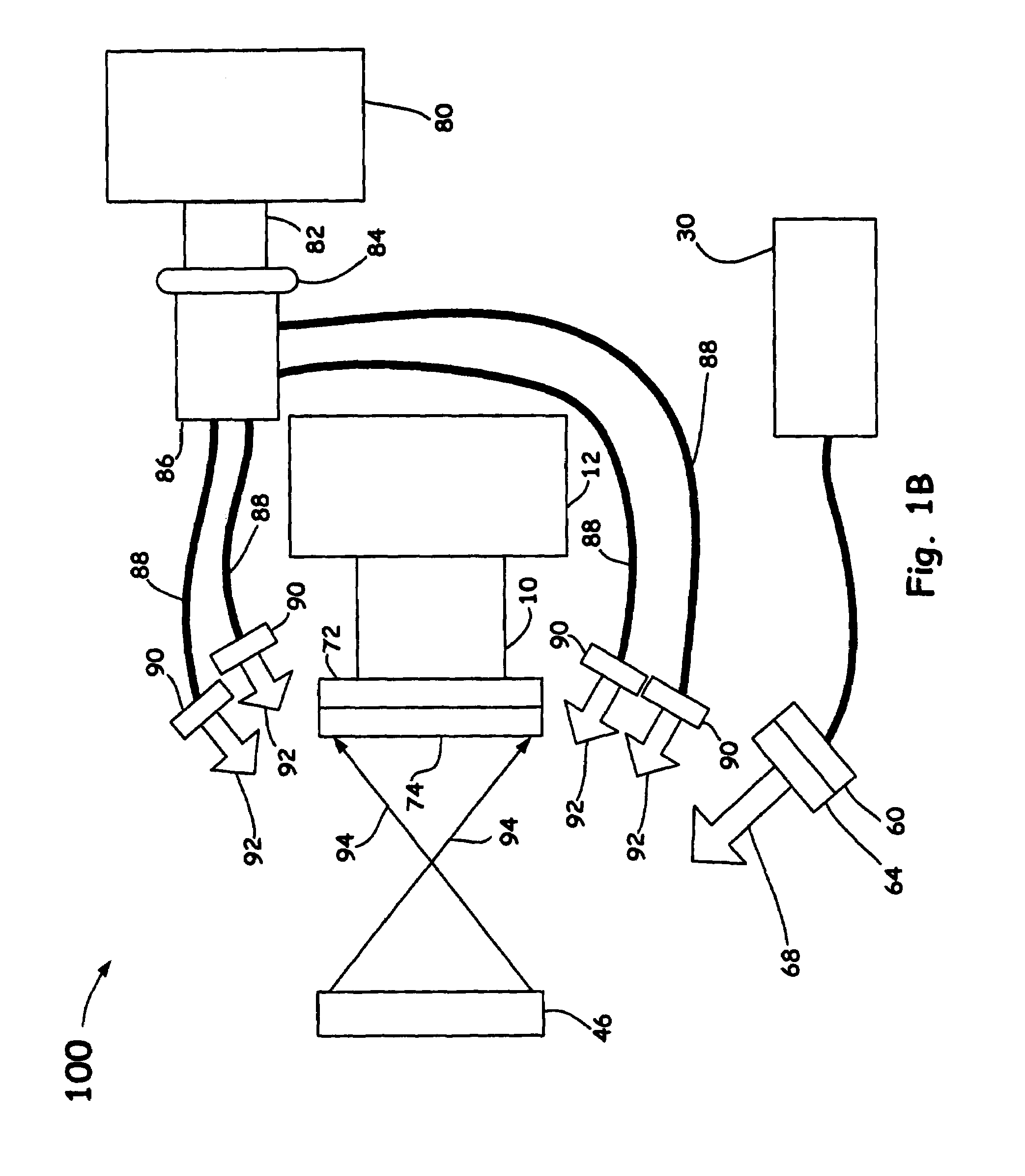
[0018]The present invention combines monochromatic laser sources, a broadband light source, optical filtering, a computer, optical imaging, and computer software capable of image analysis that includes inter-image operations. A useful feature of the present invention is that fresh surgical resections collected from patients may be measured in-vitro (e.g., in an artificial environment) and in-vivo (e.g., during medical biopsy or intervention procedures) immediately upon collection. In addition, the system has particular utility as a tissue component interrogation tool for human tissue specimens such as but not limited to kidney, uterine, bladder, breast, liver, adipose, abnormal (i.e., contrary to normal structure), normal,...


PUM

PropertyMeasurementUnit
wavelengthaaaaaaaaaa
wavelengthsaaaaaaaaaa
output poweraaaaaaaaaa

Abstract

Near infrared imaging using elastic light scattering and tissue autofluorescence are explored for medical applications. The approach involves imaging using cross-polarized elastic light scattering and tissue autofluorescence in the Near Infra-Red (NIR) coupled with image processing and inter-image operations to differentiate human tissue components.

Description

[0001]The United States Government has rights in this invention pursuant to Contract No. W-7405-ENG-48 between the United States Department of Energy and the University of California for the operation of Lawrence Livermore National Laboratory.BACKGROUND OF THE INVENTION[0002]1. Field of the Invention[0003]The present invention relates to a medical diagnostic for the examination of tissue components. Specifically, the present invention relates to an apparatus and a method of polarized elastic light scattering, Near-infra-Red (NIR) autofluorescence emission combined with image processing and inter-image operations as an interrogation means for investigating tissue components for medical applications.[0004]2. Description of Related Art[0005]Diagnostic medical equipment typically includes time-consuming, bulky, expensive apparatus that often exposes human tissue components to potentially harmful radiation and or chemicals. Optical methods and systems for the identification of objects th...

Claims


Application Information

Patent Timeline
Patent Type & Authority Patents(United States)
IPC IPC(8): A61B6/00G01N21/49G01N21/64
CPCA61B1/043A61B1/0638A61B1/307A61B5/0071A61B1/0646A61B5/0086G01N21/49G01N21/6445A61B1/063A61B5/0075A61B1/313
Inventor DEMOS, STAVROSSTAGGS, MICHAEL C.
Owner LAWRENCE LIVERMORE NAT SECURITY LLC
Why Eureka
Try Eureka